. 2020 Oct 15;21(20):7613.
doi: 10.3390/ijms21207613.

Establishment of Acquired Cisplatin Resistance in Ovarian Cancer Cell Lines Characterized by Enriched Metastatic Properties with Increased Twist Expression

Affiliations

Establishment of Acquired Cisplatin Resistance in Ovarian Cancer Cell Lines Characterized by Enriched Metastatic Properties with Increased Twist Expression

Entaz Bahar et al. Int J Mol Sci. .

Abstract

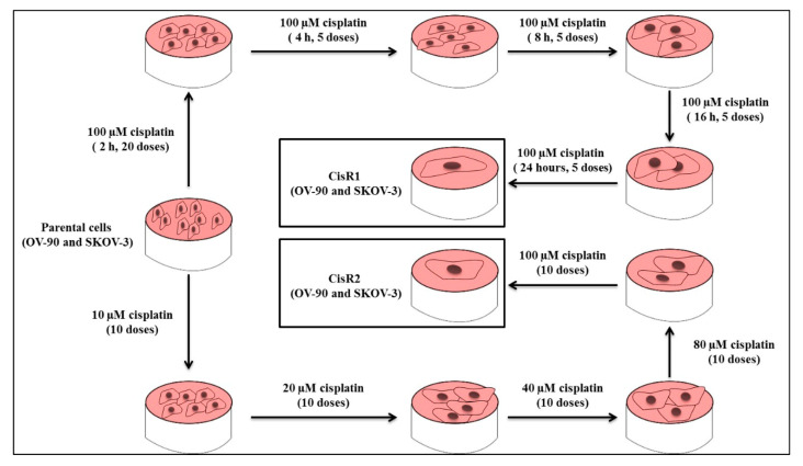
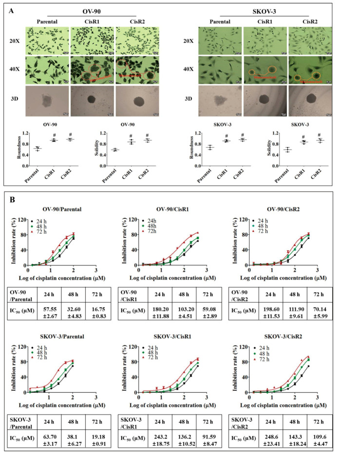
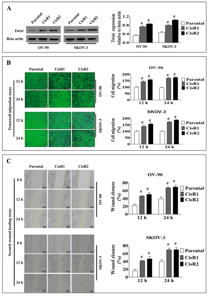
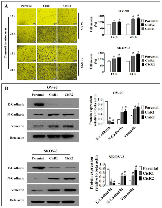
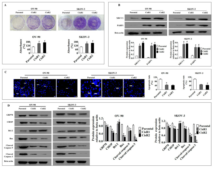
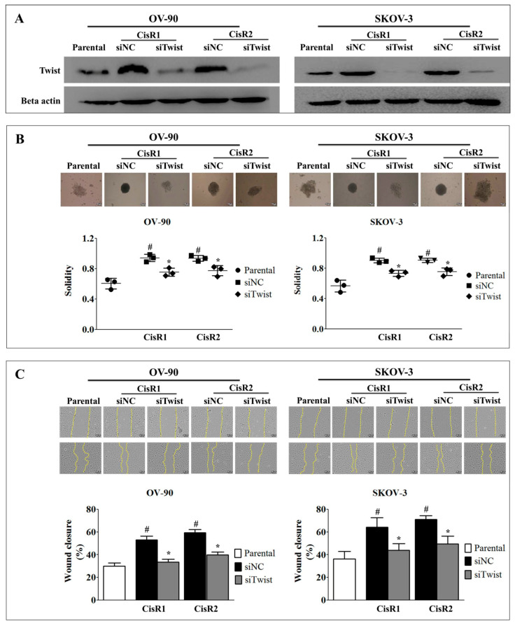
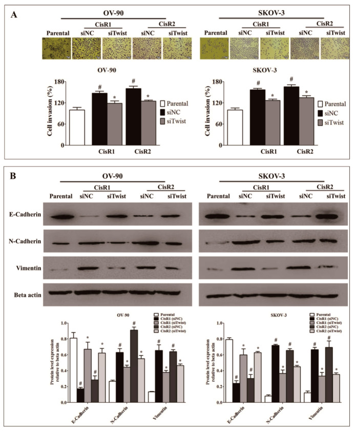
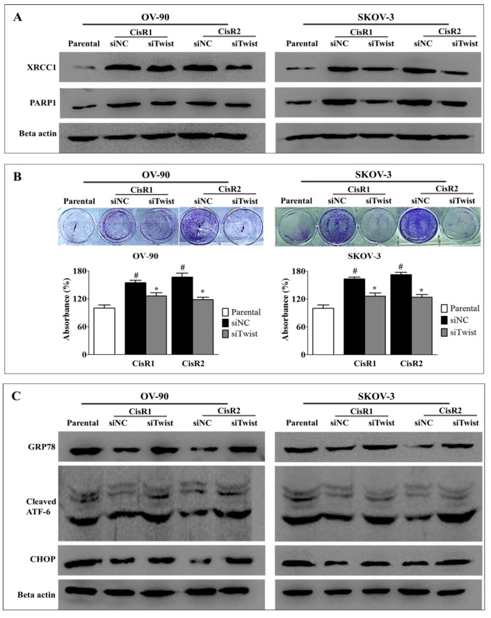
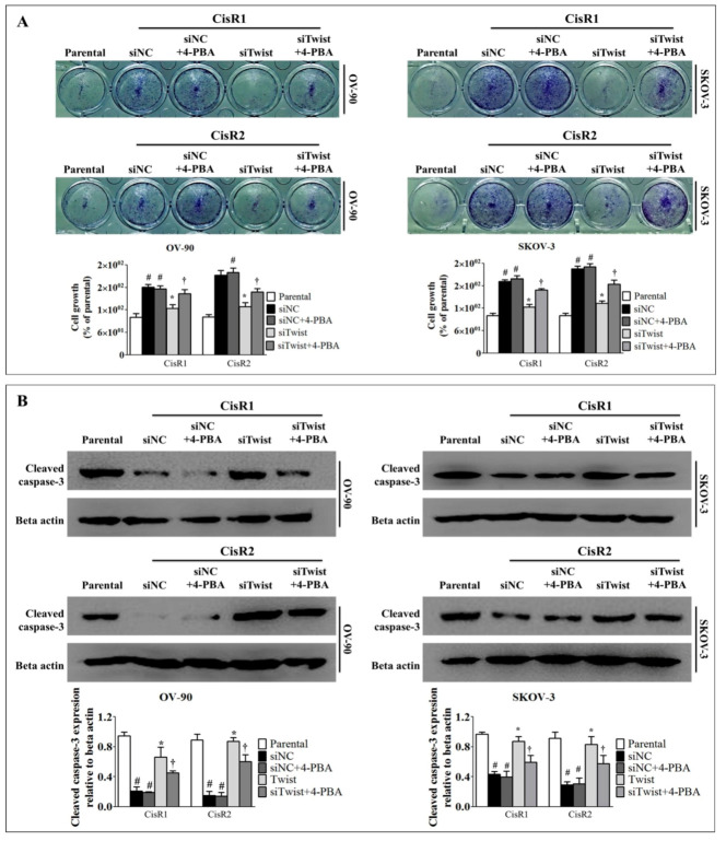
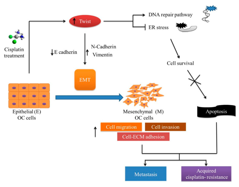
 Ovarian cancer (OC) is the most lethal of the gynecologic cancers, and platinum-based treatment is a part of the standard first-line chemotherapy regimen. However, rapid development of acquired cisplatin resistance remains the main cause of treatment failure, and the underlying mechanism of resistance in OC treatment remains poorly understood. Faced with this problem, our aim in this study was to generate cisplatin-resistant (CisR) OC cell models in vitro and investigate the role of epithelial-mesenchymal transition (EMT) transcription factor Twist on acquired cisplatin resistance in OC cell models. To achieve this aim, OC cell lines OV-90 and SKOV-3 were exposed to cisplatin using pulse dosing and stepwise dose escalation methods for a duration of eight months, and a total of four CisR sublines were generated, two for each cell line. The acquired cisplatin resistance was confirmed by determination of 50% inhibitory concentration (IC50) and clonogenic survival assay. Furthermore, the CisR cells were studied to assess their respective characteristics of metastasis, EMT phenotype, DNA repair and endoplasmic reticulum stress-mediated cell death. We found the IC50 of CisR cells to cisplatin was 3-5 times higher than parental cells. The expression of Twist and metastatic ability of CisR cells were significantly greater than those of sensitive cells. The CisR cells displayed an EMT phenotype with decreased epithelial cell marker E-cadherin and increased mesenchymal proteins N-cadherin and vimentin. We observed that CisR cells showed significantly higher expression of DNA repair proteins, X-ray repair cross-complementing protein 1 (XRCC1) and poly (ADP-ribose) polymerases 1 (PARP1), with significantly reduced endoplasmic reticulum (ER) stress-mediated cell death. Moreover, Twist knockdown reduced metastatic ability of CisR cells by suppressing EMT, DNA repair and inducing ER stress-induced cell death. In conclusion, we highlighted the utilization of an acquired cisplatin resistance model to identify the potential role of Twist as a therapeutic target to reverse acquired cisplatin resistance in OC.

Keywords: Twist; cisplatin resistance; epithelial–mesenchymal transition; metastasis; ovarian cancer.


Conflict of interest statement

The authors declare no conflict of interest.

Figures

Figure 1
Figure 1
Generation of acquired cisplatin resistance OV-90 and SKOV-3 ovarian cancer (OC) cell lines. Two sublines were generated from each cell line, using pulse and intermittent treatment method, namely CisR1 (SKOV-3/CisR1 and OV-90/CisR1) and CisR2 (SKOV-3/CisR2 and OV-90/CisR2).
Figure 2
Figure 2
Characterization of acquired CisR OC cells by measurement of 50% inhibitory concentration (IC50). (A) Morphological evaluation of CisR and parental cell lines by 2-dimensional (2D) (Magnification, upper panel 20×, scale bar 50 µm; middle panel 40×, scale bar 20 µm) and 3-dimensional (3D) (Magnification, lower panel 10×, scale bar 100 µm) cell culture. (B) The IC50 values were evaluated for parental and CisR OC cells by measuring 50% inhibition of cisplatin at 24, 48 and 72 h. Values were represented as mean ± SD (n = 3). # p < 0.05, compared with the parental group.
Figure 3
Figure 3
Metastasis behavior of cisplatin-resistant and parental OC cells. (A) Twist expression in CisR and parental OC cells. (B) Migration ability of cisplatin-resistant and parental OC cells determined by transwell migration assay (Magnification, 10×, scale bar 100 µm). (C) Wound healing capability of CisR and parental OC cells (Magnification, 10×, scale bar 100 µm). Values were represented as mean ± SD (n = 3). # p < 0.05, compared with the parental group.
Figure 4
Figure 4
The involvement of epithelial–mesenchymal transition (EMT) in acquired CisR and parental OC cells. (A) Transwell invasion assay of parental versus CisR OV-90 and SKOV-3 OC cells (Magnification, 10×, scale bar 100 µm). (B) Western blot analysis of EMT markers, E-cadherin, N-cadherin and Vimentin. Values were represented as mean ± SD (n = 3). * p < 0.05, # p < 0.01, compared with the parental group.
Figure 5
Figure 5
The CisR OC activated DNA repair pathways and suppressed endoplasmic reticulum (ER)-stress mediated cell death. (A) The clonogenic growth rate analysis reveals a significantly faster clonogenic growth of CisR cells compared to parental cells. (B) Western blot analysis of DNA repair proteins, XRCC1 and PARP1, demonstrating significantly higher expression of DNA repair proteins in CisR cells compared to parental cells. (C) Apoptosis rate evaluated by Hoechst33342 staining shows significantly lower apoptosis in CisR cells compared to parental cells (Magnification, 40×, scale bar 20 µm) (D) the Western blot analysis for GRP78, CHOP, Bcl-2, Bax, cleaved caspase-9 and cleaved caspase-3 shows significantly decreased levels of pro-apoptotic proteins and significantly increased levels of anti-apoptotic protein in CisR compared to parental cells. Values were represented as mean ± SD. * p < 0.05, # p < 0.01, compared with the parental group.
Figure 6
Figure 6
Twist knockdown result in reduction of metastasis properties in CisR OC cells. (A) Confirmation of Twist knockdown by western blot analysis. (B) 3-dimensional spheroid formation analysis of Twist knockdown CisR OC cells (Magnification, 10×, scale bar 100 µm). (C) Wound healing potential of Twist knockdown CisR OC cells (Magnification, 10×, scale bar 100 µm). Parental: Non-transfected parental cells; siNC: Cisplatin resistance cells transfected with non-targeting negative control siRNA; siTwist: Cisplatin resistance cells transfected with Twist siRNA. Values were represented as mean ± SD. # p < 0.05, compared with the parental group and * p < 0.05, compared with siNC group.
Figure 7
Figure 7
Twist knockdown reverses invasion and EMT phenotype. (A) Invasion ability of Twist knockdown CisR OC cells (Magnification, 10×, scale bar 100 µm). (B) The effect of Twist knockdown on expression of EMT-related proteins, E-cadherin, N-cadherin and vimentin. Parental: non-transfected parental cells; siNC: cisplatin resistance cells transfected with non-targeting negative control siRNA; siTwist: cisplatin resistance cells transfected with Twist siRNA. Values were represented as mean ± SD. # p < 0.05, compared with the parental group and * p < 0.05, compared with siNC group.
Figure 8
Figure 8
Twist knockdown reduces cell growth potential in CisR OC cells. (A) Western blot analysis for DNA repair proteins, PARP1 and XRCC1. (B) The cell survival analysis assayed by clonogenic assay. (C) Western blot analysis for expression of ER stress signaling proteins, GRP78, cleaved ATF-6 and CHOP. Parental: non-transfected parental cells; siNC: cisplatin resistance cells transfected with non-targeting negative control siRNA; siTwist: cisplatin resistance cells transfected with Twist siRNA. Values were represented as mean ± SD. # p < 0.05, compared with the parental group and * p < 0.05, compared with siNC group.
Figure 9
Figure 9
Twist knockdown induces cell death in CisR OC cells. (A) The relative cell viability. (B) Western blot analysis for anti-apoptotic protein, Bcl-2 and apoptotic proteins, Bax, cleaved caspase-9 and cleaved caspase-3. (C) Tunicamycin-induced ER stress response in CisR cells assayed by Western blot analysis of GRP78, cleaved ATF-6 and CHOP. Parental: non-transfected parental cells; siNC: cisplatin resistance cells transfected with non-targeting negative control siRNA; siTwist: cisplatin resistance cells transfected with Twist siRNA. Values were represented as mean ± SD. # p < 0.05, compared with the parental group, * p < 0.05, compared with siNC group and p < 0.05, compared with 0 h of CisR group.
Figure 10
Figure 10
ER stress inhibition reversed the Twist knockdown-induced cell death. (A) The cell growth analysis. (B) Western blot analysis apoptotic protein, cleaved caspase-3. The CisR cells were treated with 2.5 mM 4-PBA for 5 days. Parental: non-transfected parental cells; siNC: cisplatin resistance cells transfected with non-targeting negative control siRNA; siTwist: cisplatin resistance cells transfected with Twist siRNA. Values were represented as mean ± SD. # p < 0.05, compared with the parental group, * p < 0.05, compared with siNC group and p < 0.05, compared with siTwist group.
Figure 11
Figure 11
The possible molecular mechanism of acquired cisplatin resistance in ovarian cancer (OC) cells. Prolong or high dose of cisplatin lead to increase expression EMT transcription factor Twist followed by enriched metastasis and acquired chemotherapy resistance. The CisR cells reserved the metastasis properties, including cell proliferation, migration, invasion and cell adhesion. The CisR cells acquire the ability to repair DNA and reduce ER-stress-mediated cell death. The EMT-related phenotypes are strongly expressed in the CisR, which could lead to enhanced metastasis and acquired cisplatin resistance.

References

    1. Momenimovahed Z., Tiznobaik A., Taheri S., Salehiniya H. Ovarian cancer in the world: Epidemiology and risk factors. Int. J. Womens Health. 2019;11:287–299. doi: 10.2147/IJWH.S197604. - DOI - PMC - PubMed
    1. Budiana I.N.G., Angelina M., Pemayun T.G.A. Ovarian cancer: Pathogenesis and current recommendations for prophylactic surgery. J. Turk. Ger. Gynecol. Assoc. 2019;20:47–54. doi: 10.4274/jtgga.galenos.2018.2018.0119. - DOI - PMC - PubMed
    1. Pokhriyal R., Hariprasad R., Kumar L., Hariprasad G. Chemotherapy resistance in advanced ovarian cancer patients. Biomark Cancer. 2019;11:1179299X19860815. doi: 10.1177/1179299X19860815. - DOI - PMC - PubMed
    1. Zhang D.F., Dou P.H., Zhao D.X., Li J., Hu Y.H. Weekly cisplatin for the treatment of patients with ovarian cancer: A protocol for a systematic review of randomized controlled trial. Medicine. 2019;98:e15001. doi: 10.1097/MD.0000000000015001. - DOI - PMC - PubMed
    1. Ma J., Maliepaard M., Kolker H.J., Verweij J., Schellens J.H. Abrogated energy-dependent uptake of cisplatin in a cisplatin-resistant subline of the human ovarian cancer cell line IGROV-1. Cancer Chemother. Pharmacol. 1998;41:186–192. doi: 10.1007/s002800050727. - DOI - PubMed

MeSH terms