

Bilateral Phyllodes Giant Tumor. A Case Report Analyzed by Array-CGH
- PMID: 33076253
- PMCID: PMC7602371
- DOI: 10.3390/diagnostics10100825
Bilateral Phyllodes Giant Tumor. A Case Report Analyzed by Array-CGH
Abstract
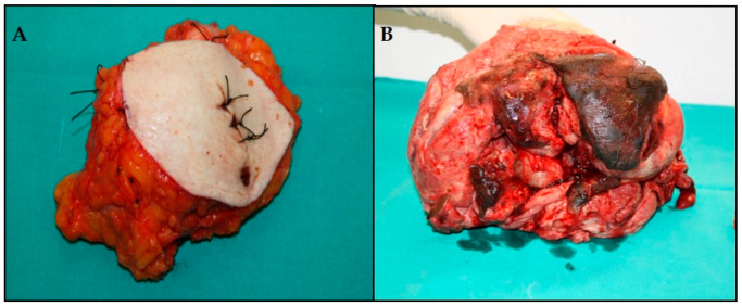
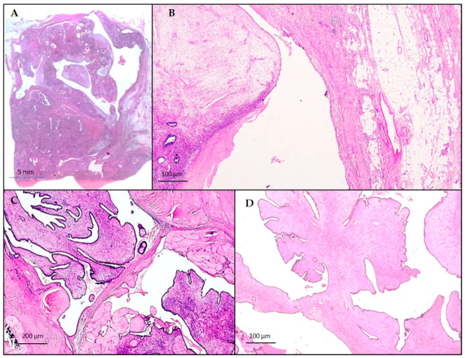
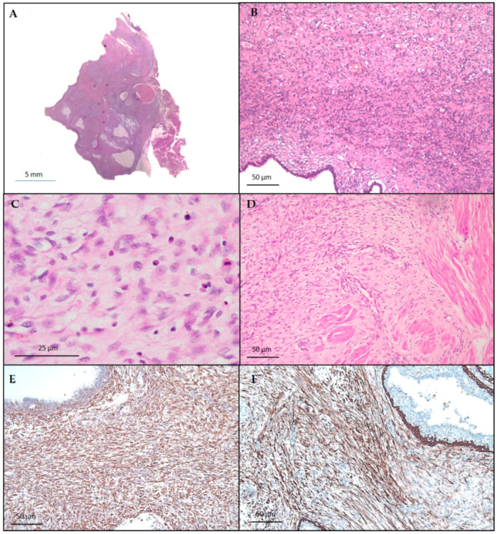
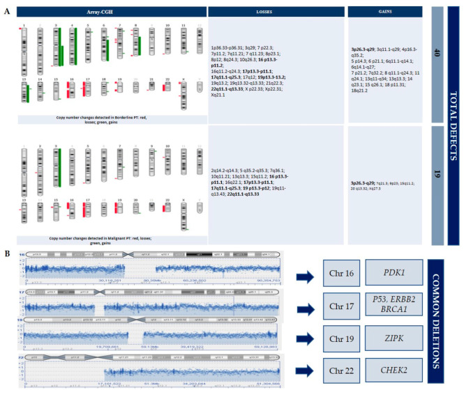
The breast phyllodes tumor is a biphasic tumor that accounts for less than of 1% of all breast neoplasms. It is classified as benign, borderline, or malignant, and can mimic benign masses. Some recurrent alterations have been identified. However, a precise molecular classification of these tumors has not yet been established. Herein, we describe a case of a 43-year-old woman that was admitted to the emergency room for a significant bleeding from the breast skin. A voluminous ulcerative mass of the left breast and multiple nodules with micro-calcifications on the right side were detected at a physical examination. A left total mastectomy and a nodulectomy of the right breast was performed. The histological diagnosis of the surgical specimens reported a bilateral giant phyllodes tumor, showing malignant features on the left and borderline characteristics associated with a fibroadenoma on the right. A further molecular analysis was carried out by an array-Comparative Genomic Hybridization (CGH) to characterize copy-number alterations. Many losses were detected in the malignant mass, involving several tumor suppressor genes. These findings could explain the malignant growth and the metastatic risk. In our study, genomic profiling by an array-CGH revealed a greater chromosomal instability in the borderline mass (40 total defects) than in the malignant (19 total defects) giant phyllodes tumor, reflecting the tumor heterogeneity. Should our results be confirmed with more sensitive and specific molecular tests (DNA sequencing and FISH analysis), they could allow a better selection of patients with adverse pathological features, thus optimizing and improving patient's management.
Keywords: array-CGH; breast tumor; phyllodes tumor.
Conflict of interest statement
The authors declare no conflict of interest.
Figures
References
-
- Punzo C., Fortarezza F., De Ruvo V., Minafra M., Laforgia R., Casamassima G., Pezzuto F., Punzi A., Caporusso C., Angelelli G., et al. Primitive squamous cell carcinoma of the breast (SCCB): Case report of an uncommon variant of metaplastic carcinoma. G. Chir. 2017;38:139–142. doi: 10.11138/gchir/2017.38.3.139. - DOI - PMC - PubMed
-
- Breast Tumours . WHO Classification of Tumours. 5th ed. IARC Press; Lyon, France: 2019.
Publication types
LinkOut - more resources
Full Text Sources
