. 2020 Oct 15;12(10):2989.
doi: 10.3390/cancers12102989.

Targeting Vesicular LGALS3BP by an Antibody-Drug Conjugate as Novel Therapeutic Strategy for Neuroblastoma

Affiliations

Targeting Vesicular LGALS3BP by an Antibody-Drug Conjugate as Novel Therapeutic Strategy for Neuroblastoma

Emily Capone et al. Cancers (Basel). .

Abstract

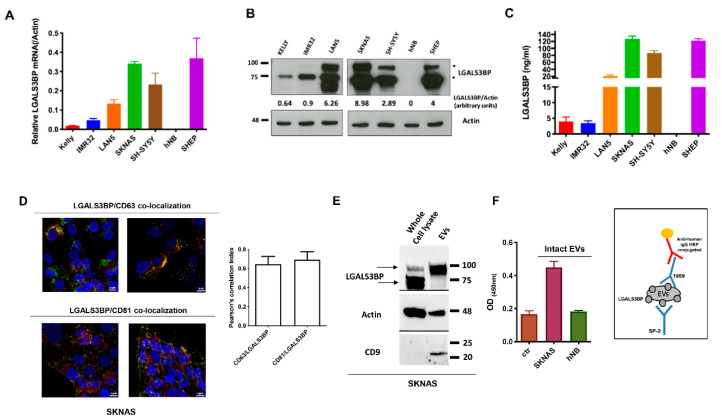
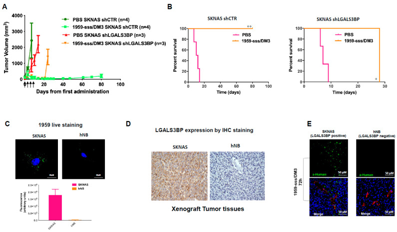
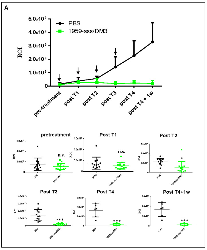
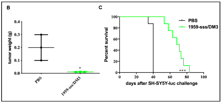
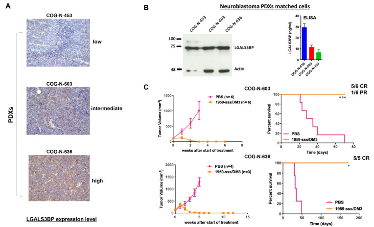
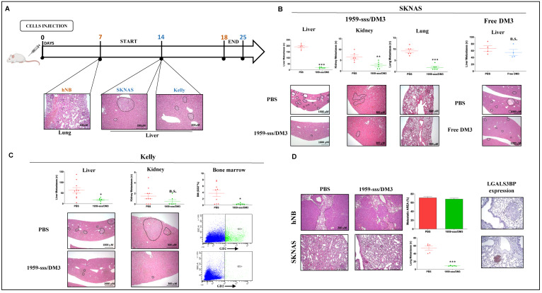
Neuroblastoma is the most common extra-cranial solid tumor in infants and children, which accounts for approximately 15% of all cancer-related deaths in the pediatric population. New therapeutic modalities are urgently needed. Antibody-Drug Conjugates (ADC)s-based therapy has been proposed as potential strategy to treat this pediatric malignancy. LGALS3BP is a highly glycosylated protein involved in tumor growth and progression. Studies have shown that LGALS3BP is enriched in extracellular vesicles (EV)s derived by most neuroblastoma cells, where it plays a critical role in preparing a favorable tumor microenvironment (TME) through direct cross talk between cancer and stroma cells. Here, we describe the development of a non-internalizing LGALS3BP ADC, named 1959-sss/DM3, which selectively targets LGALS3BP expressing neuroblastoma. 1959-sss/DM3 mediated potent therapeutic activity in different types of neuroblastoma models. Notably, we found that treatments were well tolerated at efficacious doses that were fully curative. These results offer preclinical proof-of-concept for an ADC targeting exosomal LGALS3BP approach for neuroblastomas.

Keywords: Antibody-Drug Conjugates (ADC)s; LGALS3BP; neuroblastoma; targeted therapy.


Conflict of interest statement

Stefano Iacobelli and Gianluca Sala are share older of Mediapharma srl.

Figures

Figure 1
Figure 1
LGALS3BP is expressed and secreted in neuroblastoma. (AC) LGALS3BP mRNA, intracellular and secreted protein levels in a panel of human neuroblastoma cell lines were analyzed using quantitative RT-PCR, WB and ELISA, respectively. Band intensities (B) were quantified and indicated in figure as arbitrary units. (D) Confocal imaging showing LGALS3BP (red) co-localization with CD63 and CD81 (green) in SKNAS neuroblastoma cells. Cells nuclei were stained by DRAQ5 (blue). Images taken at 63X magnification. Scale bar: 10 μM. (E) Immunoblot showing LGALS3BP and the exosomal markers Actin and CD9 expression in EVs isolated from SKNAS cell culture supernatant compared to whole cell lysate. (F) Sandwich ELISA performed on intact EVs isolated from (LGALS3BP+) SKNAS and (LGALS3BP-) hNB supernatants (left panel). Scheme of sandwich ELISA is illustrated (right panel). Ctr: ELISA blank control. Arrows (B,E) indicate the intracellular (70 kDa) and the fully mature (90 kDa) forms of LGALS3BP.
Figure 2
Figure 2
1959-sss/DM3 induces tumor shrinkage in a target-dependent manner. (A) CD1 nude mice harboring silenced shLGALS3BP or control vector-infected (shCTR) SKNAS xenografts were treated with vehicle (PBS) or 1959-sss/DM3 at the dose of 10 mg/kg twice weekly. (B) Kaplan-Meier survival curves are shown. Log-rank (Mantel-Cox) Test. * p = 0.02; ** p = 0.006. (C) Confocal images of live neuroblastoma cells labelled with humanized 1959 anti-LGALS3BP antibody for 90 min at 37 °C followed by AlexaFluor 488 conjugated secondary anti-human IgG antibody (green). Cells nuclei were stained by DRAQ5 (blue). Quantification of total immunofluorescence staining per cell is shown in arbitrary units. (D) IHC staining of neuroblastoma xenografts with 1A4.22 anti-LGALS3BP antibody. (E) 1959-sss/DM3 in vivo accumulation (green) in LGALS3BP positive (SKNAS) and negative (hNB) neuroblastoma xenografts. Blood vessels were stained using anti CD31/CD105 antibodies (red); cells nuclei were stained by DRAQ5 (blue).
Figure 3
Figure 3
Therapeutic activity of 1959-sss/DM3 in SH-SY5Y-LUC orthotopic xenograft model. (A) Orthotopic SH-SY5Y-LUC neuroblastoma xenograft showing potent antitumor activity of intravenous 1959-sss/DM3 administration (arrows) at the dose of 10 mg/kg twice weekly. Growth curve obtained was expressed as tumor region of interest (ROI). In the below panels, single animals ROI are shown. Unpaired T Test. * p = 0.01; *** p < 0.0001. (B) Tumor weight of animals sacrificed 24 h after the last treatment. Unpaired T Test. * p = 0.03. (C) Survival curves evaluated by Kaplan-Meier and analyzed by Log-rank (Mantel-Cox). *** p = 0.0001. n.s. (not significant).
Figure 3
Figure 3
Therapeutic activity of 1959-sss/DM3 in SH-SY5Y-LUC orthotopic xenograft model. (A) Orthotopic SH-SY5Y-LUC neuroblastoma xenograft showing potent antitumor activity of intravenous 1959-sss/DM3 administration (arrows) at the dose of 10 mg/kg twice weekly. Growth curve obtained was expressed as tumor region of interest (ROI). In the below panels, single animals ROI are shown. Unpaired T Test. * p = 0.01; *** p < 0.0001. (B) Tumor weight of animals sacrificed 24 h after the last treatment. Unpaired T Test. * p = 0.03. (C) Survival curves evaluated by Kaplan-Meier and analyzed by Log-rank (Mantel-Cox). *** p = 0.0001. n.s. (not significant).
Figure 4
Figure 4
1959-sss/DM3 induces tumor shrinkage in neuroblastoma PDX models. (A) LGALS3BP expression was evaluated by IHC in three neuroblastoma PDXs by IHC and in matched cell lines (B) by WB and ELISA. (C) Subcutaneous COG-N-603 and COG-N-636 PDXs growth curves of intravenous 1959-sss/DM3 administration at the dose of 10 mg/kg twice weekly for 4 doses. Survival curves were evaluated by Kaplan-Meier and analyzed by the log-rank test using Graphpad Prism 5 software. * p = 0.01; *** p = 0.0005. CR: complete remission; PR: partial response.
Figure 5
Figure 5
Targeting LGALS3BP by 1959-sss/DM3 inhibits metastatic spreading. (A) Schematic representation of experimental metastasis assays performed in SKNAS (B), Kelly (C) and hNB (D) neuroblastoma cell models. PBS, 1959-sss/DM3 (10 mg/kg), or free SH-DM3 (0.1 mg/kg) intravenous injections were performed every three days for a total of 3 doses. Metastatic lesions were analyzed and plotted on graphs as number of lesions (SKNAS and Kelly) or as percentage of metastatic area (hNB). Representative images of H&E staining are shown below. Bone marrow metastasis (C) were analyzed by FACS analysis and plotted on graphs as percentage of GD2+ cells. Representative dot plots are shown below. Expression of LGALS3BP on lung metastatic lesions was evaluated by IHC staining (D). Unpaired T Test. * p = 0.01; ** p = 0.006; *** p < 0.0001. n.s. (not significant).

References

    1. Applebaum M.A., Desai A.V., Glade Bender J.L., Cohn S.L. Emerging and investigational therapies for neuroblastoma. Expert Opin. Orphan Drugs. 2017;5:355–368. doi: 10.1080/21678707.2017.1304212. - DOI - PMC - PubMed
    1. Maris J.M., Hogarty M.D., Bagatell R., Cohn S.L. Neuroblastoma. Lancet. 2007;369:2106–2120. doi: 10.1016/S0140-6736(07)60983-0. - DOI - PubMed
    1. Pastor E.R., Mousa S.A. Current management of neuroblastoma and future direction. Crit. Rev. Oncol. Hematol. 2019;138:38–43. doi: 10.1016/j.critrevonc.2019.03.013. - DOI - PubMed
    1. Sait S., Modak S. Anti-GD2 immunotherapy for neuroblastoma. Expert Rev. Anticancer. 2017;17:889–904. doi: 10.1080/14737140.2017.1364995. - DOI - PMC - PubMed
    1. Armideo E., Callahan C., Madonia L. Immunotherapy for High-Risk Neuroblastoma: Management of Side Effects and Complications. J. Adv. Pr. Oncol. 2017;8:44–55. doi: 10.6004/jadpro.2017.8.1.4. - DOI - PMC - PubMed

LinkOut - more resources