. 2020 Oct 15;21(20):7635.
doi: 10.3390/ijms21207635.

Dissecting Total Plasma and Protein-Specific Glycosylation Profiles in Congenital Disorders of Glycosylation

Affiliations

Dissecting Total Plasma and Protein-Specific Glycosylation Profiles in Congenital Disorders of Glycosylation

Agnes L Hipgrave Ederveen et al. Int J Mol Sci. .

Abstract

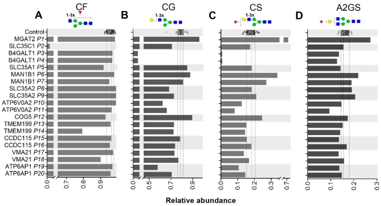
Protein N-glycosylation is a multifactorial process involved in many biological processes. A broad range of congenital disorders of glycosylation (CDGs) have been described that feature defects in protein N-glycan biosynthesis. Here, we present insights into the disrupted N-glycosylation of various CDG patients exhibiting defects in the transport of nucleotide sugars, Golgi glycosylation or Golgi trafficking. We studied enzymatically released N-glycans of total plasma proteins and affinity purified immunoglobulin G (IgG) from patients and healthy controls using mass spectrometry (MS). The applied method allowed the differentiation of sialic acid linkage isomers via their derivatization. Furthermore, protein-specific glycan profiles were quantified for transferrin and IgG Fc using electrospray ionization MS of intact proteins and glycopeptides, respectively. Next to the previously described glycomic effects, we report unprecedented sialic linkage-specific effects. Defects in proteins involved in Golgi trafficking (COG5-CDG) and CMP-sialic acid transport (SLC35A1-CDG) resulted in lower levels of sialylated structures on plasma proteins as compared to healthy controls. Findings for these specific CDGs include a more pronounced effect for α2,3-sialylation than for α2,6-sialylation. The diverse abnormalities in glycomic features described in this study reflect the broad range of biological mechanisms that influence protein glycosylation.

Keywords: congenital disorders of glycosylation; glycomics; mass spectrometry; sialic acid linkage isomers.


Conflict of interest statement

The authors declare no conflict of interest.

Figures

Figure 1
Figure 1
N-glycan structural features. The three major classes of human N-glycans are schematically presented (A) and the major structural features of complex-type N-glycans are introduced (B). Adaptation from [19].
Figure 2
Figure 2
Total plasma N-glycome (TPNG) of a COG5-CDG patient. (A) Levels of α2,3-sialylation per galactose (left) and α2,6-linked sialylation per galactose (right) on triantennary glycans (A3GS) of TPNG. The box plot shows the median (dashed line) with the interquartile range (dotted lines) for healthy controls (n = 10) whilst the bars give the values determined for the COG5-CDG patient. (B) and (C) show the TPNG MALDI-TOF-MS profiles of healthy control 5 and the COG5-CDG patient, respectively. All glycans were detected as [M+Na]+. For glycan annotation see Figure 1.
Figure 3
Figure 3
IgG1 Fc glycosylation for SLC35C1-CDG (P2). (A) Fucosylation (CF) of the IgG1 Fc portions. The box plot shows the median (dashed line) with the interquartile range (dotted lines) for healthy controls (n = 10) whilst the bar gives the value determined for the patient. (B) Extracted ion chromatograms (EIC) of selected glycoforms of the different IgG subclasses and (C) sum spectrum of the indicated window (1.3–1.8 min; IgG1 cluster) from the LC-MS analysis of the patient SLC35C1-CDG tryptic IgG Fc glycopeptides. The proposed glycopeptide structures are based on fragmentation and literature. All glycopeptide signals annotated in (C) were [M+3H]3+. For glycan structure schemes see Figure 1.
Figure 4
Figure 4
Total plasma N-glycome (TPNG) structural features determined for CDG patients. Levels of α2,3-sialylation per galactose (left) and α2,6-linked sialylation per galactose (right) are displayed for non-fucosylated (A) diantennary, (B) triantennary and (C) tetraantennary glycans as well as for fucosylated (D) diantennary, (E) triantennary and (F) tetraantennary glycans. The box plot shows the median (dashed line) with the interquartile range (dotted lines) for healthy controls (n = 10) whilst the bars give the individual values determined for the CDG patients.
Figure 5
Figure 5
IgG1 Fc glycosylation of CDG patients. Levels of IgG1 Fc fucosylation (A), galactosylation (B) sialylation (C) and sialylation per galactose (D) as determined at the glycopeptide level are displayed for 20 CDG patients. Reference values as determined for healthy controls (n = 10) are shown by box plot with median (dashed line) and interquartile range (dotted lines).

References

    1. Varki A. Biological roles of glycans. Glycobiology. 2017;27:3–49. doi: 10.1093/glycob/cww086. - DOI - PMC - PubMed
    1. Lauc G., Pezer M., Rudan I., Campbell H. Mechanisms of disease: The human N-glycome. Biochim. Biophys. Acta. 2016;1860:1574–1582. doi: 10.1016/j.bbagen.2015.10.016. - DOI - PubMed
    1. Thaysen-Andersen M., Packer N.H. Site-specific glycoproteomics confirms that protein structure dictates formation of N-glycan type, core fucosylation and branching. Glycobiology. 2012;22:1440–1452. doi: 10.1093/glycob/cws110. - DOI - PubMed
    1. Reily C., Stewart T.J., Renfrow M.B., Novak J. Glycosylation in health and disease. Nat. Rev. Nephrol. 2019;15:346–366. doi: 10.1038/s41581-019-0129-4. - DOI - PMC - PubMed
    1. Freeze H.H., Chong J.X., Bamshad M.J., Ng B.G. Solving glycosylation disorders: Fundamental approaches reveal complicated pathways. Am. J. Hum. Genet. 2014;94:161–175. doi: 10.1016/j.ajhg.2013.10.024. - DOI - PMC - PubMed

MeSH terms

LinkOut - more resources