Review
. 2020 Oct 15;9(10):999.
doi: 10.3390/antiox9100999.

Vasoconstrictor Mechanisms in Chronic Hypoxia-Induced Pulmonary Hypertension: Role of Oxidant Signaling

Affiliations
Review

Vasoconstrictor Mechanisms in Chronic Hypoxia-Induced Pulmonary Hypertension: Role of Oxidant Signaling

Simin Yan et al. Antioxidants (Basel). .

Abstract

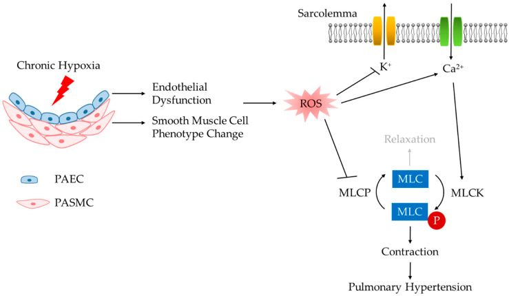
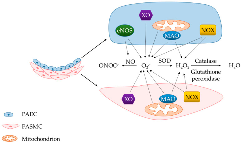
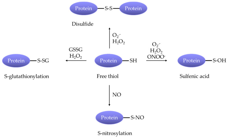
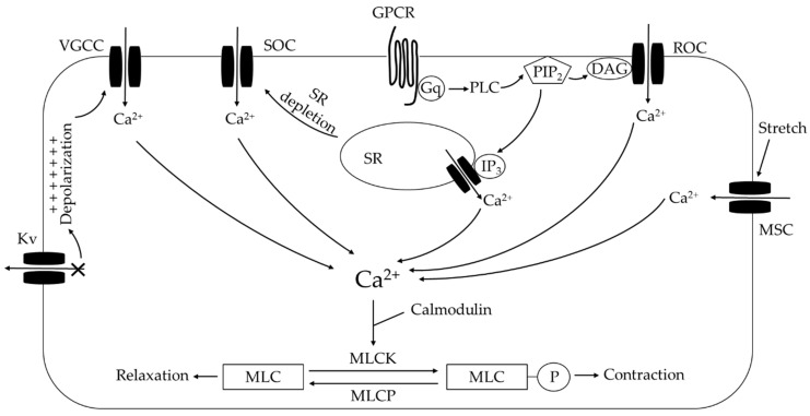
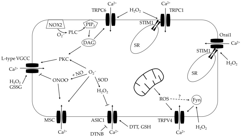
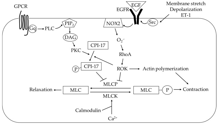
Elevated resistance of pulmonary circulation after chronic hypoxia exposure leads to pulmonary hypertension. Contributing to this pathological process is enhanced pulmonary vasoconstriction through both calcium-dependent and calcium sensitization mechanisms. Reactive oxygen species (ROS), as a result of increased enzymatic production and/or decreased scavenging, participate in augmentation of pulmonary arterial constriction by potentiating calcium influx as well as activation of myofilament sensitization, therefore mediating the development of pulmonary hypertension. Here, we review the effects of chronic hypoxia on sources of ROS within the pulmonary vasculature including NADPH oxidases, mitochondria, uncoupled endothelial nitric oxide synthase, xanthine oxidase, monoamine oxidases and dysfunctional superoxide dismutases. We also summarize the ROS-induced functional alterations of various Ca2+ and K+ channels involved in regulating Ca2+ influx, and of Rho kinase that is responsible for myofilament Ca2+ sensitivity. A variety of antioxidants have been shown to have beneficial therapeutic effects in animal models of pulmonary hypertension, supporting the role of ROS in the development of pulmonary hypertension. A better understanding of the mechanisms by which ROS enhance vasoconstriction will be useful in evaluating the efficacy of antioxidants for the treatment of pulmonary hypertension.

Keywords: calcium influx; calcium sensitization; chronic hypoxia; pulmonary hypertension; pulmonary vasoconstriction; reactive oxygen species.


Conflict of interest statement

The authors declare no conflict of interest.

Figures

Figure 1
Figure 1
Enhanced vasoconstriction resulting from chronic hypoxia-induced functional alterations of endothelial and smooth muscle cells contributes to pulmonary hypertension. PAEC, pulmonary arterial endothelial cell; PASMC, pulmonary arterial smooth muscle cell; ROS, reactive oxygen species; MLCK, myosin light chain kinase; MLCP, myosin light chain phosphatase; MLC, myosin light chain.
Figure 2
Figure 2
O2.− is generated from various enzymatic sources and converted to other forms of ROS, including H2O2 and ONOO. H2O2 can also be produced from NOX4 and MAOs. PAEC, pulmonary arterial endothelial cell; PASMC, pulmonary arterial smooth muscle cell; ROS, reactive oxygen species; O2.−, superoxide; H2O2, hydrogen peroxide; ONOO, peroxynitrite; H2O, water; NOX, NADPH oxidase; eNOS, endothelial nitric oxide synthase; XO, xanthine oxidase; MAO, monoamine oxidase; SOD, superoxide dismutase.
Figure 3
Figure 3
ROS facilitate pulmonary arterial constriction in pulmonary hypertension by make various posttranslational modifications at cysteine residues of ion channels and molecules.
Figure 4
Figure 4
Summary of Ca2+-dependent influx and release mechanisms in pulmonary arterial smooth muscle cells following chronic hypoxia. See text for details. KV, voltage-gated K+ channel; VGCC, voltage-gated Ca2+ channel; SOC, store-operated channel; ROC, receptor-operated channel; MSC, mechanosensitive channel; GPCR, G protein-coupled receptor; PLC, phospholipase C; PIP2, phosphatidylinositol 4,5-bisphosphate; DAG, diacylglycerol; IP3, inositol triphosphate; SR, sarcoplasmic reticulum; MLCK, myosin light chain kinase; MLCP, myosin light chain phosphatase; MLC, myosin light chain.
Figure 5
Figure 5
ROS modulation of Ca2+ influx. See text for details. L-type VGCC, L-type voltage-gated Ca2+ channel; TRPC, canonical transient receptor potential channel; TRPV4, transient receptor potential vanilloid 4; ASIC 1, acid sensing ion channel 1; MSC, mechanosensitive channel; NOX2, NADPH oxidase 2; PLC, phospholipase C; PIP2, phosphatidylinositol 4,5-bisphosphate; DAG, diacylglycerol; PKC, protein kinase C; SR, sarcoplasmic reticulum; STIM1, stromal interaction molecule 1; ROS, reactive oxygen species; O2.−, superoxide; H2O2, hydrogen peroxide; ONOO, peroxynitrite; SOD, superoxide dismutase; NO, nitric oxide; Fyn, Fyn kinase; GSSG, oxidized glutathione; GSH, reduced glutathione; DTT, dithiothreitol; DTNB, 5,5′-Dithiobis(2-nitrobenzoic acid).
Figure 6
Figure 6
Summary of Ca2+ sensitization in pulmonary arterial smooth muscle cells. Myofilament Ca2+ sensitization is facilitated by ROS following CH. In particular, membrane stretch and endothelin 1 (ET-1) activate Src kinase-epidermal growth factor receptor (EGFR)-NADPH oxidase 2 (NOX2) signaling axis that contributes to CH-induced augmentation of Ca2+-independent pulmonary vasoconstriction and pulmonary hypertension. See text for details. GPCR, G protein-coupled receptor; PLC, phospholipase C; PIP2, phosphatidylinositol 4,5-bisphosphate; DAG, diacylglycerol; PKC, protein kinase C; O2.−, superoxide; ROK, Rho kinase; MLCP, myosin light chain phosphatase; MLCK, myosin light chain kinase; MLC, myosin light chain.

Similar articles

Cited by

References

    1. Simonneau G., Montani D., Celermajer D.S., Denton C.P., Gatzoulis M.A., Krowka M., Williams P.G., Souza R. Haemodynamic definitions and updated clinical classification of pulmonary hypertension. Eur. Respir. J. 2019;53:1801913. doi: 10.1183/13993003.01913-2018. - DOI - PMC - PubMed
    1. Brown L.M., Chen H., Halpern S., Taichman D., McGoon M.D., Farber H.W., Frost A.E., Liou T.G., Turner M., Feldkircher K., et al. Delay in recognition of pulmonary arterial hypertension: Factors identified from the REVEAL Registry. Chest. 2011;140:19–26. doi: 10.1378/chest.10-1166. - DOI - PMC - PubMed
    1. Jernigan N.L., Naik J.S., Weise-Cross L., Detweiler N.D., Herbert L.M., Yellowhair T.R., Resta T.C. Contribution of reactive oxygen species to the pathogenesis of pulmonary arterial hypertension. PLoS ONE. 2017;12:e0180455. doi: 10.1371/journal.pone.0180455. - DOI - PMC - PubMed
    1. Stenmark K.R., Fagan K.A., Frid M.G. Hypoxia-Induced Pulmonary Vascular Remodeling. Circ. Res. 2006;99:675–691. doi: 10.1161/01.RES.0000243584.45145.3f. - DOI - PubMed
    1. Young J.M., Williams D.R., Thompson A.A.R. Thin Air, Thick Vessels: Historical and Current Perspectives on Hypoxic Pulmonary Hypertension. Front. Med. (Lausanne) 2019;6:93. doi: 10.3389/fmed.2019.00093. - DOI - PMC - PubMed

LinkOut - more resources