. 2020 Oct 15;9(10):3316.
doi: 10.3390/jcm9103316.

Changes in Gene Expression Associated with Collagen Regeneration and Remodeling of Extracellular Matrix after Percutaneous Electrolysis on Collagenase-Induced Achilles Tendinopathy in an Experimental Animal Model: A Pilot Study

Affiliations

Changes in Gene Expression Associated with Collagen Regeneration and Remodeling of Extracellular Matrix after Percutaneous Electrolysis on Collagenase-Induced Achilles Tendinopathy in an Experimental Animal Model: A Pilot Study

José Luis Sánchez-Sánchez et al. J Clin Med. .

Abstract

Percutaneous electrolysis is an emerging intervention proposed for the management of tendinopathies. Tendon pathology is characterized by a significant cell response to injury and gene expression. No study investigating changes in expression of those genes associated with collagen regeneration and remodeling of extracellular matrix has been conducted. The aim of this pilot study was to investigate gene expression changes after the application of percutaneous electrolysis on experimentally induced Achilles tendinopathy with collagenase injection in an animal model. Fifteen Sprague Dawley male rats were randomly divided into three different groups (no treatment vs. percutaneous electrolysis vs. needling). Achilles tendinopathy was experimentally induced with a single bolus of collagenase injection. Interventions consisted of 3 sessions (one per week) of percutaneous electrolysis or just needling. The rats were euthanized, and molecular expression of genes involved in tendon repair and remodeling, e.g., Cox2, Mmp2, Mmp9, Col1a1, Col3a1, Vegf and Scx, was examined at 28 days after injury. Histological tissue changes were determined with hematoxylin-eosin and safranin O analyses. The images of hematoxylin-eosin and Safranin O tissue images revealed that collagenase injection induced histological changes compatible with a tendinopathy. No further histological changes were observed after the application of percutaneous electrolysis or needling. A significant increase in molecular expression of Cox2, Mmp9 and Vegf genes was observed in Achilles tendons treated with percutaneous electrolysis to a greater extent than after just needling. The expression of Mmp2, Col1a1, Col3a1, or Scx genes also increased, but did not reach statistical significance. This animal study demonstrated that percutaneous electrolysis applied on an experimentally induced Achilles tendinopathy model could increase the expression of some genes associated with collagen regeneration and remodeling of extracellular matrix. The observed gene overexpression was higher with percutaneous electrolysis than with just needling.

Keywords: Achilles; gene expression; percutaneous electrolysis; tendinopathy.


Conflict of interest statement

The authors declare no conflict of interest.

Figures

Figure 1
Figure 1
Application of Percutaneous Electrolysis (EPI© device) on the Achilles tendon of a rat. The positive electrode was placed on the tail of the rat with a cinch (figure on the left top corner).
Figure 2
Figure 2
Flow Diagram summarizing the Experiment Groups.
Figure 3
Figure 3
Stained longitudinal images with hematoxylin–eosin of a normal Achilles tendon (A) and of an injured Achilles tendon of rats euthanized after one week after injection of collagenase (B). The scale bar is at 50 μm.
Figure 4
Figure 4
Stained longitudinal images with Safranin O of a normal Achilles tendon (A) and of an injured Achilles tendon of rats euthanized after one week after injection of collagenase showing the presence of fibrocartilaginous tissue (B). The scale bar is at 50 μm.
Figure 5
Figure 5
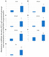
Levels of expression of Cox2 (A), Mmp2 (B), Mmp9 (C), Col1a1 (D), Col3a1 (E), Vegf (F) and Scx (G) genes in an healthy control Achilles tendon (C) and experimentally induced tendinopathy tendon (T1). Data were quantified and normalized by using the RT-qPCR procedure. Each bar represents the fold change (FC) ± standard deviation (SD) of the normalized values. *** p < 0.001; ** p < 0.01; * p < 0.05.
Figure 6
Figure 6
Stained longitudinal images with hematoxylin–eosin of injured Achilles tendon with collagenase injection by group: no intervention (A), treated with percutaneous electrolysis (B,C), or treated needling (D). Arrows in C shows several leucocyte cells. The scale bar is at 50 μm (A,B,D) and 10 μm (C).
Figure 7
Figure 7
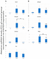
Levels of expression of Cox2 (A), Mmp2 (B), Mmp9 (C), Col1a1 (D), Col3a1 (E), Vegf (F) and Scx (G) genes in an injured Achilles tendon with collagenase injection by group: no intervention (T2), percutaneous electrolysis (T2 + PE) and needling (T2 + N). Data were quantified and normalized by using the RT-qPCR procedure. Each bar represents the fold change (FC) ± standard deviation (SD). *** p < 0.001; ** p < 0.01; * p < 0.05.

References

    1. Sobhani S., Dekker R., Postema K., Dijkstra P.U. Epidemiology of ankle and foot overuse injuries in sports: A systematic review. Scand. J. Med. Sci. Sports. 2013;23:669–686. doi: 10.1111/j.1600-0838.2012.01509.x. - DOI - PubMed
    1. Waldecker U., Hofmann G., Drewitz S. Epidemiologic investigation of 1394 feet: Coincidence of hindfoot malalignment and Achilles tendon disorders. Foot Ankle Surg. 2012;18:119–123. doi: 10.1016/j.fas.2011.04.007. - DOI - PubMed
    1. Cook J.L., Rio E., Purdam C.R., Docking S.I. Revisiting the continuum model of tendon pathology: What is its merit in clinical practice and research? Br. J. Sports Med. 2016;50:1187–1191. doi: 10.1136/bjsports-2015-095422. - DOI - PMC - PubMed
    1. Xu Y., Murrell G.A. The Basic Science of Tendinopathy. Clin. Orthop. Relat. Res. 2008;466:1528–1538. doi: 10.1007/s11999-008-0286-4. - DOI - PMC - PubMed
    1. Wu F., Nerlich M., Docheva D. Tendon injuries: Basic science and new repair proposals. EFORT. Open. Rev. 2017;2:332–342. doi: 10.1302/2058-5241.2.160075. - DOI - PMC - PubMed