. 2020 Oct 19;18(1):162.
doi: 10.1186/s12964-020-00656-0.

Sensory neurons from dorsal root ganglia regulate endothelial cell function in extracellular matrix remodelling

Affiliations

Sensory neurons from dorsal root ganglia regulate endothelial cell function in extracellular matrix remodelling

Alice Leroux et al. Cell Commun Signal. .

Abstract

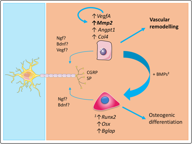
Background: Recent physiological and experimental data highlight the role of the sensory nervous system in bone repair, but its precise role on angiogenesis in a bone regeneration context is still unknown. Our previous work demonstrated that sensory neurons (SNs) induce the osteoblastic differentiation of mesenchymal stem cells, but the influence of SNs on endothelial cells (ECs) was not studied.

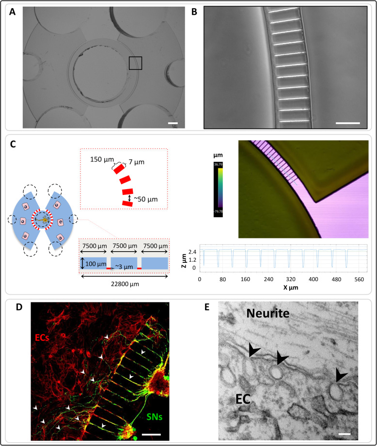
Methods: Here, in order to study in vitro the interplay between SNs and ECs, we used microfluidic devices as an indirect co-culture model. Gene expression analysis of angiogenic markers, as well as measurements of metalloproteinases protein levels and enzymatic activity, were performed.

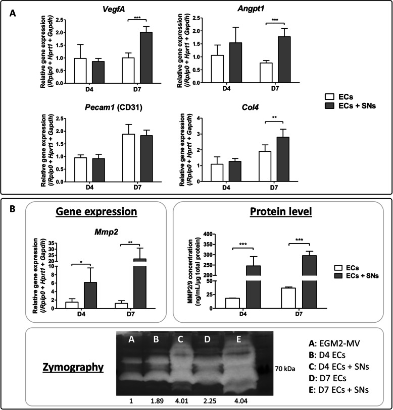
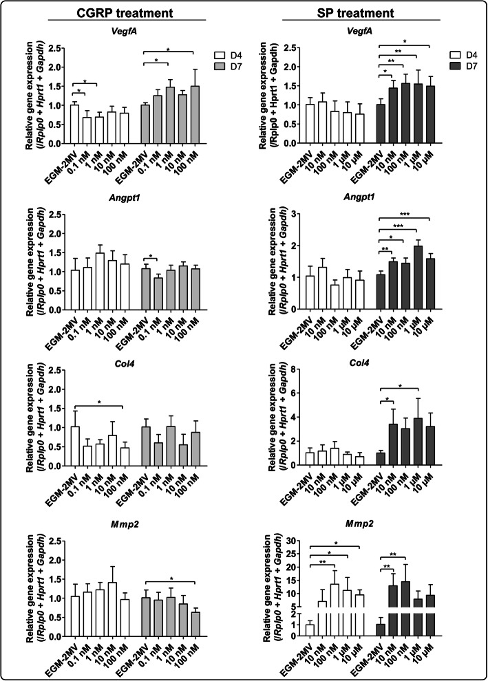
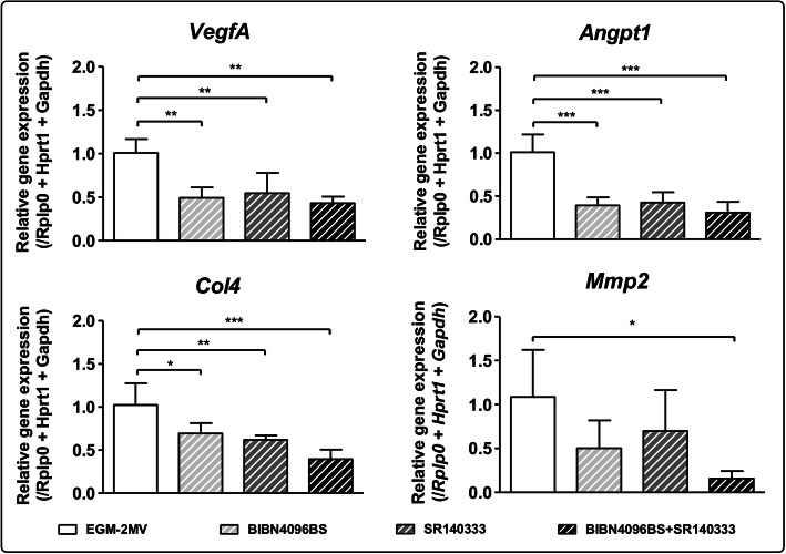
Results: We were able to demonstrate that two sensory neuropeptides, calcitonin gene-related peptide (CGRP) and substance P (SP), were involved in the transcriptional upregulation of angiogenic markers (vascular endothelial growth factor, angiopoietin 1, type 4 collagen, matrix metalloproteinase 2) in ECs. Co-cultures of ECs with SNs also increased the protein level and enzymatic activity of matrix metalloproteinases 2 and 9 (MMP2/MMP9) in ECs.

Conclusions: Our results suggest a role of sensory neurons, and more specifically of CGRP and SP, in the remodelling of endothelial cells extracellular matrix, thus supporting and enhancing the angiogenesis process. Video abstract.

Keywords: Angiogenesis; Cellular communication; Extracellular matrix Remodelling; Innervation; Matrix Metalloproteinases; Neurovascular interplay.


Conflict of interest statement

The authors declare that they have no competing interests.

Figures

Fig. 1
Fig. 1
Microfluidic devices validation. ab Conventional photolithography techniques were used to create the microfluidic devices with precise and specific dimensions. Scale bars = 100 μM. c Scheme of the microfluidic devices dimensions (left). Profilometry was performed to confirm the microchannels dimensions (right). d Sensory neurons (SNs) derived from rat dorsal root ganglia were seeded in the central compartment of the devices. Rat bone marrow derived endothelial cells (ECs) were cultivated in the lateral compartments. After 4 days of culture, cells were fixed and stained with β-III tubulin (SNs in green) and phalloidin (ECs in red). The neurites emitted by the sensory neurons were able to cross the microchannels and go from the central compartment to the lateral ones (white arrowheads) where they closely interact with endothelial cells. Scale bar = 100 μm. e Transmission Electron Microscopy was performed to confirm this close interaction and showed vesicles in ECs (black arrowheads). Scale bar = 100 nm
Fig. 2
Fig. 2
Transcriptional and translational regulation of ECs by SNs. a Gene expression analysis of angiogenic markers expressed by endothelial cells cultivated in the microfluidic devices in presence (grey bars) or absence (white bars) of sensory neurons in the central compartment. After 4 and 7 days of culture, cells were harvested, RNA was extracted and RT-qPCR was performed. Gene expression of Angpt1, VegfA and Col4 is upregulated in ECs when co-cultivated with sensory neurons after 7 days of culture. The graphs represent mean values ± SD (n = 4 independent experiments, unpaired t test, ** p < 0.01, *** p < 0.001). b Mmp2 gene expression is upregulated at both time points in presence of sensory neurons (n = 4 independent experiments, unpaired t test, * p < 0.05, ** p < 0.01). Concentration of MMP2 and MMP9 was measured in ECs’ supernatants cultivated with (grey bars) or without (white bars) sensory neurons. At both time points, MMP2/MMP9 concentration is increased in ECs when they are co-cultivated in presence of SNs (n = 5 microfluidic devices, unpaired t test, *** p < 0.001). Representative picture of enzymatic activity of matrix metalloproteinases measured by zymography. The band intensity was quantified using ImageJ software. In presence of sensory neurons, ECs show an increase of MMP2/MMP9 enzymatic activity at day 4 and day 7 (pool of n = 6 microfluidic devices)
Fig. 3
Fig. 3
Effect of CGRP and SP on ECs transcriptional profiles. ECs cultivated in 48-wells plates were treated either with CGRP (from 0,1 nM to 100 nM) or SP (from 10 nM to 10 μM) for 4 days (white bars) or 7 days (grey bars) of culture. Total RNA was extracted followed by Vegfa, Angpt1, Col4, and Mmp2 gene expression analysis by RT-qPCR. VegfA expression is upregulated by CGRP after 7 days of culture whereas VegfA, Angpt1, Col4 and Mmp2 expression is upregulated by SP. (n = 3 independent experiments, One-way ANOVA followed by post hoc Dunnett’s test, * p < 0.05, ** p < 0.01, *** p < 0.001)
Fig. 4
Fig. 4
Effect of the inhibition of CGRP and SP on ECs transcriptional profiles. ECs were cultivated for 7 days in the microfluidic devices in presence of sensory neurons. Endothelial cells were either not treated (EGM-2MV) or treated with CGRP antagonist (BIBN4096BS), SP antagonist (SR140333) or both, at a concentration of 10 μM each. Total RNA was extracted and gene expression of VegfA, Angpt1, Col4 and Mmp2 was analyzed by RT-qPCR. CGRP and SP antagonists downregulate ECs gene expression of Angpt1, VegfA and Col4, both separately and together. Mmp2 gene expression is only significantly downregulated when ECs were treated with both antagonists. (n = 6 microfluidic devices, One-way ANOVA followed by post hoc Dunnett’s test, * p < 0.05, ** p < 0.01, *** p < 0.001)
Fig. 5
Fig. 5
Role of sensory neurons in osteogenic differentiation and vascular remodelling. SNs enhance the osteogenic differentiation of MSCs through the upregulation of genes such as Runx2 (runt-related transcription factor 2), Sp7 (osterix) or Bglap (osteocalcin) [according to Silva et al., 2017 [46]]. Through the secretion of CGRP and SP, SNs also play a role in vascular remodeling by upregulating the expression of some angiogenic markers in ECs. More specifically, they strongly upregulate the expression of Mmp2 as well as MMP2/MMP9 protein level and enzymatic activity, suggesting a role in angiogenesis through extracellular matrix remodeling. Besides, ECs are known to have an osteoinductive effect on MSC through the release of bone morphogenetic proteins (BMPs) [according to Grellier et al., 2009 [2]], thus the interplay between these three cell types should be considered for the development of bone regeneration strategies

References

    1. Brandi ML, Collin-Osdoby P. Vascular biology and the skeleton. J Bone Miner Res. 2006;21(2):183–192. doi: 10.1359/JBMR.050917. - DOI - PubMed
    1. Grellier M, Bordenave L, Amédée J. Cell-to-cell communication between osteogenic and endothelial lineages: implications for tissue engineering. Trends Biotechnol. 2009;27(10):562–571. doi: 10.1016/j.tibtech.2009.07.001. - DOI - PubMed
    1. Lafage-Proust M-H, Roche B, Langer M, Cleret D, Vanden Bossche A, Olivier T, et al. Assessment of bone vascularization and its role in bone remodeling. BoneKEy Rep. 2015;8:4. - PMC - PubMed
    1. Hurrell DJ. The nerve supply of bone. J Anat. 1937;72(Pt 1):54–61. - PMC - PubMed
    1. Miller MR, Kasahara M. Observations on the innervation of human long bones. Anat Rec. 1963;145(1):13–23. doi: 10.1002/ar.1091450104. - DOI

Publication types

MeSH terms