. 2020 Oct 20;18(1):137.
doi: 10.1186/s12915-020-00876-3.

Gut bacterial deamination of residual levodopa medication for Parkinson's disease

Affiliations

Gut bacterial deamination of residual levodopa medication for Parkinson's disease

Sebastiaan P van Kessel et al. BMC Biol. .

Abstract

Background: Parkinson's disease (PD) is a progressive neurodegenerative disorder characterized by both motor and non-motor symptoms. Gastrointestinal tract dysfunction is one of the non-motor features, where constipation is reported as the most common gastrointestinal symptom. Aromatic bacterial metabolites are attracting considerable attention due to their impact on gut homeostasis and host's physiology. In particular, Clostridium sporogenes is a key contributor to the production of these bioactive metabolites in the human gut.

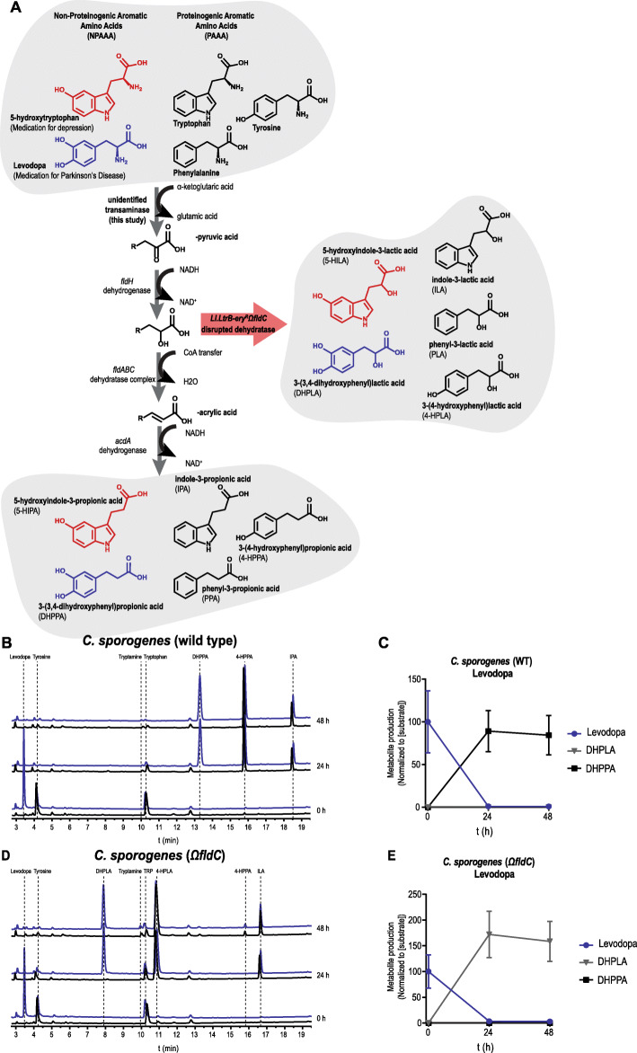
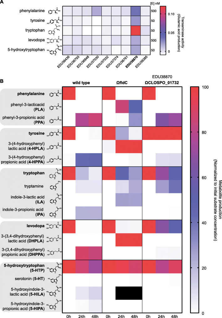
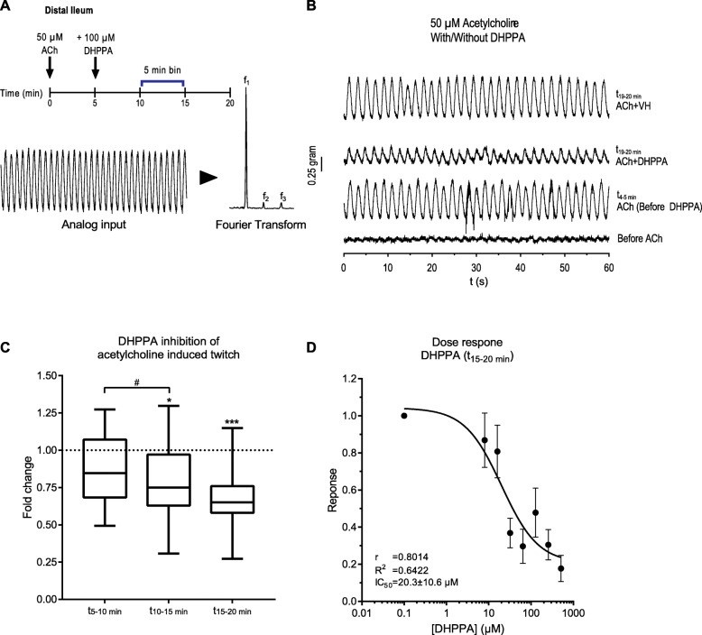
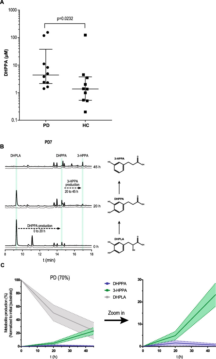
Results: Here, we show that C. sporogenes deaminates levodopa, the main treatment in Parkinson's disease, and identify the aromatic aminotransferase responsible for the initiation of the deamination pathway. The deaminated metabolite from levodopa, 3-(3,4-dihydroxyphenyl)propionic acid, elicits an inhibitory effect on ileal motility in an ex vivo model. We detected 3-(3,4-dihydroxyphenyl)propionic acid in fecal samples of Parkinson's disease patients on levodopa medication and found that this metabolite is actively produced by the gut microbiota in those stool samples.

Conclusions: Levodopa is deaminated by the gut bacterium C. sporogenes producing a metabolite that inhibits ileal motility ex vivo. Overall, this study underpins the importance of the metabolic pathways of the gut microbiome involved in drug metabolism not only to preserve drug effectiveness, but also to avoid potential side effects of bacterial breakdown products of the unabsorbed residue of medication.

Keywords: Aminotransferase; Bioactive metabolites; Clostridium sporogenes; Drug side effects; Gastrointestinal motility; Non-motor symptoms.


Conflict of interest statement

The authors declare no competing interests.

Figures

Fig. 1
Fig. 1
Levodopa is deaminated by Clostridium sporogenes. a Full reductive deamination pathway of C. sporogenes is depicted resulting in the full deamination (R-propionic acid) of (non)-proteinogenic aromatic amino acids ((N)PAAAs). The red arrow indicates a disrupted deamination pathway of C. sporogenes, where the dehydratase subunit fldC is mutagenized, resulting in a pool of partially deaminated metabolites (R-lactic acid) by C. sporogenes. b HPLC-ED curves from supernatant of a C. sporogenes batch culture conversion of levodopa (3-(3,4-dihydroxyphenyl)alanine) over time. At the beginning of growth (timepoint 0 h), 100 μM of levodopa (blue) is added to the culture medium; the black line in the chromatogram depicts the control samples. In 24 h, levodopa is completely converted to DHPPA (3-(3,4-dihydroxyphenyl)propionic acid), the deaminated product of levodopa. Other aromatic amino acids from the medium, tryptophan and tyrosine (which are detectable with ED), are converted to the deaminated products IPA (indole-3-propionic acid) and 4-HPPA (3-(4-hydroxyphenyl)propionic acid). c Quantification (n = 3) of levodopa conversion to DHPPA by C. sporogenes wild type (also see Additional File 1: Table S1). d Analysis of the supernatant of CSΩfldC shows that levodopa is not deaminated to DHPPA but to its intermediate product DHPLA (3-(3,4-dihydroxyphenyl)lactic acid) within 24 h. Tryptophan and tyrosine are converted to their intermediates ILA (indole-3-lactic acid) and 4-HPLA (3-(4-hydroxyphenyl)lactic acid), respectively. e Quantification (n = 3) of levodopa conversion to DHPLA by C. sporogenes ΩfldC (also see Additional File 1: Table S1). All experiments were performed in 3 independent biological replicates, and means with error bars representing the SEM are depicted
Fig. 2
Fig. 2
Identification of the aromatic amino transferase initiating the deamination pathway. In order to identify which aminotransferase is responsible for the initial transaminase reaction, all class I/II aminotransferases were cloned and purified to test the activity against (N)PAAAs. a Transaminase activity (production of glutamate) for all substrates is depicted. EDU38870 (CLOSPO_01732) was involved in all transaminase reactions. EDU37030 showed similar activity as EDU38870, for phenylalanine. Experiment was performed in technical duplicates to screen for candidate genes for mutagenesis in C. sporogenes. b Targeted metabolic quantification of deamination products from CSWT, CSΩfldC, and CSΩCLOSPO_01732 reveals that EDU38870 is involved in the transamination of all for all tested (N)PAAAs. All quantified deamination products are normalized to their initial substrate concentration, and the data represents 3 independent biological replicates. Corresponding values are reported, and metabolite concentration differences between WT and ΩfldC or ΩCLOSPO_01732 were tested for significance using Student’s t test, in Additional File 1: Table S1. Black squares indicate that quantification was not possible because of a coeluting HPLC-ED peak. As no commercial standards are available for 5-HILA and 5-HIPA, the peaks were quantified assuming a similar ED-detector response as for 5-HTP
Fig. 3
Fig. 3
DHPPA inhibits the acetylcholine-induced twitch from mouse ileum. a Experimental setup, where 5 min after adding 50 μM acetylcholine, 100 μM DHPPA is added. The panel below indicates how the amplitude of the frequencies of the observed oscillations (from 5 min bins) is extracted by a Fourier transform of the analog input. b A representative 1-min recording trace before and after the addition of acetylcholine and DHPPA or vehicle (VH) is shown. ACh, acetylcholine; VH, vehicle (0.05% ethanol). c Inhibition of DHPPA on acetylcholine-induced twitch binned in intervals of 5 min shows a decrease in contractility over time (n = 6 biological replicates and experiments were repeated 1–4 times per tissue). Significance was tested using repeated measures (RM) 1-way ANOVA followed by Tukey’s test (*p < 0.0021, ***p < 0.0002, #p < 0.0021). Box represents the median with interquartile range, and whiskers represent the maxima and minima. d Dose response curve of DHPPA on the acetylcholine-induced twitch at the t15–20 minute bin (n = 4 biological replicates) with a half maximal inhibitory concentration (IC50) of 20.3 ± 10.6 μM
Fig. 4
Fig. 4
Higher DHPPA levels in PD patients and active levodopa deamination pathway in PD fecal suspensions. a DHPPA was extracted from fecal samples of PD patients (n = 10) and age-matched healthy controls (n = 10) using activated alumina beads, and concentrations were quantified using a standard curve of DHPPA on the HPLC-ED with 3,4-dihydroxybenzylamine as internal standard. DHPPA concentrations are depicted on the logarithmic y-axis, and individual levels are indicated and compared between Parkinson’s disease (PD) patients and age-matched healthy controls (HC). The cross-header represents the median (PD, 4.36 μM; HC, 1.37 μM) and the interquartile range (PD, 2.15–37.90 μM; HC, 0.53–3.75 μM). Significance was tested using an unpaired nonparametric Mann-Whitney test (p = 0.0232). b A representative HPLC-ED chromatogram of fecal suspension from PD7 where DHPPA is produced from DHPLA (black) after 20 h and is further metabolized to 3-HPPA after 45 h of incubation. The control, without the addition of DHPLA, is indicated in gray. The green bars indicate the retention time of the standards indicated. c Metabolite profiles of the PD fecal suspensions that produced DHPPA/3-HPPA within 20–45 h (70%) are merged as replicates. Lines represent the mean and the shadings the SEM; a zoom in graph of DHPPA and 3-HPPA is depicted on the right

References

    1. Donia MS, Fischbach MA. Small molecules from the human microbiota. Science. 2015;349:1254766. doi: 10.1126/science.1254766. - DOI - PMC - PubMed
    1. Bansal T, Alaniz RC, Wood TK, Jayaraman A. The bacterial signal indole increases epithelial-cell tight-junction resistance and attenuates indicators of inflammation. Proc Natl Acad Sci. 2010;107:228–233. doi: 10.1073/pnas.0906112107. - DOI - PMC - PubMed
    1. Schiering C, Wincent E, Metidji A, Iseppon A, Li Y, Potocnik AJ, et al. Feedback control of AHR signalling regulates intestinal immunity. Nature. 2017;542:242–245. doi: 10.1038/nature21080. - DOI - PMC - PubMed
    1. Yano JM, Yu K, Mazmanian SK, Hsiao Correspondence EY, Donaldson GP, Shastri GG, et al. Indigenous bacteria from the gut microbiota regulate host serotonin biosynthesis. Cell. 2015;161:264–276. doi: 10.1016/j.cell.2015.02.047. - DOI - PMC - PubMed
    1. Bhattarai Y, Williams BB, Battaglioli EJ, Whitaker WR, Till L, Grover M, et al. Gut microbiota-produced tryptamine activates an epithelial G-protein-coupled receptor to increase colonic secretion. Cell Host Microbe. 2018;23:775–785. doi: 10.1016/j.chom.2018.05.004. - DOI - PMC - PubMed

Publication types

Supplementary concepts

LinkOut - more resources