. 2020 Nov 3;117(44):27676-27684.
doi: 10.1073/pnas.2014463117. Epub 2020 Oct 19.

Liquid-liquid phase separation promotes animal desiccation tolerance

Affiliations

Liquid-liquid phase separation promotes animal desiccation tolerance

Clinton Belott et al. Proc Natl Acad Sci U S A. .

Abstract

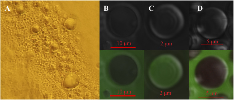
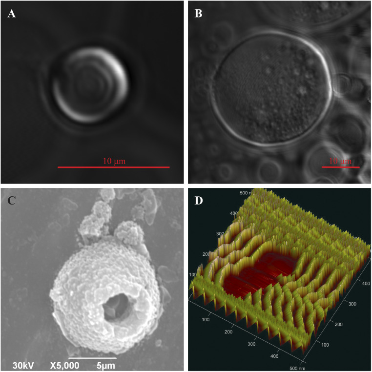
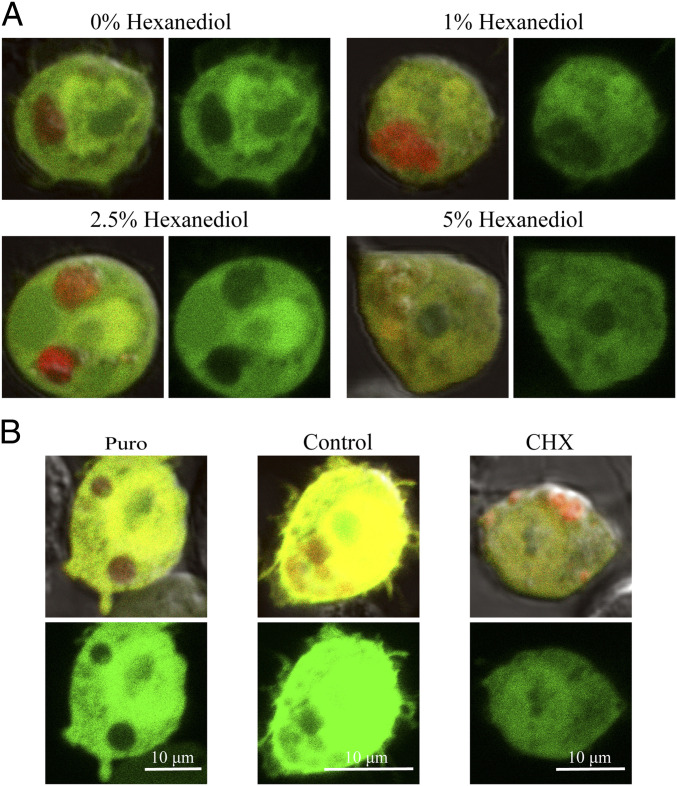
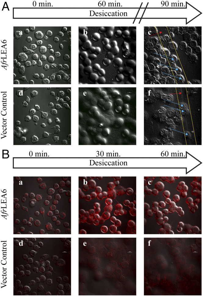
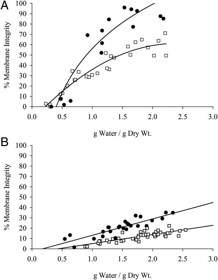
Proteinaceous liquid-liquid phase separation (LLPS) occurs when a polypeptide coalesces into a dense phase to form a liquid droplet (i.e., condensate) in aqueous solution. In vivo, functional protein-based condensates are often referred to as membraneless organelles (MLOs), which have roles in cellular processes ranging from stress responses to regulation of gene expression. Late embryogenesis abundant (LEA) proteins containing seed maturation protein domains (SMP; PF04927) have been linked to storage tolerance of orthodox seeds. The mechanism by which anhydrobiotic longevity is improved is unknown. Interestingly, the brine shrimp Artemia franciscana is the only animal known to express such a protein (AfrLEA6) in its anhydrobiotic embryos. Ectopic expression of AfrLEA6 (AWM11684) in insect cells improves their desiccation tolerance and a fraction of the protein is sequestered into MLOs, while aqueous AfrLEA6 raises the viscosity of the cytoplasm. LLPS of AfrLEA6 is driven by the SMP domain, while the size of formed MLOs is regulated by a domain predicted to engage in protein binding. AfrLEA6 condensates formed in vitro selectively incorporate target proteins based on their surface charge, while cytoplasmic MLOs formed in AfrLEA6-transfected insect cells behave like stress granules. We suggest that AfrLEA6 promotes desiccation tolerance by engaging in two distinct molecular mechanisms: by raising cytoplasmic viscosity at even modest levels of water loss to promote cell integrity during drying and by forming condensates that may act as protective compartments for desiccation-sensitive proteins. Identifying and understanding the molecular mechanisms that govern anhydrobiosis will lead to significant advancements in preserving biological samples.

Keywords: cryptobiosis; late embryogenesis abundant; liquid-liquid phase separation; membraneless organelle; water stress.


Conflict of interest statement

The authors declare no competing interest.

Figures

Fig. 1.
Fig. 1.
AfrLEA6 is predicted to have three distinct regions. (A) The N-terminal SMP repeats (purple) exhibit alpha-helical propensity. The C-terminal domain (green) exhibits fuzzy self-interactions. These two domains are linked by an intrinsically disordered spacer enriched in proline, glycine, and aromatic residues (blue). I-Tasser structural prediction of AfrLEA6 is based on hierarchical stability of known crystal structures, thus associating this structure with a possible conformation in the dried state. (B) SmartEMBLE identifies two N-terminal SMP domains in AfrLEA6 (purple). (C) The protein is overall negatively charged, with alternating charges (green) at the C terminus promoting interactions with other proteins or itself.
Fig. 2.
Fig. 2.
AfrLEA6 undergoes a liquid-liquid phase separation that sequesters GFP based on surface charge. (A) AfrLEA6 (0.17 mg/mL) separates from solution into a liquid phase in a buffer mimicking the intracellular milieu of A. franciscana. (B) Standard GFP (stGFP-7) is partitioned outside of the AfrLEA6 droplet. (C) Positive GFP (pGFP+36) is selectively partitioned and enriched within the droplet, and (D) highly natively charged GFP (nGFP-30) is also excluded.
Fig. 3.
Fig. 3.
AfrLEA6 undergoes phase transitions during desiccation. (A) Confocal microscopy shows that in vitro AfrLEA6 condensates are spherical and heterogenous at low to moderate dehydration. (B) AfrLEA6 condensates in vitro increase in viscosity and form a gel-like matrix at moderate to severe desiccation. (C) SEM imaging shows that some AfrLEA6 condensates maintain a spherical structure in the desiccated state. (D) AFM imaging reveals a series of mobile parallel proteins, aligning into a hydrogel structure.
Fig. 4.
Fig. 4.
AfrLEA6 condensates behave like stress granules in vivo. Colocalization of GFP with or exclusion from AfrLEA6 condensates is visualized by comparing images that overlay red (mCherry) and green (GFP) fluorescence with images showing only the green fluorescence signal (Right). (A) Cells expressing AfrLEA6 tagged with mCherry concurrently with GFP were incubated for 1 h with 0 to 5% (wt/vol) of 1,6-hexanediol in DPBS. (Images to the Left show overlay of red and green fluorescence and images to the Right show green fluorescence only.) (B) Cells expressing AfrLEA6-mCherry were subjected to either puromycin (25 µg/mL) or CHX (100 µg/mL) for 2 to 3 h. (Top images shows overlay of red and green fluorescence and Bottom image shows green fluorescence only.)
Fig. 5.
Fig. 5.
In vitro LLPS of AfrLEA6 is dependent on the SMP domain, while condensates fusion is facilitated by the predicted protein-binding domain. Kc167 cells expressing truncated versions of AfrLEA6 were imaged using confocal microscopy. Colocalization of GFP with or exclusion from AfrLEA6 condensates is visualized by comparing images that overlay red (mCherry) and green (GFP) fluorescence (Left) with images showing only the green fluorescence signal (Right). (A) LLPS of AfrLEA6 was absent when both SMP domains were removed. (B) LLPS of AfrLEA6 occurred when the predicted protein-binding domain was removed, but condensates fusion was hindered. (C) Removing only one of two SMP repeats had no apparent effect on the LLPS of AfrLEA6.
Fig. 6.
Fig. 6.
AfrLEA6 expression increases the structural integrity and intracellular viscosity of Kc167 cells during desiccation. (A) Kc167 cells expressing AfrLEA6 (A, a to c) were desiccated concurrently with vector control cells (A, d to f). Cells expressing AfrLEA6 retained more of their native spherical morphology (A and B) than vector control cells (A, e). To demonstrate differences in cell height, samples were scratched multiple times with a pipette tip (A, c and f). Yellow lines indicate positions of performed scratches. Red arrows indicate areas where the scratch passed through salt deposits, and blue arrows indicate areas where the scratch passed through cells. (B) Fresh cells from both lines were incubated with Nile Red at a final concentration of 0.1 µg/mL for 5 min. Increasing red fluorescence is indicative of increasing intracellular viscosity. The decrease in red fluorescence between (B, b) and (B, c) may be due to the intracellular environment changing from a viscous gel to a glassy state, where Nile Red starts to precipitate out of solution and become nonfluorescent. Presented images are representative images from one of three separate trials. (Scale bars: 10 µm.)
Fig. 7.
Fig. 7.
Desiccation tolerance is improved by AfrLEA6. Control Kc167 cells (open squares) and cells expressing AfrLEA6 (solid circles) were desiccated at an RH of 0% (A) and 75.5% (B). Cells expressing AfrLEA6 were more desiccation-tolerant at either RH level. (A) n = 18 to 28; F-statistic, 68.8 on 2 and 45 df; P < 0.01, ANCOVA. (B) n = 26 to 47; F-statistic, 121.9 on 2 and 72 df; P < 0.01, ANCOVA.
Fig. 8.
Fig. 8.
AfrLEA6 expression increases osmotic stress tolerance toward NaCl and sucrose. Nontransfected control (NTC), vector control (VC), and cells expressing AfrLEA6 were cultured for 48 h in culture medium supplemented with +400 mOsmol/kg of either NaCl (white bars) or sucrose (gray bars). In both cases, expression of AfrLEA6 significantly increased the osmotic stress tolerance of Kc167 cells. n = 9 to 18; P < 0.05, ANOVA. Different capital letters denote significant differences between cell lines in response to NaCl, while lowercase letters denote significant differences in response to sucrose.

Similar articles

Cited by

References

    1. Ball P., Water is an active matrix of life for cell and molecular biology. Proc. Natl. Acad. Sci. U.S.A. 114, 13327–13335 (2017). - PMC - PubMed
    1. Benner S. A., Ricardo A., Carrigan M. A., Is there a common chemical model for life in the universe? Curr. Opin. Chem. Biol. 8, 672–689 (2004). - PubMed
    1. Finney J. L., Water? What’s so special about it? Philos. Trans. R Soc. Lond. B Biol. Sci. 359, 1145–1163; discussion 1163–1145, 1323–1148 (2004). - PMC - PubMed
    1. Yancey P. H., Organic osmolytes as compatible, metabolic and counteracting cytoprotectants in high osmolarity and other stresses. J. Exp. Biol. 208, 2819–2830 (2005). - PubMed
    1. McCluney K. E.et al. ., Shifting species interactions in terrestrial dryland ecosystems under altered water availability and climate change. Biol. Rev. Camb. Philos. Soc. 87, 563–582 (2012). - PubMed

Publication types

MeSH terms