. 2020 Dec 18;203(2):e00463-20.
doi: 10.1128/JB.00463-20. Print 2020 Dec 18.

Bacteriophage SP01 Gene Product 56 Inhibits Bacillus subtilis Cell Division by Interacting with FtsL and Disrupting Pbp2B and FtsW Recruitment

Affiliations

Bacteriophage SP01 Gene Product 56 Inhibits Bacillus subtilis Cell Division by Interacting with FtsL and Disrupting Pbp2B and FtsW Recruitment

Amit Bhambhani et al. J Bacteriol. .

Abstract

Previous work identified gene product 56 (gp56), encoded by the lytic bacteriophage SP01, as being responsible for inhibition of Bacillus subtilis cell division during its infection. Assembly of the essential tubulin-like protein FtsZ into a ring-shaped structure at the nascent site of cytokinesis determines the timing and position of division in most bacteria. This FtsZ ring serves as a scaffold for recruitment of other proteins into a mature division-competent structure permitting membrane constriction and septal cell wall synthesis. Here, we show that expression of the predicted 9.3-kDa gp56 of SP01 inhibits later stages of B. subtilis cell division without altering FtsZ ring assembly. Green fluorescent protein-tagged gp56 localizes to the membrane at the site of division. While its localization does not interfere with recruitment of early division proteins, gp56 interferes with the recruitment of late division proteins, including Pbp2b and FtsW. Imaging of cells with specific division components deleted or depleted and two-hybrid analyses suggest that gp56 localization and activity depend on its interaction with FtsL. Together, these data support a model in which gp56 interacts with a central part of the division machinery to disrupt late recruitment of the division proteins involved in septal cell wall synthesis.IMPORTANCE Studies over the past decades have identified bacteriophage-encoded factors that interfere with host cell shape or cytokinesis during viral infection. The phage factors causing cell filamentation that have been investigated to date all act by targeting FtsZ, the conserved prokaryotic tubulin homolog that composes the cytokinetic ring in most bacteria and some groups of archaea. However, the mechanisms of several phage factors that inhibit cytokinesis, including gp56 of bacteriophage SP01 of Bacillus subtilis, remain unexplored. Here, we show that, unlike other published examples of phage inhibition of cytokinesis, gp56 blocks B. subtilis cell division without targeting FtsZ. Rather, it utilizes the assembled FtsZ cytokinetic ring to localize to the division machinery and to block recruitment of proteins needed for septal cell wall synthesis.

Keywords: Bacillus subtilis; FtsZ; SP01; cell division.


Figures

FIG 1
FIG 1
gp56 inhibits B. subtilis cell division. (A and C) Representative micrographs of live JH642 (WT), DPH102 (amyE::orf56), and DPH175 (amyE::orf56Δ65) cells from mid-log-phase cultures (see Materials and Methods) grown in LB medium with or without 1 mM IPTG, as indicated, for induction of chromosomally placed orf56 or orf56Δ65 at the amyE locus. DIC shows cells in bright-field (top row), and FM4-64 fluorescence staining (bottom row) shows cell membranes to differentiate between undivided individual cell filaments and multiple chained cells that form in the JH642 background with septal FM4-64 staining. Scale bar = 5 μm. (B and D) CFU per milliliter from plating efficiency assays using dilutions of strains as in panels A and C, taken from mid-log-phase cultures grown in LB medium with or without 1 mM IPTG, as indicated, and plated on either LB agar (left) or LB agar with 1mM IPTG (right). Values are averages from three separate trials, with standard deviations shown in error bars.
FIG 2
FIG 2
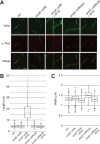
gp56 does not inhibit FtsZ ring assembly in B. subtilis. (A) Representative false-colored immunofluorescence micrographs of glutaraldehyde/paraformaldehyde-fixed JH642 (WT), DPH102 (amyE::orf56), and DPH175 (amyE::orf56Δ65) cells taken from mid-log-phase cultures with or without 1 mM IPTG as indicated. Fluorescent wheat germ agglutinin (WGA) staining (top row) shows the cell wall, and anti-FtsZ (middle row) shows the signal from fluorescence marker-conjugated secondary antibody to the primary antibody against FtsZ. Merge (bottom row) shows a merge of the WGA and anti-FtsZ signals. (B) Scatter-box plots of cell length quantification (n > 100) of strains from panel A. Box borders denote upper and lower quartiles, with the horizontal line in the box depicting the median and X depicting the mean. Whiskers show upper and lower deviations of data. (C) Scatter-box plots of cell width quantification (n > 100) of strains from panel A, with data shown as in panel B.
FIG 3
FIG 3
gp56 prevents recruitment of late, essential division proteins needed for B. subtilis septal cell wall synthesis. (A) Representative false-colored fluorescence micrographs of live cells of strains expressing EzrA-GFP (PL847 and DPH111), GFP-DivIB (DPH79 and DPH97), GFP-DivIC (DPH617 and DPH618), GFP-FtsL (DPH1108 and DPH584), GFP-Pbp2B (DPH414 and DPH415), or GFP-FtsW (DPH408 and DPH409) in the absence (left) or presence (right) of gp56. Each field of view includes a DIC image showing cells in bright-field, FM4-64 showing fluorescently stained membranes, and the GFP fluorescence signal showing localization of the indicated fusion construct. Scale bar = 5 μm. (B) Merged panels, on the same scale, of WGA and GFP signals from panel A.
FIG 4
FIG 4
GFP-gp56 localizes to the B. subtilis site of division in an FtsZ-dependent manner. (A) Representative false-colored fluorescence micrographs of live DPH50 (amyE::gfp-orf56) and DPH170 (amyE::gfp-orf56Δ65) cells taken from mid-log-phase cultures with 0.1% xylose present to induce fusion protein expression. The GFP signal (top) shows fusion protein localization, and the FM4-64 fluorescence signal (bottom) shows cell membranes. Scale bar = 5 μm. (B) Representative false-colored fluorescence micrographs of live DPH504 (ftsZ::spc xylA::tet thrC::Pxyl-ftsZ amyE::Pspac-gfp-orf56) cells taken from mid-log-phase cultures with 1 mM IPTG present to induce GFP-gp56 in the presence (left) or absence (right) of xylose to express or to deplete FtsZ. The GFP signal (top) shows GFP-gp56 localization, and the FM4-64 fluorescence signal (bottom) shows cell membranes. Scale as in panel A.
FIG 5
FIG 5
GFP-gp56 localization to the B. subtilis site of division requires DivIC/FtsL. (A) Representative false-colored fluorescence micrographs of live cells taken from mid-log-phase cultures containing (left) or missing (right) FtsA (DPH503), EzrA (JH642 or DPH55), DivIB (JH642 or DPH177), DivIC (DPH302), FtsL and Pbp2B in combination (DPH1121), or Pbp2B alone (DPH1119) through deletion/depletion. Each field of view includes a DIC image showing cells in bright-field, FM4-64 showing fluorescently stained membranes, and the GFP fluorescence signal showing localization of GFP-gp56. Scale bar = 5 μm. (B) Merged panels, on the same scale, of WGA and GFP signals from panel A.
FIG 6
FIG 6
SP01 gp56 interacts FtsL and its activity is suppressed by divIC overexpression. (A) Photos of LB Amp100 Kan50 agar plates with 1 mM IPTG and 50 g/ml X-Gal, showing growth of DHM1 BACTH background E. coli strains harboring pUT18C-orf56 (left), pUT18C-orf56Δ65 (center), or pUT18C-ftsL (right) and a panel of indicated genes cloned in pKT25. (B) β-Galactosidase activities of the indicated BACTH plasmid combinations in strains as in panel A. Values are an average of three independent trials, with error bars representing standard deviations. (C) Representative false-colored fluorescence micrographs of live cells taken from mid-log-phase cultures, with DIC (left) showing cells in bright-field and FM4-64 (right) showing fluorescently stained membranes. DPH660 (amyE::Pspac-divIC pPW19) and DPH661 (amyE::Pspac-divIC pPW19-orf56) cells were grown with 1 mM IPTG to overexpress divIC and to express gene 56 from pPW19 (top two rows). DPH1152 (amyE::Pspac-divIC thrC::Pxyl-orf56) cells were grown with 0.1% xylose to express gene 56 in the absence (third row) or presence (bottom row) of 1 mM IPTG to overexpress divIC. Scale bar = 5 μm.
FIG 7
FIG 7
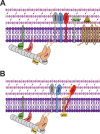
SP01 gp56 interacts with B. subtilis FtsL to disrupt the division machinery and to prevent recruitment of the Pbp2B and FtsW that are essential for septal cell wall synthesis. (A) Under normal conditions, the B. subtilis division machinery assembles into a cytokinesis-competent apparatus allowing for proper membrane constriction and septal wall synthesis. FtsZ polymers assemble at the membrane through interactions with membrane-associated FtsA, SepF, and EzrA. Stabilized by nonessential ZapA, assembled FtsZ and its membrane-associated partners allow recruitment of DivIB-FtsL-DivIC complexes that in turn help recruit Pbp2B and FtsW. (B) In the presence of SP01 gp56, the phage peptide interacts with FtsL, mildly disrupting its normal localization and its interaction with DivIC, preventing the normal recruitment of Pbp2B and FtsW. While lack of this late recruitment would also normally lead to rapid loss of the DivIB-DivIC-FtsL complex, its interaction with gp56 prevents their loss, effectively freezing the division machinery in a midassembled state that is unable to constrict or to build the septal cell wall, thereby leading to cell filamentation and death.

References

    1. Haeusser DP, Margolin W. 2016. Splitsville: structural and functional insights into the dynamic bacterial Z ring. Nat Rev Microbiol 14:305–319. doi:10.1038/nrmicro.2016.26. - DOI - PMC - PubMed
    1. Trueba FJ. 1982. On the precision and accuracy achieved by Escherichia coli cells at fission about their middle. Arch Microbiol 131:55–59. doi:10.1007/BF00451499. - DOI - PubMed
    1. Yu X-C, Margolin W. 1999. FtsZ ring clusters in min and partition mutants: role of both the Min system and the nucleoid in regulating FtsZ ring localization. Mol Microbiol 32:315–326. doi:10.1046/j.1365-2958.1999.01351.x. - DOI - PubMed
    1. Du S, Lutkenhaus J. 2019. At the heart of bacterial cytokinesis: the Z ring. Trends Microbiol 27:781–791. doi:10.1016/j.tim.2019.04.011. - DOI - PMC - PubMed
    1. Tripathy S, Sahu SK. 2019. FtsZ inhibitors as a new genera of antibacterial agents. Bioorg Chem 91:103169. doi:10.1016/j.bioorg.2019.103169. - DOI - PubMed

Publication types

MeSH terms

LinkOut - more resources