. 2020 Oct 19;10(1):17712.
doi: 10.1038/s41598-020-74677-7.

A pioneer calf foetus microbiome

Affiliations

A pioneer calf foetus microbiome

Cesar E Guzman et al. Sci Rep. .

Abstract

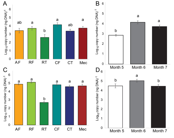
Foetus sterility until parturition is under debate due to reports of microorganisms in the foetal environment and meconium. Sufficient controls to overcome sample contamination and provide direct evidence of microorganism viability in the pre-rectal gastrointestinal tract (GIT) have been lacking. We conducted molecular and culture-based analyses to investigate the presence of a microbiome in the foetal GIT of calves at 5, 6 and 7 months gestation, while controlling for contamination. The 5 components of the GIT (ruminal fluid, ruminal tissue, caecal fluid, caecal tissue and meconium) and amniotic fluid were found to contain a pioneer microbiome of distinct bacterial and archaeal communities. Bacterial and archaeal richness varied between GIT components. The dominant bacterial phyla in amniotic fluid differed to those in ruminal and caecal fluids and meconium. The lowest bacterial and archaeal abundances were associated with ruminal tissues. Viable bacteria unique to the ruminal fluids, which were not found in the controls from 5, 6 and 7 months gestation, were cultured, subcultured, sequenced and identified. We report that the foetal GIT is not sterile but is spatially colonised before birth by a pioneer microbiome.


Conflict of interest statement

The authors declare no competing interests.

Figures

Figure 1
Figure 1
Quantitative PCR estimation (mean ± SE) of bacterial (A,B) and archaeal (C,D) 16S rRNA gene copy number in each GIT component and amniotic fluid (A,C) and at each gestational age (B,D). Amniotic fluid (AF), ruminal fluid (RF, n = 12), ruminal tissue (RT, n = 4), caecal fluid (CF, n = 8), caecal tissue (CT, n = 10), meconium (Mec, n = 11). Gestational age: 5 months (n = 16), 6 months (n = 18), 7 months (n = 18). Different letters above bars indicate a significant difference (p < 0.05) between means as determined via Tukey’s HSD and Dunn’s post hoc tests for bacterial and archaeal data, respectively.
Figure 2
Figure 2
Box plots of bacteria (AC) and archaea (DF) for Shannon diversity indices (A,D), observed Chao1 estimates of ESV richness (B,E), and Simpson evenness (C,F) for communities between GIT components and amniotic fluid. Amniotic fluid (AF), ruminal fluid (RF), ruminal tissue (RT), caecal fluid (CF), caecal tissue (CT), meconium (Mec). Different letters above bars indicate a significant difference via Kruskal–Wallis testing (p < 0.05).
Figure 3
Figure 3
Non-metric multidimensional scaling ordination of bacterial (A,C) and archaeal (B,D) 16S rRNA profiles using weighted (A,B) and unweighted (C,D) UniFrac distance metrics. Colour is indicative of the sample type from which the community was sampled: orange formula image , amniotic fluid; light green formula image , ruminal fluid; dark green formula image , ruminal tissue; cyan formula image , caecal fluid; blue formula image , caecal tissue; red formula image , meconium. Two-dimension stress ranged from 0.06 to 0.08.
Figure 4
Figure 4
Mean relative abundance of bacterial phylum level (A) and order level (B) ESVs in each sample type (GIT component and amniotic fluid) at each gestational age (5, 6 and 7 months). Significant phylum-level and order-level changes in relative abundance between sample types are denoted next to the taxonomic key. For each pair of sample types, the arrow indicates the direction of change in abundance in the second sample type relative to the first. ESVs are clustered by colour to the levels of phylum and order. ESVs with taxonomic assignments at the phylum level and order level with confidence values lower than 90% are denoted as ‘Unknown’. Levels of gDNA in caecal fluid from foetuses at 5 months gestation were below the concentration threshold (0.5 ng/µL) for samples to be included in the sequencing run. Amniotic fluid (AF), ruminal fluid (RF), ruminal tissue (RT), caecal fluid (CF), caecal tissue (CT), meconium (Mec).
Figure 5
Figure 5
Mean relative abundance of archaeal ESVs in each sample type (GIT component and amniotic fluid) at each gestational age (5, 6 and 7 months). ESVs are clustered by colour to the level of order. ESVs with taxonomic assignments at the order level with confidence values lower than 90% are denoted as ‘Unknown’. The gDNA from caecal tissues at 5 months gestation was below the concentration threshold (0.5 ng/µL) for samples to be included in the sequencing run. Amniotic fluid (AF), ruminal fluid (RF), ruminal tissue (RT), caecal fluid (CF), caecal tissue (CT), meconium (Mec).

References

    1. Grönlund M, Arvilommi H, Kero P, Lehtonen O, Isolauri E. Importance of intestinal colonisation in the maturation of humoral immunity in early infancy: a prospective follow up study of healthy infants aged 0–6 months. Arch. Dis. Childhood Fetal Neonat. Ed. 2000;83:F186–F192. - PMC - PubMed
    1. Chong C, Bloomfield F, O’Sullivan J. Factors affecting gastrointestinal microbiome development in neonates. Nutrients. 2018;10:274. - PMC - PubMed
    1. Gritz EC, Bhandari V. The human neonatal gut microbiome: a brief review. Front. Pediatr. 2015 doi: 10.3389/fped.2015.00017. - DOI - PMC - PubMed
    1. Bäckhed F, et al. Dynamics and stabilization of the human gut microbiome during the first year of life. Cell Host Microbe. 2015;17:690–703. - PubMed
    1. Grölund M-M, Lehtonen O-P, Eerola E, Kero P. Fecal microflora in healthy infants born by different methods of delivery: permanent changes in intestinal flora after cesarean delivery. J. Pediatr. Gastroenterol. Nutr. 1999;28:19–25. - PubMed

Publication types