Review
. 2021 Jan;17(1):46-64.
doi: 10.1038/s41581-020-00357-4. Epub 2020 Oct 19.

Immunity, endothelial injury and complement-induced coagulopathy in COVID-19

Affiliations
Review

Immunity, endothelial injury and complement-induced coagulopathy in COVID-19

Luca Perico et al. Nat Rev Nephrol. 2021 Jan.

Abstract

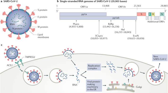
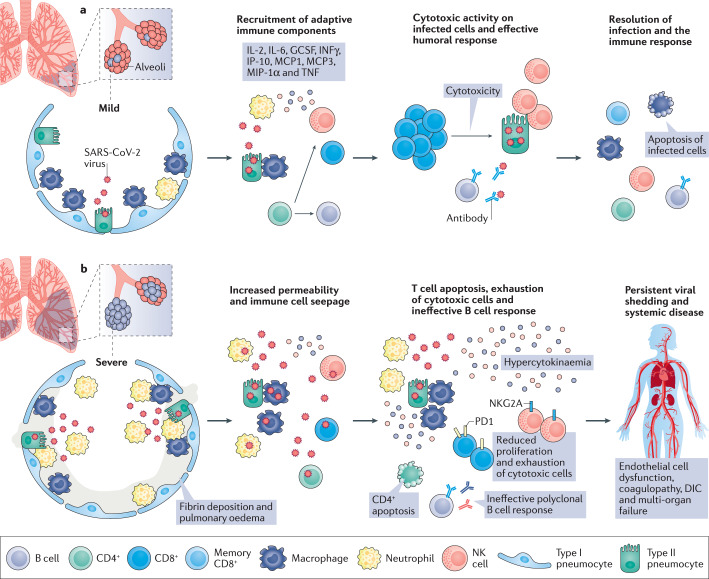
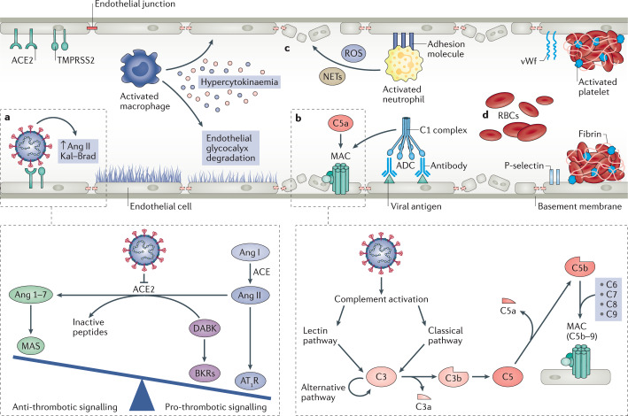
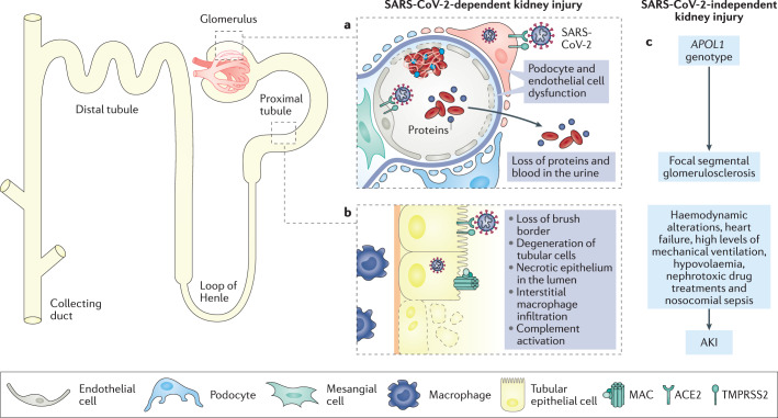
In December 2019, a novel coronavirus was isolated from the respiratory epithelium of patients with unexplained pneumonia in Wuhan, China. This pathogen, named severe acute respiratory syndrome coronavirus 2 (SARS-CoV-2), causes a pathogenic condition that has been termed coronavirus disease 2019 (COVID-19) and has reached pandemic proportions. As of 17 September 2020, more than 30 million confirmed SARS-CoV-2 infections have been reported in 204 different countries, claiming more than 1 million lives worldwide. Accumulating evidence suggests that SARS-CoV-2 infection can lead to a variety of clinical conditions, ranging from asymptomatic to life-threatening cases. In the early stages of the disease, most patients experience mild clinical symptoms, including a high fever and dry cough. However, 20% of patients rapidly progress to severe illness characterized by atypical interstitial bilateral pneumonia, acute respiratory distress syndrome and multiorgan dysfunction. Almost 10% of these critically ill patients subsequently die. Insights into the pathogenic mechanisms underlying SARS-CoV-2 infection and COVID-19 progression are emerging and highlight the critical role of the immunological hyper-response - characterized by widespread endothelial damage, complement-induced blood clotting and systemic microangiopathy - in disease exacerbation. These insights may aid the identification of new or existing therapeutic interventions to limit the progression of early disease and treat severe cases.


Conflict of interest statement

The authors declare no competing interests.

Figures

Fig. 1
Fig. 1. SARS-CoV-2 structure, genome composition and life cycle.
a | Severe acute respiratory syndrome coronavirus 2 (SARS-CoV-2) is a typical betacoronavirus belonging the Orthocoronavirinae family. Each SARS-CoV-2 virion has a diameter of approximately 100–200 nm. Like other coronaviruses, the SARS-CoV-2 envelope comprises a lipid membrane and three structural components: the spike (S) glycoprotein, the envelope (E) protein and the membrane (M) protein. Within the viral envelope, the nucleocapsid (N) protein holds the positive-sense single-stranded RNA, which is 29,903 bases in length. b | The SARS-CoV-2 genome is composed of ten open reading frames (ORFs). At least two-thirds of the viral genome are contained in ORF1a and ORF1b, which together encode a polyprotein, pp1ab, which is further cleaved into 16 non-structural proteins that are involved in genome transcription and replication. Of these proteins, papain-like protease (PLpro) and 3C-like protease (3CLpro) are encoded by ORF1a, whereas RNA-dependent RNA polymerase (RdRp), helicase (Hel) and exonuclease (ExoN) are encoded by ORF1b. The remaining ORFs encode the structural S glycoproteins, and the E protein, M protein and N protein, as well as several accessory proteins with unknown functions. c | SARS-CoV-2 enters the host cell using the endosomal pathway and the cell surface non-endosomal pathway. In the setting of endosomal entry, the SARS-CoV-2 virion attaches to its target cells by direct binding of the S glycoprotein to the host receptor angiotensin-converting enzyme 2 (ACE2). Upon binding, the transmembrane protease serine 2 (TMPRSS2) cleaves and primes S glycoprotein, leading to the fusion of the viral and cell membranes. In addition to canonical viral entry via the endosomal pathway, non-endosomal entry at the plasma membrane may be an additional infection route for SARS-CoV-2. Within the target cells, SARS-CoV-2 is disassembled to release nucleocapsid and viral RNA into the cytoplasm for translation and replication. Translated viral proteins are then assembled in the endoplasmic reticulum (ER) to form the new virions, which are then released from the Golgi membrane system by exocytosis into the extracellular compartment.
Fig. 2
Fig. 2. Pathogenesis and outcomes of COVID-19.
a | Following infection of the lungs, (severe acute respiratory syndrome coronavirus 2 (SARS-CoV-2) induces the death of epithelial cells, in particular, type II pneumocytes, as part of the viral replication cycle. Macrophages and neutrophils elicit a specific innate immune response to eradicate the pathogen and kill virus-infected cells. The increase in pro-inflammatory cytokines within the lung leads to the recruitment of leukocytes, further propagating the local inflammatory response. Among these cytokines, interleukin-2 (IL-2), IL-6, granulocyte-colony-stimulating factor (GCSF), interferon-γ (IFNγ), IFNγ inducible protein 10 (IP-10), monocyte chemoattractant protein 1 and 3 (MCP1 and 3), macrophage inflammatory protein 1α (MIP-1α) and tumour necrosis factor (TNF) stimulate the adaptive immune response. At this stage, infiltration of lymphocytes (CD4+ and CD8+ T cells) and natural killer (NK) cells is required to ensure an optimal defence response against SARS-CoV-2. CD4+ T cells mediate antibody production by B cells and also enhance effector CD8+ T cell and NK responses during viral infections. This orchestrated immune response leads to viral eradication and resolution of the disease. In these patients, coronavirus disease 2019 (COVID-19) seems to manifest as a mild disease with symptoms similar to the common flu that resolve spontaneously. b | For unknown reasons, but possibly individual predisposition or differences in viral loads during primary SARS-CoV-2 infection, some patients experience more severe disease. A maladaptive immune response, characterized by lymphopenia and suppression of CD4+ T cells, is likely accountable for the poor prognosis of these patients. In the absence of robust CD4+ T cell activation, B cells generate a polyclonal antibody response that may be ineffective in neutralizing SARS-CoV-2. Increased numbers of exhausted T cells that express high levels of programmed cell death protein 1 (PD1), suggest decreased proliferation and activity of CD8+ T cells. Similarly, NK cells exhibit increased levels of the inhibitory CD94–NK group 2 member A (NKG2A). Impaired cytotoxic activity results in persistent viral shedding that amplifies macrophage and neutrophil activation, leading to the massive production of cytokines (a process referred to as hypercytokinaemia). In these patients, COVID-19 manifests as a severe disease, consisting of advanced pneumonia and acute respiratory distress syndrome. The generation of excess cytokines and persistent viral infection leads to systemic vascular damage, disseminated intravascular coagulation (DIC) and the failure of vital organs, including the kidney and the heart.
Fig. 3
Fig. 3. Effect of SARS-CoV-2 infection on endothelial cell function, systemic coagulation and thrombosis.
a | Severe acute respiratory syndrome coronavirus 2 (SARS-CoV-2) directly infects endothelial cells owing to their high expression levels of angiotensin-converting enzyme 2 (ACE2) and transmembrane protease serine 2 (TMPRSS2). After binding by SARS-CoV-2, ACE2 is internalized, and the lack of ACE2 on endothelial cells favours the progression of inflammatory and thrombotic processes triggered by local angiotensin II (Ang II) hyperactivity. Inhibition of ACE2 by binding of SARS-CoV-2 reduces the ACE2-mediated conversion of Ang II to Ang 1–7, the vasoactive ligand of the MAS receptor. The reduction of MAS receptor activation induces a pro-inflammatory phenotype through increased activation of type 1 angiotensin receptors (AT1R). Additionally, the reduction in levels of ACE2 limits the degradation of des-Arg9 bradykinin (DABK) into inactive peptides, ultimately leading to increased pro-thrombotic signalling via the activation of bradykinin receptors (BKRs). b | SARS-CoV-2 also activates the complement system — an integral component of the innate immune response. The complement cascade can be activated by three different pathways, the classical, the lectin and the alternative pathway, that resolve around the formation of the C3 convertases that cleave C3, generating the pro-inflammatory peptide C3a and large amount of C3b that opsonizes pathogens. C3b also forms the C5 convertase, which leads to release of the potent anaphylatoxin C5a, as well as the fragment C5b, responsible for the formation of the membrane attack complex (MAC) C5b–9 on target cells, which is considered to be the terminal event of complement activation. In addition, complexes of SARS-CoV-2-specific antibodies and viral antigens might induce endothelial cell injury through activation of the C1 complex of the classical pathway and induction of antibody-dependent cytotoxicity (ADC). c | Pro-inflammatory cytokines and chemokines released by activated macrophages amplify the vicious cycle of vascular integrity disruption, vessel coagulation and thrombosis by degrading the endothelial glycocalyx, activating the coagulation system and dampening anticoagulant mechanisms. The adhesive phenotype of endothelial cells induced by inflammatory cytokines and chemokines promotes infiltration of neutrophils, which produce large amounts of histotoxic mediators, including reactive oxygen species (ROS) and neutrophil extracellular traps (NETs), ultimately leading to injury of endothelial cells. d | Activated endothelial cells initiate coagulation by expressing P-selectin, von Willebrand factor (vWf) and fibrinogen, leading to massive platelet binding, fibrin formation and clotting of red blood cells (RBCs), ultimately resulting in systemic thrombosis and disseminated intravascular coagulation.
Fig. 4
Fig. 4. The effects of COVID-19 on the kidney.
The kidney expresses high levels of angiotensin-converting enzyme 2 (ACE2) and transmembrane protease serine 2 (TMPRSS2) and has been identified as a target organ for severe acute respiratory syndrome coronavirus 2 (SARS-CoV-2) infection by several studies. a | Within the glomerulus, podocytes and endothelial cells have been identified as specific sites for viral infection. SARS-CoV-2-induced podocyte dysfunction is likely to induce impairment of glomerular filtration, leading to proteinuria and haematuria, which are often observed in patients with coronavirus disease 2019 (COVID-19). Infection of endothelial cells alters glomerular capillary haemostasis and induces the formation of fibrin thrombi. b | SARS-CoV-2 has also been found in proximal tubular cells, which exhibit high levels of ACE2 in the apical brush border. SARS-CoV-2 infection leads to loss of the brush border and vacuolar degeneration in tubular epithelial cells, with luminal debris composed of necrotic epithelium with evidence of complement activation and membrane attack complex (MAC) (C5b–9) deposition on tubular cells and massive macrophage infiltration in the kidney interstitium. c | In addition, a non-viral-dependent mechanism might also be accountable for kidney dysfunction in the context of COVID-19, including a possible contribution of the APOL1 ‘risk’ genotype in inducing focal segmental glomerulosclerosis following SARS-CoV-2 infection, as well as the role of haemodynamic factors, cardiac dysfunction, high levels of mechanical ventilation, hypovolaemia, nephrotoxic drug treatments and nosocomial sepsis resulting in COVID-19-associated acute kidney injury (AKI).

References

    1. Andersen KG, Rambaut A, Lipkin WI, Holmes EC, Garry RF. The proximal origin of SARS-CoV-2. Nat. Med. 2020;6:450–452. - PMC - PubMed
    1. Zhou P, et al. A pneumonia outbreak associated with a new coronavirus of probable bat origin. Nature. 2020;579:270–273. - PMC - PubMed
    1. Shi J, et al. Susceptibility of ferrets, cats, dogs, and other domesticated animals to SARS-coronavirus 2. Science. 2020;368:1016–1020. - PMC - PubMed
    1. Zhang YZ, Holmes EC. A genomic perspective on the origin and emergence of SARS-CoV-2. Cell. 2020;181:223–227. - PMC - PubMed
    1. Zhou H, et al. A novel bat coronavirus closely related to SARS-CoV-2 contains natural insertions at the S1/S2 cleavage site of the spike protein. Curr. Biol. 2020;30:2196–2203.e3. - PMC - PubMed

Publication types

MeSH terms

Substances