. 2021 Feb;5(2):169-178.
doi: 10.1038/s41551-020-00632-6. Epub 2020 Oct 19.

Restoration of visual function in adult mice with an inherited retinal disease via adenine base editing

Affiliations

Restoration of visual function in adult mice with an inherited retinal disease via adenine base editing

Susie Suh et al. Nat Biomed Eng. 2021 Feb.

Erratum in

Abstract

Cytosine base editors and adenine base editors (ABEs) can correct point mutations predictably and independent of Cas9-induced double-stranded DNA breaks (which causes substantial indel formation) and homology-directed repair (which typically leads to low editing efficiency). Here, we show, in adult mice, that a subretinal injection of a lentivirus expressing an ABE and a single-guide RNA targeting a de novo nonsense mutation in the Rpe65 gene corrects the pathogenic mutation with up to 29% efficiency and with minimal formation of indel and off-target mutations, despite the absence of the canonical NGG sequence as a protospacer-adjacent motif. The ABE-treated mice displayed restored RPE65 expression and retinoid isomerase activity, and near-normal levels of retinal and visual functions. Our findings motivate the further testing of ABEs for the treatment of inherited retinal diseases and for the correction of pathological mutations with non-canonical protospacer-adjacent motifs.


Conflict of interest statement

Competing interests

D.R.L. is a consultant and co-founder of Beam Therapeutics, Prime Medicine, Editas Medicine and Pairwise Plants, companies that use genome editing.

Figures

Fig. 1 |
Fig. 1 |. In vitro validation of Rpe65 mutation correction by the ABE.
a, The rd12 mouse model has a homozygous C•G to T•A nonsense mutation in exon 3 of the Rpe65 gene, changing arginine (green) to a stop codon (red). b, The ABE efficiently converts A to G in the genome that corresponds to a window of positions ~4-8 (yellow) in the guide RNA, counting the NGG PAM as positions 21-23. c, Validation of rd12 (left lane) and WT (right lane) reporter cell lines by western blot analysis. The RPE65 band (65 kDa) is detected from the WT cell lysate but not from the rd12 cell lysate. α-Tubulin (52 kDa) was used as a loading control. d, Five sgRNAs were designed to place the target mutation (red letter) within the ABE activity window. e, Western blot analysis of rd12 cells following ABEmax and sgRNA plasmid transfection, showing the rescue of RPE65 protein with sgRNA-A5 and sgRNA-A6. rd12 cells transfected with pcDNA3.1 encoding WT mouse RPE65 were used as a positive control (+Ctrl). Non-transfected rd12 cells were used as a negative control (−Ctrl). ABE (200 kDa) and β-actin (42 kDa) were used as loading controls. An additional band at 50 kDa has unknown identity but is irrelevant to RPE65 expression. MM, molecular mass. f, Immunocytochemistry of rd12 cells transfected with sgRNA-A5 and sgRNA-A6 (n = 3 images from independent experiments), showing RPE65+ cells (red). Green indicates GRP78 and blue indicates DAPI staining. GRP78 served as a marker for endoplasmic reticulum localization. Scale bars, 50 μm. g, Deep sequencing of the target locus in rd12 cells at 48 h post-transfection (n = 4 biologically independent replicates). Means ± s.e.m. are shown. The types of allelic variants are shown as DNA and translated amino acid sequences in the table. Allelic variants occurring at a frequency of <0.1% were not included in the analysis.
Fig. 2 |
Fig. 2 |. Subretinal delivery of ABE corrects the mutation and restores RPE65 expression in rd12 mice.
a, Schematic of three lentiviral vector genomes for subretinal delivery (top) and an outline of the in vivo experiments (bottom). LV-ABE-A5 and -A6 express the codon-optimized ABE (ABEmax) and sgRNA-A5 and sgRNA-A6, respectively. LV-ABE-NT is a control virus expressing ABEmax and non-targeting sgRNA. CMV, cytomegalovirus; LTR, long terminal repeat. b, Western blot analysis to detect RPE65 (65 kDa) expression from mouse RPE tissue lysate after treatment. Each lane represents a single eye with ~100% eGFP coverage. ABE (200 kDa) and β-actin (42 kDa) were used as loading controls. c,d, Immunofluorescence analysis of representative eye cross-sections (c) and RPE flatmounts (d) of treated mice. Green indicates RPE65, blue indicates DAPI staining and red shows ZO-1 (a protein marker for tight junctions). Scale bars, 50 μm. INL, inner nuclei layer; ONL, outer nuclei layer. n = 2 images in c and n = 3 images in d. e, Quantification of RPE65+ cells from a single representative RPE flatmount, based on immunofluorescence (n = 10 images representing different areas). Means ± s.e.m. are shown. f, Deep-sequencing analysis of the rd12 locus in genomic DNA isolated from the RPE tissue of untreated (n = 3 eyes), NT-treated (n = 4 eyes), A5-treated (n = 8 eyes) and A6-treated mice (n = 8 eyes). Means ± s.e.m. are shown. g, Pie charts showing the composition of allelic variants in one representative eye, for the A5 treatment (left) and the A6 treatment (right). Fifteen nucleotides spanning the target mutation (T7) are shown as a reference using the unedited rd12 mouse sequence. Allelic variants with <0.05% reads were excluded. Unless otherwise noted, all post-treatment analyses were performed 5 weeks after subretinal injection using the mouse eyes that had >70% eGFP coverage.
Fig. 3 |
Fig. 3 |. Restoration of a visual cycle and retinal function in rd12 mice after base editing.
a, Schematic of the visual cycle, demonstrating the enzymatic role of RPE65. RDH5, retinol dehydrogenase 5. b,c, Retinoid profiles of dark-adapted mouse eyes (b) and 0.5-s flash-bleached mouse eyes (c). Peak a, all-trans-retinyl esters; peak b, 11-cis-retinal; peak c, all-trans-retinal. Each chromatogram represents the homogenate from two eyes. d, Schematic of the visual pathway in the mouse. LGN, lateral geniculate nucleus; SC, superior colliculus; V1, primary visual cortex. e, Scotopic ERG waveforms of five mice from each group upon light stimulus of −0.3 log[cd s m−2]. A representative waveform from one WT mouse is shown at the top. f, Measurements of scotopic a- and b-wave amplitudes by the ERG recordings in e (n = 8 mice in each group). Means ± s.e.m. are shown. ***P < 0.001 (one-way analysis of variance with Bonferroni test). g, Schematic of the OMR apparatus. A mouse is placed on an elevated platform where it can freely move and track the virtual rotating pattern stimulus displayed on the screen. Evaluation of head movements synchronous to the stimulation is automated. h,i, Summary of OMRs at various pattern contrasts (h) and a comparison of average responses at 10, 20 and 50% pattern contrasts (i). An OMR index of 1 indicates head movements by chance. Statistically significant tracking was inferred using a one-tailed t-test based on a null hypothesis mean of 1 (h). Thin lines represent responses from individual animals. Thick lines represent average responses from each group. Numbers of mice were as follows: n = 6 (untreated); n = 7 (NT treated); n = 8 (A5 treated); and n = 8 (WT). The dashed horizontal line indicates an OMR index of 1. Means ± s.e.m. are shown. ***P < 0.001 (one-way analysis of variance with Bonferroni test).
Fig. 4 |
Fig. 4 |. Base editing restores neuronal activity of the primary visual cortex (V1) in response to visual stimuli.
a,b, Representative example of VEPs from a single mouse (a) and the average of normalized VEPs from each group (b). Shading indicates s.e.m. N, number of mice; n, number of recording sites. c,d, Population summaries of normalized VEP amplitudes (c) and response latency (d). Means ± s.e.m. are shown. ***P < 0.001 (two-tailed Mann-Whitney U-test). e, Flash-evoked single-neuron recordings from WT, untreated, NT-treated and A5-treated mice are shown as raster plots for single neurons (top) and as population averages (solid lines; bottom). Shading indicates s.e.m. The 500-ms duration of the flash stimulus is indicated by light grey background shading. Numbers of neurons (n) and mice (N) were as follows: n = 84 and N = 10 (WT); n = 62 and N = 4 mice (untreated); n = 75 and N = 6 (NT treated); and n = 81 and N = 8 mice (A5 treated). f, Summary of single-neuron firing rates from each group in response to flash stimuli. Means ± s.e.m. are shown. ***P < 0.001 (two-tailed Mann-Whitney U-test). g, Proportion of visually responsive and non-responsive single neurons in V1. Sample sizes for f and g match the numbers of neurons in e.
Fig. 5 |
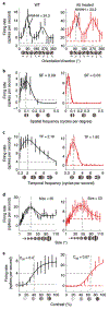
Fig. 5 |. V1 neurons in A5-treated mice show selectivity to stimulus parameters.
ae, Comparisons of single-cell responses to different stimulus parameters between WT and A5-treated mice, including tuning curves for orientation (a), spatial frequency (b), temporal frequency (c), size (d) and contrast (e). Horizontal dashed lines indicate background activity, whereas vertical dashed lines indicate optimum stimulus parameters. C50, percentage contrast at half of the peak response; HWHH, half-width at half height; SF, preferred spatial frequency; TF, optimal temporal frequency; size, optimal stimulus diameter. Mean ± s.e.m. are shown.

Comment in

References

    1. Cremers FP, van den Hurk JA & den Hollander AI Molecular genetics of Leber congenital amaurosis. Hum. Mol. Genet. 11, 1169–1176 (2002). - PubMed
    1. Den Hollander AI, Roepman R, Koenekoop RK & Cremers FPM Leber congenital amaurosis: genes, proteins and disease mechanisms. Prog. Retin. Eye Res. 27, 391–419 (2008). - PubMed
    1. Miraldi Utz V, Coussa RG, Antaki F & Traboulsi EI Gene therapy for RPE65-related retinal disease. Ophthalmic Genet. 39, 671–677 (2018). - PubMed
    1. Maguire AM et al. Efficacy, safety, and durability of voretigene neparvovec-rzyl in RPE65 mutation-associated inherited retinal dystrophy: results of phase 1 and 3 trials. Ophthalmology 126, 1273–1285 (2019). - PubMed
    1. Bainbridge JW et al. Long-term effect of gene therapy on Leber’s congenital amaurosis. N. Engl. J. Med. 372, 1887–1897 (2015). - PMC - PubMed

Publication types