

Natural history of persistent subretinal fluid following the successful repair of rhegmatogenous retinal detachment
- PMID: 33078114
- PMCID: PMC7511378
- DOI: 10.18240/ijo.2020.10.17
Natural history of persistent subretinal fluid following the successful repair of rhegmatogenous retinal detachment
Abstract
Aim: To provide a detailed description of the natural history of persistent subretinal fluid (SRF) after successful repair of rhegmatogenous retinal detachment (RRD) and its association with visual outcome.
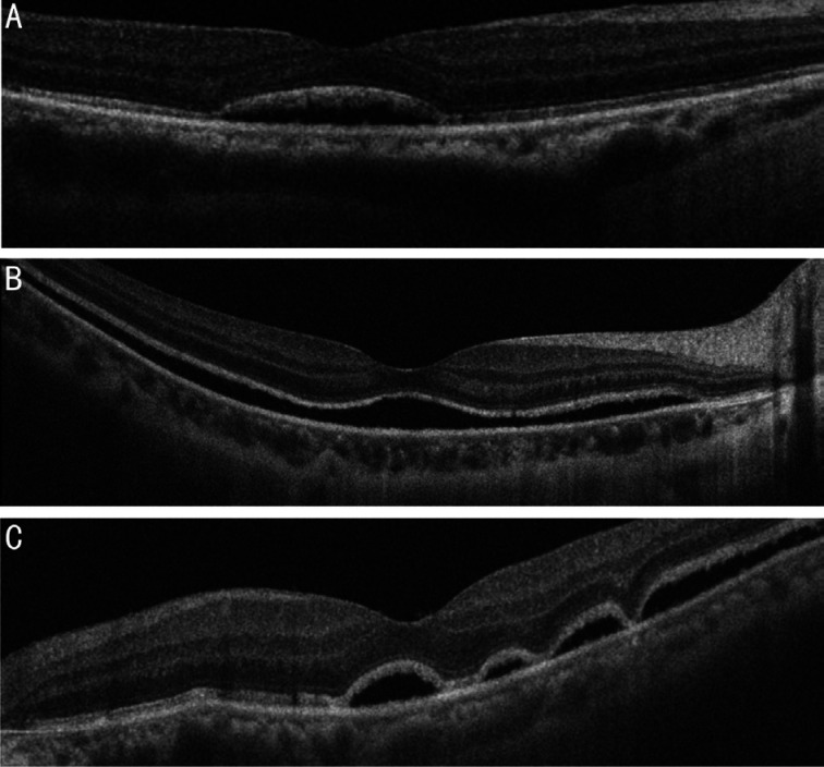
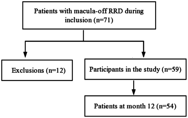
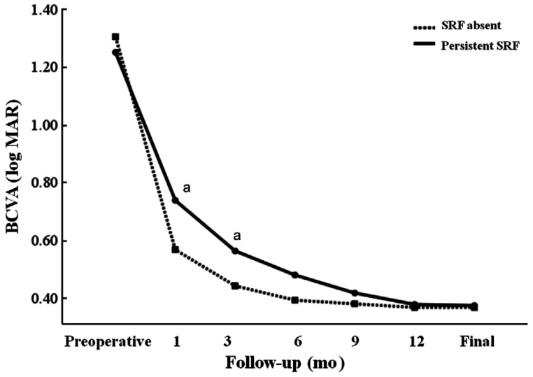
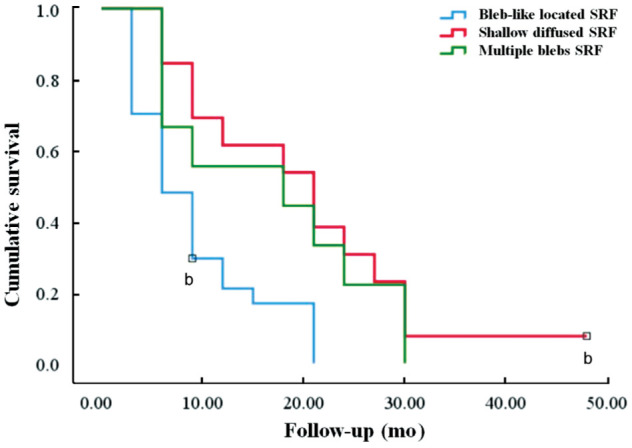
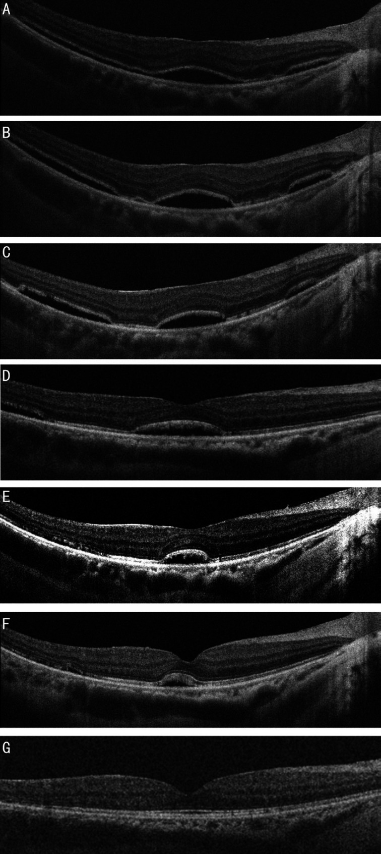
Methods: This was a prospective long-term follow-up for eyes undergoing scleral buckling (SB) surgery for macula-off RRD. Examinations were carried out preoperatively and postoperatively at 1, 3, 6, 9 and 12mo, until persistent SRF had completely resolved. One month postoperatively, optical coherence tomography (OCT) was used to classify SRF into three patterns: bleb-like loculated (BL), shallow-diffused (SD), and multiple blebs (MB). Serial OCT imaging was used to evaluate morphological changes in SRF until its complete disappearance. Patients were divided into two groups depending on the presence or absence of persistent SRF.
Results: A total of 59 patients (59 eyes) were included. There were no statistical differences between two groups at baseline, except for the proportion of patients with high myopia and a younger age. One month after surgery, OCT detected persistent SRF in 49 eyes (83.1%). The 3 morphological patterns of SRF were observed in 27 eyes (55.1%) with BL, 13 eyes (26.5%) with SD, and 9 eyes (18.4%) with MB. The mean time for complete absorption differed significantly across the three SRF patterns (F=8.097, P=0.001), which was 8.8±6.1, 20.1±12.1, and 16.7±10.2mo in BL, SD, and MB, respectively. In 9 of the 13 eyes with SD, the pattern transformed into MB type. In cases involving MB, the size and number of blebs decreased gradually until they had been completely absorbed. Eyes with persistent SRF were more likely to demonstrate disruption of the ellipsoid zone (49.0% vs 10%, P=0.034). The final best-corrected visual acuity of two groups was 0.37±0.11 (with SRF) vs 0.34±0.12 (without SRF) logMAR (P=0.499), respectively.
Conclusion: High preoperative myopia and younger age are associated with persistent SRF. BL is the most commonly observed pattern with the shortest duration and gradually disappeared. Most cases involving SD SRF transform into MB type during resolution. The size and number of the MBs decrease gradually until they were completely absorbed. The absence of persistent SRF may contribute to slow visual recovery in the short-term but does not influence the final visual outcome.
Keywords: optical coherence tomography; rhegmatogenous retinal detachment; subretinal fluid; visual acuity.
International Journal of Ophthalmology Press.
Figures
Similar articles
-
Recovery course of persistent posterior subretinal fluid after successful repair of rhegmatogenous retinal detachment.Eur J Ophthalmol. 2024 Jul;34(4):1217-1227. doi: 10.1177/11206721231210693. Epub 2023 Oct 30. Eur J Ophthalmol. 2024. PMID: 37901895
-
Spectral-domain optical coherence tomography analysis of persistent subretinal fluid after scleral buckling surgery for macula-off retinal detachment.Eye (Lond). 2015 Sep;29(9):1186-93. doi: 10.1038/eye.2015.113. Epub 2015 Jul 3. Eye (Lond). 2015. PMID: 26139048 Free PMC article.
-
Persistent subretinal fluid after successful scleral buckle surgery for macula-off retinal detachment.Chin Med J (Engl). 2011 Dec;124(23):4007-11. Chin Med J (Engl). 2011. PMID: 22340333
-
[Logistic regression analysis of risk factors for subretinal fluid after rhegmatogenous retinal detachment repair].Zhonghua Yan Ke Za Zhi. 2021 Jun 11;57(6):426-432. doi: 10.3760/cma.j.cn112142-20201225-00908. Zhonghua Yan Ke Za Zhi. 2021. PMID: 34098691 Chinese.
-
Persistent subretinal fluid after surgery for rhegmatogenous retinal detachment: hypothesis and review.Graefes Arch Clin Exp Ophthalmol. 2012 Jun;250(6):795-802. doi: 10.1007/s00417-011-1870-y. Epub 2012 Jan 11. Graefes Arch Clin Exp Ophthalmol. 2012. PMID: 22234351 Review.
Cited by
-
Reversing the paradigm on the urgency of acute retinal detachments defined by their foveal status: when off may be more urgent than on.BMJ Open Ophthalmol. 2024 Apr 19;9(1):e001668. doi: 10.1136/bmjophth-2024-001668. BMJ Open Ophthalmol. 2024. PMID: 38683951 Free PMC article. No abstract available.
-
Subfoveal Choroidal Thickness After Successful Retinal Detachment Repair with Persistent Subretinal Fluid at the Macula.Clin Ophthalmol. 2025 Jun 11;19:1847-1854. doi: 10.2147/OPTH.S520076. eCollection 2025. Clin Ophthalmol. 2025. PMID: 40599498 Free PMC article.
-
Effect of High Myopia on Delayed Absorption of Subretinal Fluid after Scleral Buckling Surgery.J Clin Med. 2022 Jul 5;11(13):3906. doi: 10.3390/jcm11133906. J Clin Med. 2022. PMID: 35807191 Free PMC article.
-
Effect of external subretinal fluid drainage on persistent subretinal fluid after scleral buckle surgery in macula-involving rhegmatogenous retinal detachment.Sci Rep. 2023 Dec 13;13(1):22176. doi: 10.1038/s41598-023-49719-5. Sci Rep. 2023. PMID: 38093092 Free PMC article.
-
How should we report the foveal status in eyes with "macula-off" retinal detachment?Eye (Lond). 2023 Feb;37(2):228-234. doi: 10.1038/s41433-022-02074-7. Epub 2022 May 3. Eye (Lond). 2023. PMID: 35505112 Free PMC article. Review.
References
-
- Kim YK, Woo SJ, Park KH, Yu YS, Chung H. Comparison of persistent submacular fluid in vitrectomy and scleral buckle surgery for macula-involving retinal detachment. Am J Ophthalmol. 2010;149(4):623–629.e1. - PubMed
-
- Ricker LJ, Noordzij LJ, Goezinne F, Cals DW, Berendschot TT, Liem AT, Hendrikse F, La Heij EC. Persistent subfoveal fluid and increased preoperative foveal thickness impair visual outcome after macula-off retinal detachment repair. Retina. 2011;31(8):1505–1512. - PubMed
-
- Chantarasorn Y, Oellers P, Eliott D. Choroidal thickness is associated with delayed subretinal fluid absorption after rhegmatogenous retinal detachment surgery. Ophthalmol Retina. 2019;3(11):947–955. - PubMed
-
- Benson SE, Schlottmann PG, Bunce C, Xing W, Charteris DG. Optical coherence tomography analysis of the macula after scleral buckle surgery for retinal detachment. Ophthalmology. 2007;114(1):108–112. - PubMed
LinkOut - more resources
Full Text Sources
Medical
Miscellaneous