

CBCT image quality QA: Establishing a quantitative program
- PMID: 33078562
- PMCID: PMC7701111
- DOI: 10.1002/acm2.13062
CBCT image quality QA: Establishing a quantitative program
Abstract
Purpose: Routine quality assurance (QA) of cone-beam computed tomography (CBCT) scans used for image-guided radiotherapy is prescribed by the American Association of Physicists in Medicine Task Group (TG)-142 report. For CBCT image quality, TG-142 recommends using clinically established baseline values as QA tolerances. This work examined how image quality parameters vary both across machines of the same model and across different CBCT techniques. Additionally, this work investigated how image quality values are affected by imager recalibration and repeated exposures during routine QA.
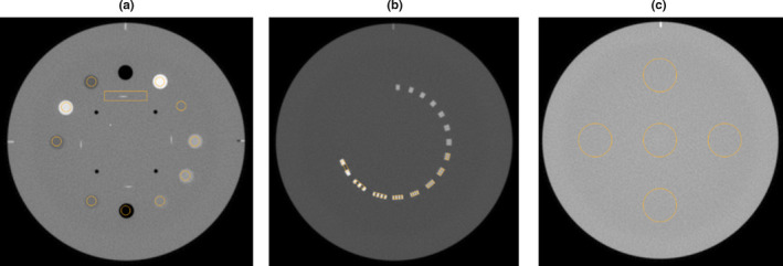
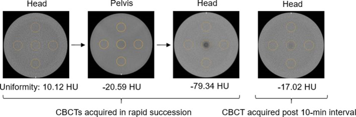
Methods: Cone-beam computed tomography scans of the Catphan 604 phantom were taken on four TrueBeam® and one Edge™ linear accelerator using four manufacturer-provided techniques. TG-142 image quality parameters were calculated for each CBCT scan using SunCHECK Machine™. The variability of each parameter with machine and technique was evaluated using a two-way ANOVA test on a dataset consisting of 200 CBCT scans. The impact of imager calibration on image quality parameters was examined for a subset of three machines using an unpaired Student's t-test. The effect of artifacts appearing on CBCTs taken in rapid succession was characterized and an approach to reduce their appearance was evaluated. Additionally, a set of baselines and tolerances for all image quality metrics was presented.
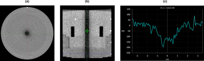
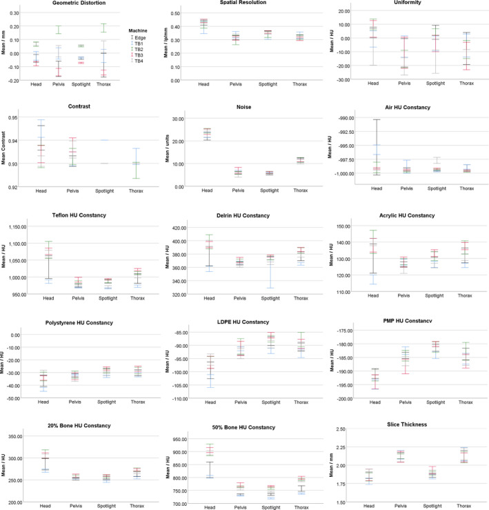
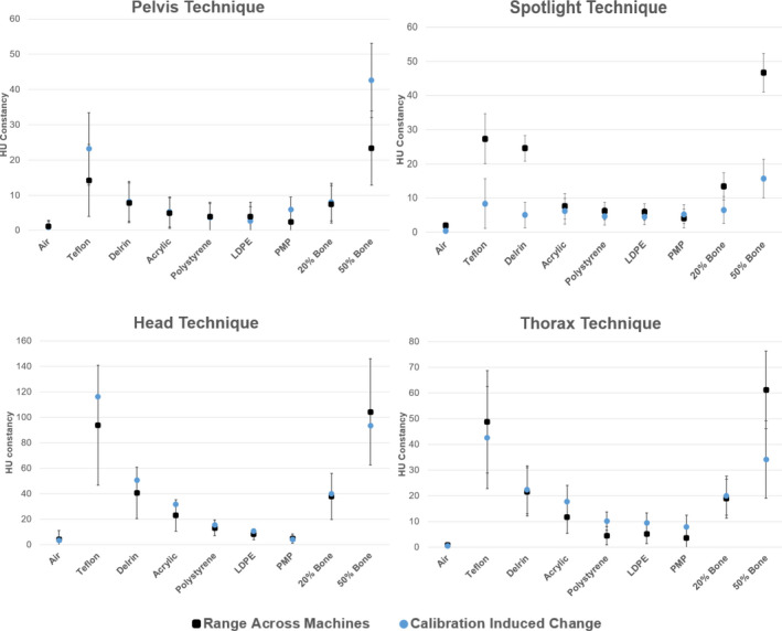
Results: All imaging parameters except geometric distortion varied with technique (P < 0.05) and all imaging parameters except slice thickness varied with machine (P < 0.05). Imager calibration can change the expected value of all imaging parameters, though it does not consistently do so. While changes are statistically significant, they may not be clinically significant. Finally, rapid acquisition of CBCT scans can introduce image artifacts that degrade CBCT uniformity.
Conclusions: This work characterized the variability of acquired CBCT data across machines and CBCT techniques along with the impact of imager calibration and rapid CBCT acquisition on image quality.
Keywords: cone-beam computed tomography; image quality; institutional baselines; linear accelerator quality assurance.
© 2020 The Authors. Journal of Applied Clinical Medical Physics published by Wiley Periodicals, LLC on behalf of American Association of Physicists in Medicine.
Conflict of interest statement
David L. Barbee reports Sun Nuclear Corporation paid for conference travel to speak. None of the other authors has any conflict of interest to disclose.
Figures
References
-
- Jaffray DA, Drake DG, Moreau M, Martinez AA, Wong JW. A radiographic and tomographic imaging system integrated into a medical linear accelerator for localization of bone and soft‐tissue targets. Int J Radiat Oncol Biol Phys. 1999;45:773–789. - PubMed
-
- Ozdemir O, Russell RL, Berlin AA. A 3D probabilistic deep learning system for detection and diagnosis of lung cancer using low‐dose CT scans. IEEE Trans Med Imaging. 2020;39:1419–1429. - PubMed
-
- Gao Z, Wang X, Sun S, et al. Learning physical properties in complex visual scenes: an intelligent machine for perceiving blood flow dynamics from static CT angiography imaging. Neur Netw. 2020;2020:82–93. - PubMed
-
- Van De Leemput SC, Prokop M, Van Ginneken B, Manniesing R. Stacked bidirectional convolutional LSTMS for deriving 3D non‐contrast CT from spatiotemporal 4D CT. IEEE Trans Med Imaging. 2020;39:985–996. - PubMed
-
- Jaffray DA, Siewerdsen JH, Wong JW, Martinez AA. Flat‐panel cone‐beam computed tomography for image‐guided radiation therapy. Int J Radiat Oncol Biol Phys. 2002;53:1337–1349. - PubMed
MeSH terms
LinkOut - more resources
Full Text Sources
Miscellaneous
