. 2020 Oct 30;40(10):BSR20201761.
doi: 10.1042/BSR20201761.

MicroRNA-183-5p contributes to malignant progression through targeting PDCD4 in human hepatocellular carcinoma

Affiliations

MicroRNA-183-5p contributes to malignant progression through targeting PDCD4 in human hepatocellular carcinoma

Xiaohui Duan et al. Biosci Rep. .

Abstract

Hepatocellular carcinoma (HCC) remains one of the most common malignant tumors worldwide. The present study aimed to investigate the biological role of microRNA-183-5p (miR-183-5p), a novel tumor-related microRNA (miRNA), in HCC and illuminate the possible molecular mechanisms. The expression patterns of miR-183-5p in clinical samples were characterized using qPCR analysis. Kaplan-Meier survival curve was applied to evaluate the correlation between miR-183-5p expression and overall survival of HCC patients. Effects of miR-183-5p knockdown on HCC cell proliferation, apoptosis, migration and invasion capabilities were determined via Cell Counting Kit-8 (CCK8) assays, flow cytometry, scratch wound healing assays and Transwell invasion assays, respectively. Mouse neoplasm transplantation models were established to assess the effects of miR-183-5p knockdown on tumor growth in vivo. Bioinformatics analysis, dual-luciferase reporter assays and rescue assays were performed for mechanistic researches. Results showed that miR-183-5p was highly expressed in tumorous tissues compared with adjacent normal tissues. Elevated miR-183-5p expression correlated with shorter overall survival of HCC patients. Moreover, miR-183-5p knockdown significantly suppressed proliferation, survival, migration and invasion of HCC cells compared with negative control treatment. Consistently, miR-183-5p knockdown restrained tumor growth in vivo. Furthermore, programmed cell death factor 4 (PDCD4) was identified as a direct target of miR-183-5p. Additionally, PDCD4 down-regulation was observed to abrogate the inhibitory effects of miR-183-5p knockdown on malignant phenotypes of HCC cells. Collectively, our data suggest that miR-183-5p may exert an oncogenic role in HCC through directly targeting PDCD4. The current study may offer some new insights into understanding the role of miR-183-5p in HCC.

Keywords: PDCD4; hepatocellular carcinoma; malignant progression; microRNA-183-5p; poor prognosis.


Conflict of interest statement

The authors declare that there are no competing interests associated with the manuscript.

Figures

Figure 1
Figure 1. Up-regulation of miR-183-5p is observed in HCC
(A) miR-183-5p expression was assessed by quantitative RT-PCR analysis in cancerous tissues and adjacent normal tissues from a cohort of HCC patients (n=50). (B) miR-183-5p was up-regulated in 46 of 50 HCC patients. (C) miR-183-5p expression levels were compared by quantitative RT-PCR analysis in tumorous tissues of patients from different TNM stages. (D) Overall survival of HCC patients was evaluated using log-rank test and Kaplan–Meier survival analysis. (E) miR-183-5p expression was measured in normal human liver L02 cells and HCC cell lines. *P<0.05; **P<0.01. Student’s t test was used to compare the difference between two groups. *P<0.05.
Figure 2
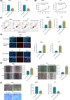
Figure 2. miR-183-5p knockdown suppresses HCC cell proliferation, survival and invasion
(A) Transfection efficiency in Huh-6 and Li-7 cells was determined using quantitative RT-PCR at 48 h post-transfection with NC inhibitor or miR-183-5p inhibitor. (B) Proliferation ability of Huh-6 and Li-7 cells was measured using CCK8 assays after transfection with NC inhibitor or miR-183-5p inhibitor. (C) Apoptosis of Huh-6 and Li-7 cells was evaluated by flow cytometry after transfection with NC inhibitor or miR-183-5p inhibitor. (D) Apoptosis of Huh-6 and Li-7 cells was visualized via DAPI/PI double staining after transfection with NC inhibitor or miR-183-5p inhibitor. (E) Migration ability of Huh-6 and Li-7 cells was assessed by wound healing assays after transfection with NC inhibitor or miR-183-5p inhibitor. (F) Invasion capability of Huh-6 and Li-7 cells was determined by Transwell invasion assays after transfection with NC inhibitor or miR-183-5p inhibitor. **P<0.01. Student’s t test was used to compare the difference between two groups.
Figure 3
Figure 3. PDCD4 is a direct target of miR-183-5p
(A) A schematic diagram of the binding site between the seed sequence of miR-183-5p and the 3′UTR of PDCD4 mRNA. (B) The relative luciferase activity generated by the luciferase reporter vectors containing wildtype or mutant PDCD4 mRNA 3′UTR was determined examined in 293T cells after transfection of miR-183-5p mimics or negative control mimics. (C) PDCD4 mRNA expression levels in cancerous tissues and matched normal tissues of HCC patients (n=50) were detected by quantitative RT-PCR analysis. (D) The correlation of miR-183-5p expression and PDCD4 mRNA expression was determined by Pearson’s correlation analysis. (E) PDCD4 mRNA expression levels were examined by quantitative RT-PCR analysis after transfection with NC inhibitor or miR-183-5p inhibitor. (F) PDCD4 protein expression levels were measured by Western blotting analysis. **P<0.01. Student’s t test was used to compare the difference between two groups.
Figure 4
Figure 4. PDCD4 down-regulation alleviates suppressing effects of miR-183-5p knockdown on malignant phenotypes of HCC cells
(A) PDCD4 protein expression was determined by Western blotting analysis in miR-183-5p inhibitor-treated Li-7 cells after transfection with si-PDCD4. (B) Proliferation of miR-183-5p inhibitor-treated Li-7 cells was analyzed via CCK8 assays after transfection with si-PDCD4. (C) Apoptosis of miR-183-5p inhibitor-treated Li-7 cells was visualized by DAPI/PI double staining after transfection with si-PDCD4. (D) Apoptosis of miR-183-5p inhibitor-treated Li-7 cells was analyzed using flow cytometry after transfection with si-PDCD4. (E) Migration capability of miR-183-5p inhibitor-treated Li-7 cells was assessed via wound healing assays after transfection with si-PDCD4. (F) Invasion ability of miR-183-5p inhibitor-treated Li-7 cells was detected by Transwell invasion assays after transfection with si-PDCD4. **P<0.01. One-way ANOVA followed by Dunnett’s multiple comparison was used to determine the difference among three different groups.
Figure 5
Figure 5. miR-183-5p knockdown inhibits tumor growth in murine xenograft models
(A) Neoplasm transplantation models were established via subcutaneous injection of Li-7 cells in the flank of BALB/c nude mice (n=5). (B) The volumes of tumors collected from negative control group and miR-183-5p inhibitor group were measured and recorded every 7 days. (C) All the mice were euthanized and killed at day 35 post-inoculation, and the tumors harvested from negative control group and miR-183-5p inhibitor group were weighed. (D) Relative expression levels of miR-183-5p. (E) Ki67 protein expression was visualized using immunohistochemical staining in the collected tumors. (F) Vimentin protein expression was analyzed by immunohistochemical staining in the harvested tumors. (G) PDCD4 protein expression was determined via immunohistochemical staining in the collected tumors. (H) PDCD4 protein expression in the collected tumors was examined using Western blotting analysis. **P<0.01. Student’s t test was used to compare the difference between two groups.

References

    1. Huang M., Wang H., Hu X. and Cao X. (2019) lncRNA MALAT1 binds chromatin remodeling subunit BRG1 to epigenetically promote inflammation-related hepatocellular carcinoma progression. Oncoimmunology 8, e1518628 10.1080/2162402X.2018.1518628 - DOI - PMC - PubMed
    1. Zhang H., Deng T., Ge S., Liu Y., Bai M., Zhu K. et al. . (2019) Exosome circRNA secreted from adipocytes promotes the growth of hepatocellular carcinoma by targeting deubiquitination-related USP7. Oncogene 38, 2844–2859 10.1038/s41388-018-0619-z - DOI - PMC - PubMed
    1. Ahodantin J., Bou-Nader M., Cordier C., Megret J., Soussan P., Desdouets C. et al. . (2019) Hepatitis B virus X protein promotes DNA damage propagation through disruption of liver polyploidization and enhances hepatocellular carcinoma initiation. Oncogene 38, 2645–2657 10.1038/s41388-018-0607-3 - DOI - PubMed
    1. Liu R., Li Y., Tian L., Shi H., Wang J., Liang Y. et al. . (2019) Gankyrin drives metabolic reprogramming to promote tumorigenesis, metastasis and drug resistance through activating beta-catenin/c-Myc signaling in human hepatocellular carcinoma. Cancer Lett. 443, 34–46 10.1016/j.canlet.2018.11.030 - DOI - PubMed
    1. Chen J., Huang X., Wang W., Xie H., Li J., Hu Z. et al. . (2018) LncRNA CDKN2BAS predicts poor prognosis in patients with hepatocellular carcinoma and promotes metastasis via the miR-153-5p/ARHGAP18 signaling axis. Aging (Albany N.Y.) 10, 3371–3381 - PMC - PubMed

Publication types

MeSH terms