Review
. 2020 Dec;297(3):503-512.
doi: 10.1148/radiol.2020200771. Epub 2020 Oct 20.

Intra-articular Corticosteroid Injections for the Treatment of Hip and Knee Osteoarthritis-related Pain: Considerations and Controversies with a Focus on Imaging- Radiology Scientific Expert Panel

Affiliations
Review

Intra-articular Corticosteroid Injections for the Treatment of Hip and Knee Osteoarthritis-related Pain: Considerations and Controversies with a Focus on Imaging- Radiology Scientific Expert Panel

Ali Guermazi et al. Radiology. 2020 Dec.

Abstract

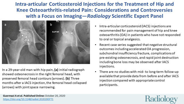
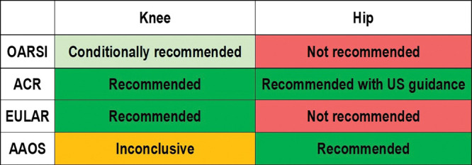
Current management of osteoarthritis (OA) is primarily focused on symptom control. Intra-articular corticosteroid (IACS) injections are often used for pain management of hip and knee OA in patients who have not responded to oral or topical analgesics. Recent case series suggested that negative structural outcomes including accelerated OA progression, subchondral insufficiency fracture, complications of pre-existing osteonecrosis, and rapid joint destruction (including bone loss) may be observed in patients who received IACS injections. This expert panel report reviews the current understanding of pain in OA, summarizes current international guidelines regarding indications for IACS injection, and considers preinterventional safety measures, including imaging. Potential profiles of those who would likely benefit from IACS injection and a suggestion for an updated patient consent form are presented. As of today, there is no established recommendation or consensus regarding imaging, clinical, or laboratory markers before an IACS injection to screen for OA-related imaging abnormalities. Repeating radiographs before each subsequent IACS injection remains controversial. The true cause and natural history of these complications are unclear and require further study. To determine the cause and natural history, large prospective studies evaluating the risk of accelerated OA or joint destruction after IACS injections are needed. However, given the relatively rare incidence of these adverse outcomes, any clinical trial would be challenging in design and a large number of patients would need to be included.


Figures

None
Graphical abstract
Osteoarthritis Research Society International (OARSI), American College of Rheumatology (ACR), European League Against Rheumatism (EULAR), and the American Association of Orthopedic Surgeons (AAOS) recommendations for intra-articular corticosteroid injections.
Figure 1:
Osteoarthritis Research Society International (OARSI), American College of Rheumatology (ACR), European League Against Rheumatism (EULAR), and the American Association of Orthopedic Surgeons (AAOS) recommendations for intra-articular corticosteroid injections.
Subchondral insufficiency fracture in a 61-year-old woman with severe knee pain unrelated to trauma referred to radiology for intra-articular corticosteroid (IACS) injection. (a) Anteroposterior radiograph of the right knee obtained the day of the IACS injection shows mild osteoarthritis (OA) with small osteophytes of the lateral tibia and femur (arrows) and no joint space narrowing. (b) Coronal fat-suppressed proton density-weighted MRI performed 1 month after the IACS injection shows subchondral insufficiency fracture (arrow) with extensive bone marrow edema of the lateral femoral condyle (*) and adjacent soft tissue edema. There is also a severe lateral meniscus extrusion (arrowhead). (c) Repeat radiograph of the right knee 3 months later shows the subchondral insufficiency fracture with collapse of the articular contour of the lateral femoral condyle (arrow) surrounded by bone sclerosis (*) and lateral tibiofemoral joint space narrowing (arrowhead) likely secondary to the severe lateral meniscal subluxation. A normal or mild OA baseline radiograph in a patient with severe joint pain as in this case should trigger a preprocedural MRI to depict occult findings of clinical relevance such as subchondral insufficiency fracture.
Figure 2a:
Subchondral insufficiency fracture in a 61-year-old woman with severe knee pain unrelated to trauma referred to radiology for intra-articular corticosteroid (IACS) injection. (a) Anteroposterior radiograph of the right knee obtained the day of the IACS injection shows mild osteoarthritis (OA) with small osteophytes of the lateral tibia and femur (arrows) and no joint space narrowing. (b) Coronal fat-suppressed proton density-weighted MRI performed 1 month after the IACS injection shows subchondral insufficiency fracture (arrow) with extensive bone marrow edema of the lateral femoral condyle (*) and adjacent soft tissue edema. There is also a severe lateral meniscus extrusion (arrowhead). (c) Repeat radiograph of the right knee 3 months later shows the subchondral insufficiency fracture with collapse of the articular contour of the lateral femoral condyle (arrow) surrounded by bone sclerosis (*) and lateral tibiofemoral joint space narrowing (arrowhead) likely secondary to the severe lateral meniscal subluxation. A normal or mild OA baseline radiograph in a patient with severe joint pain as in this case should trigger a preprocedural MRI to depict occult findings of clinical relevance such as subchondral insufficiency fracture.
Figure 2b:
Subchondral insufficiency fracture in a 61-year-old woman with severe knee pain unrelated to trauma referred to radiology for intra-articular corticosteroid (IACS) injection. (a) Anteroposterior radiograph of the right knee obtained the day of the IACS injection shows mild osteoarthritis (OA) with small osteophytes of the lateral tibia and femur (arrows) and no joint space narrowing. (b) Coronal fat-suppressed proton density-weighted MRI performed 1 month after the IACS injection shows subchondral insufficiency fracture (arrow) with extensive bone marrow edema of the lateral femoral condyle (*) and adjacent soft tissue edema. There is also a severe lateral meniscus extrusion (arrowhead). (c) Repeat radiograph of the right knee 3 months later shows the subchondral insufficiency fracture with collapse of the articular contour of the lateral femoral condyle (arrow) surrounded by bone sclerosis (*) and lateral tibiofemoral joint space narrowing (arrowhead) likely secondary to the severe lateral meniscal subluxation. A normal or mild OA baseline radiograph in a patient with severe joint pain as in this case should trigger a preprocedural MRI to depict occult findings of clinical relevance such as subchondral insufficiency fracture.
Figure 2c:
Subchondral insufficiency fracture in a 61-year-old woman with severe knee pain unrelated to trauma referred to radiology for intra-articular corticosteroid (IACS) injection. (a) Anteroposterior radiograph of the right knee obtained the day of the IACS injection shows mild osteoarthritis (OA) with small osteophytes of the lateral tibia and femur (arrows) and no joint space narrowing. (b) Coronal fat-suppressed proton density-weighted MRI performed 1 month after the IACS injection shows subchondral insufficiency fracture (arrow) with extensive bone marrow edema of the lateral femoral condyle (*) and adjacent soft tissue edema. There is also a severe lateral meniscus extrusion (arrowhead). (c) Repeat radiograph of the right knee 3 months later shows the subchondral insufficiency fracture with collapse of the articular contour of the lateral femoral condyle (arrow) surrounded by bone sclerosis (*) and lateral tibiofemoral joint space narrowing (arrowhead) likely secondary to the severe lateral meniscal subluxation. A normal or mild OA baseline radiograph in a patient with severe joint pain as in this case should trigger a preprocedural MRI to depict occult findings of clinical relevance such as subchondral insufficiency fracture.
Osteonecrosis in a 59-year-old man referred to radiology for intra-articular corticosteroid (IACS) injection. (a) Anteroposterior radiograph of the right knee shows severe osteoarthritis with bone on bone appearance (arrows) and large definite osteophytes (arrowheads). There is a subchondral sclerosis of the medial femoral condyle (*), which is expected in advanced osteoarthritis. No sign of subchondral insufficiency fracture or osteonecrosis. Patient was experiencing an acute episode of pain exacerbation at time of presentation. (b) Coronal fat-suppressed intermediate-weighted MRI was performed before the IACS injection and discloses a large area of osteonecrosis of the medial femoral condyle (arrows) with pathognomonic serpiginous demarcation and fat-equivalent center of lesion (*). No collapse of the articular contour is seen. There is attrition (ie, surface remodeling) as part of the advanced osteoarthritis process. IACS injection was not performed.
Figure 3a:
Osteonecrosis in a 59-year-old man referred to radiology for intra-articular corticosteroid (IACS) injection. (a) Anteroposterior radiograph of the right knee shows severe osteoarthritis with bone on bone appearance (arrows) and large definite osteophytes (arrowheads). There is a subchondral sclerosis of the medial femoral condyle (*), which is expected in advanced osteoarthritis. No sign of subchondral insufficiency fracture or osteonecrosis. Patient was experiencing an acute episode of pain exacerbation at time of presentation. (b) Coronal fat-suppressed intermediate-weighted MRI was performed before the IACS injection and discloses a large area of osteonecrosis of the medial femoral condyle (arrows) with pathognomonic serpiginous demarcation and fat-equivalent center of lesion (*). No collapse of the articular contour is seen. There is attrition (ie, surface remodeling) as part of the advanced osteoarthritis process. IACS injection was not performed.
Figure 3b:
Osteonecrosis in a 59-year-old man referred to radiology for intra-articular corticosteroid (IACS) injection. (a) Anteroposterior radiograph of the right knee shows severe osteoarthritis with bone on bone appearance (arrows) and large definite osteophytes (arrowheads). There is a subchondral sclerosis of the medial femoral condyle (*), which is expected in advanced osteoarthritis. No sign of subchondral insufficiency fracture or osteonecrosis. Patient was experiencing an acute episode of pain exacerbation at time of presentation. (b) Coronal fat-suppressed intermediate-weighted MRI was performed before the IACS injection and discloses a large area of osteonecrosis of the medial femoral condyle (arrows) with pathognomonic serpiginous demarcation and fat-equivalent center of lesion (*). No collapse of the articular contour is seen. There is attrition (ie, surface remodeling) as part of the advanced osteoarthritis process. IACS injection was not performed.
Rapid progressive osteoarthritis (RPOA) type 1 in a 52-year-old man referred to radiology for intra-articular corticosteroid (IACS) injection. (a) Anteroposterior radiograph of the right knee shows mild osteoarthritis with definite osteophytes (arrows) and minimal joint space narrowing of the medial tibiofemoral joint (arrowheads). (b) Baseline coronal fat-suppressed intermediate-weighted MRI scan confirms the osteophytes (black arrows) and shows diffuse cartilage loss at the medial femoral condyle (white arrow) with moderate subluxation of the medial meniscus (arrowhead). (c) Six months after the IACS injection, a repeat anteroposterior radiograph of the right knee shows severe medial tibiofemoral joint space narrowing (arrows) with loss of more than 2 mm of joint space width consistent with rapid progressive osteoarthritis type 1. (d) Coronal fat-suppressed intermediate-weighted MRI scan confirms extensive loss of cartilage at the medial femur and tibia (white arrows) and worsening of the medial meniscal subluxation (arrowhead). There is also subchondral bone marrow edema at the medial tibia and femur (black arrows).
Figure 4a:
Rapid progressive osteoarthritis (RPOA) type 1 in a 52-year-old man referred to radiology for intra-articular corticosteroid (IACS) injection. (a) Anteroposterior radiograph of the right knee shows mild osteoarthritis with definite osteophytes (arrows) and minimal joint space narrowing of the medial tibiofemoral joint (arrowheads). (b) Baseline coronal fat-suppressed intermediate-weighted MRI scan confirms the osteophytes (black arrows) and shows diffuse cartilage loss at the medial femoral condyle (white arrow) with moderate subluxation of the medial meniscus (arrowhead). (c) Six months after the IACS injection, a repeat anteroposterior radiograph of the right knee shows severe medial tibiofemoral joint space narrowing (arrows) with loss of more than 2 mm of joint space width consistent with rapid progressive osteoarthritis type 1. (d) Coronal fat-suppressed intermediate-weighted MRI scan confirms extensive loss of cartilage at the medial femur and tibia (white arrows) and worsening of the medial meniscal subluxation (arrowhead). There is also subchondral bone marrow edema at the medial tibia and femur (black arrows).
Figure 4b:
Rapid progressive osteoarthritis (RPOA) type 1 in a 52-year-old man referred to radiology for intra-articular corticosteroid (IACS) injection. (a) Anteroposterior radiograph of the right knee shows mild osteoarthritis with definite osteophytes (arrows) and minimal joint space narrowing of the medial tibiofemoral joint (arrowheads). (b) Baseline coronal fat-suppressed intermediate-weighted MRI scan confirms the osteophytes (black arrows) and shows diffuse cartilage loss at the medial femoral condyle (white arrow) with moderate subluxation of the medial meniscus (arrowhead). (c) Six months after the IACS injection, a repeat anteroposterior radiograph of the right knee shows severe medial tibiofemoral joint space narrowing (arrows) with loss of more than 2 mm of joint space width consistent with rapid progressive osteoarthritis type 1. (d) Coronal fat-suppressed intermediate-weighted MRI scan confirms extensive loss of cartilage at the medial femur and tibia (white arrows) and worsening of the medial meniscal subluxation (arrowhead). There is also subchondral bone marrow edema at the medial tibia and femur (black arrows).
Figure 4c:
Rapid progressive osteoarthritis (RPOA) type 1 in a 52-year-old man referred to radiology for intra-articular corticosteroid (IACS) injection. (a) Anteroposterior radiograph of the right knee shows mild osteoarthritis with definite osteophytes (arrows) and minimal joint space narrowing of the medial tibiofemoral joint (arrowheads). (b) Baseline coronal fat-suppressed intermediate-weighted MRI scan confirms the osteophytes (black arrows) and shows diffuse cartilage loss at the medial femoral condyle (white arrow) with moderate subluxation of the medial meniscus (arrowhead). (c) Six months after the IACS injection, a repeat anteroposterior radiograph of the right knee shows severe medial tibiofemoral joint space narrowing (arrows) with loss of more than 2 mm of joint space width consistent with rapid progressive osteoarthritis type 1. (d) Coronal fat-suppressed intermediate-weighted MRI scan confirms extensive loss of cartilage at the medial femur and tibia (white arrows) and worsening of the medial meniscal subluxation (arrowhead). There is also subchondral bone marrow edema at the medial tibia and femur (black arrows).
Figure 4d:
Rapid progressive osteoarthritis (RPOA) type 1 in a 52-year-old man referred to radiology for intra-articular corticosteroid (IACS) injection. (a) Anteroposterior radiograph of the right knee shows mild osteoarthritis with definite osteophytes (arrows) and minimal joint space narrowing of the medial tibiofemoral joint (arrowheads). (b) Baseline coronal fat-suppressed intermediate-weighted MRI scan confirms the osteophytes (black arrows) and shows diffuse cartilage loss at the medial femoral condyle (white arrow) with moderate subluxation of the medial meniscus (arrowhead). (c) Six months after the IACS injection, a repeat anteroposterior radiograph of the right knee shows severe medial tibiofemoral joint space narrowing (arrows) with loss of more than 2 mm of joint space width consistent with rapid progressive osteoarthritis type 1. (d) Coronal fat-suppressed intermediate-weighted MRI scan confirms extensive loss of cartilage at the medial femur and tibia (white arrows) and worsening of the medial meniscal subluxation (arrowhead). There is also subchondral bone marrow edema at the medial tibia and femur (black arrows).
Rapid progressive osteoarthritis type 2 in a 38-year-old woman referred to radiology for intra-articular corticosteroid (IACS) injection. (a) Baseline anteroposterior radiograph of the left hip shows mild osteoarthritis with small definite osteophytes at the lateral acetabulum and femoral head (arrows) and no definite joint space narrowing. (b) Six months after the IACS injection a repeat anteroposterior radiograph of the left hip shows a complete collapse of the head of the femur with marked bone loss of the femoral head (arrow) and surface remodeling and flattening of the acetabulum (arrowheads). (c) Coronal fat-suppressed proton density-weighted MRI scan obtained on the same day demonstrates diffuse bone marrow edema of the femur and acetabulum (black and white *) and a large hip joint effusion (#) reflecting an ongoing process of marked synovial activation.
Figure 5a:
Rapid progressive osteoarthritis type 2 in a 38-year-old woman referred to radiology for intra-articular corticosteroid (IACS) injection. (a) Baseline anteroposterior radiograph of the left hip shows mild osteoarthritis with small definite osteophytes at the lateral acetabulum and femoral head (arrows) and no definite joint space narrowing. (b) Six months after the IACS injection a repeat anteroposterior radiograph of the left hip shows a complete collapse of the head of the femur with marked bone loss of the femoral head (arrow) and surface remodeling and flattening of the acetabulum (arrowheads). (c) Coronal fat-suppressed proton density-weighted MRI scan obtained on the same day demonstrates diffuse bone marrow edema of the femur and acetabulum (black and white *) and a large hip joint effusion (#) reflecting an ongoing process of marked synovial activation.
Figure 5b:
Rapid progressive osteoarthritis type 2 in a 38-year-old woman referred to radiology for intra-articular corticosteroid (IACS) injection. (a) Baseline anteroposterior radiograph of the left hip shows mild osteoarthritis with small definite osteophytes at the lateral acetabulum and femoral head (arrows) and no definite joint space narrowing. (b) Six months after the IACS injection a repeat anteroposterior radiograph of the left hip shows a complete collapse of the head of the femur with marked bone loss of the femoral head (arrow) and surface remodeling and flattening of the acetabulum (arrowheads). (c) Coronal fat-suppressed proton density-weighted MRI scan obtained on the same day demonstrates diffuse bone marrow edema of the femur and acetabulum (black and white *) and a large hip joint effusion (#) reflecting an ongoing process of marked synovial activation.
Figure 5c:
Rapid progressive osteoarthritis type 2 in a 38-year-old woman referred to radiology for intra-articular corticosteroid (IACS) injection. (a) Baseline anteroposterior radiograph of the left hip shows mild osteoarthritis with small definite osteophytes at the lateral acetabulum and femoral head (arrows) and no definite joint space narrowing. (b) Six months after the IACS injection a repeat anteroposterior radiograph of the left hip shows a complete collapse of the head of the femur with marked bone loss of the femoral head (arrow) and surface remodeling and flattening of the acetabulum (arrowheads). (c) Coronal fat-suppressed proton density-weighted MRI scan obtained on the same day demonstrates diffuse bone marrow edema of the femur and acetabulum (black and white *) and a large hip joint effusion (#) reflecting an ongoing process of marked synovial activation.
Osteonecrosis in a 29-year-old man who presented with right hip pain. (a) Anteroposterior radiograph and (b) coronal fat-suppressed proton density-weighted MRI of the right hip obtained on the same day show osteonecrosis in the right femoral head, with preserved femoral head contours (arrows). He subsequently went to the sports medicine clinic and was administered a right hip joint intra-articular corticosteroid (IACS) injection for pain. Three months later, he was referred to our institution for repeat IACS injection due to worsening pain. (c) Repeat anteroposterior right hip radiograph shows collapse of the superior femoral head articular surface (arrows). (d) Coronal reformatted CT image of the right hip confirms the collapse of the superior femoral head articular surface (arrows) and shows new hip joint space narrowing. Patient subsequently underwent right hip joint replacement.
Figure 6a:
Osteonecrosis in a 29-year-old man who presented with right hip pain. (a) Anteroposterior radiograph and (b) coronal fat-suppressed proton density-weighted MRI of the right hip obtained on the same day show osteonecrosis in the right femoral head, with preserved femoral head contours (arrows). He subsequently went to the sports medicine clinic and was administered a right hip joint intra-articular corticosteroid (IACS) injection for pain. Three months later, he was referred to our institution for repeat IACS injection due to worsening pain. (c) Repeat anteroposterior right hip radiograph shows collapse of the superior femoral head articular surface (arrows). (d) Coronal reformatted CT image of the right hip confirms the collapse of the superior femoral head articular surface (arrows) and shows new hip joint space narrowing. Patient subsequently underwent right hip joint replacement.
Figure 6b:
Osteonecrosis in a 29-year-old man who presented with right hip pain. (a) Anteroposterior radiograph and (b) coronal fat-suppressed proton density-weighted MRI of the right hip obtained on the same day show osteonecrosis in the right femoral head, with preserved femoral head contours (arrows). He subsequently went to the sports medicine clinic and was administered a right hip joint intra-articular corticosteroid (IACS) injection for pain. Three months later, he was referred to our institution for repeat IACS injection due to worsening pain. (c) Repeat anteroposterior right hip radiograph shows collapse of the superior femoral head articular surface (arrows). (d) Coronal reformatted CT image of the right hip confirms the collapse of the superior femoral head articular surface (arrows) and shows new hip joint space narrowing. Patient subsequently underwent right hip joint replacement.
Figure 6c:
Osteonecrosis in a 29-year-old man who presented with right hip pain. (a) Anteroposterior radiograph and (b) coronal fat-suppressed proton density-weighted MRI of the right hip obtained on the same day show osteonecrosis in the right femoral head, with preserved femoral head contours (arrows). He subsequently went to the sports medicine clinic and was administered a right hip joint intra-articular corticosteroid (IACS) injection for pain. Three months later, he was referred to our institution for repeat IACS injection due to worsening pain. (c) Repeat anteroposterior right hip radiograph shows collapse of the superior femoral head articular surface (arrows). (d) Coronal reformatted CT image of the right hip confirms the collapse of the superior femoral head articular surface (arrows) and shows new hip joint space narrowing. Patient subsequently underwent right hip joint replacement.
Figure 6d:
Osteonecrosis in a 29-year-old man who presented with right hip pain. (a) Anteroposterior radiograph and (b) coronal fat-suppressed proton density-weighted MRI of the right hip obtained on the same day show osteonecrosis in the right femoral head, with preserved femoral head contours (arrows). He subsequently went to the sports medicine clinic and was administered a right hip joint intra-articular corticosteroid (IACS) injection for pain. Three months later, he was referred to our institution for repeat IACS injection due to worsening pain. (c) Repeat anteroposterior right hip radiograph shows collapse of the superior femoral head articular surface (arrows). (d) Coronal reformatted CT image of the right hip confirms the collapse of the superior femoral head articular surface (arrows) and shows new hip joint space narrowing. Patient subsequently underwent right hip joint replacement.
Suggestion of the use of imaging in the context of intra-articular corticosteroid (IACS) injection (to be tested for efficacy and cost-effectiveness). (a) First IACS injection and (b) repeat IACS injection. Obtaining weight-bearing imaging prior to repeat IACS injection is not supported by rheumatologists and orthopedic surgeons on the panel. OA = osteoarthritis, SIF = subchondral insufficiency fracture, RPOA = rapid progressive OA.
Figure 7a:
Suggestion of the use of imaging in the context of intra-articular corticosteroid (IACS) injection (to be tested for efficacy and cost-effectiveness). (a) First IACS injection and (b) repeat IACS injection. Obtaining weight-bearing imaging prior to repeat IACS injection is not supported by rheumatologists and orthopedic surgeons on the panel. OA = osteoarthritis, SIF = subchondral insufficiency fracture, RPOA = rapid progressive OA.
Figure 7b:
Suggestion of the use of imaging in the context of intra-articular corticosteroid (IACS) injection (to be tested for efficacy and cost-effectiveness). (a) First IACS injection and (b) repeat IACS injection. Obtaining weight-bearing imaging prior to repeat IACS injection is not supported by rheumatologists and orthopedic surgeons on the panel. OA = osteoarthritis, SIF = subchondral insufficiency fracture, RPOA = rapid progressive OA.

References

    1. Turkiewicz A, Petersson IF, Björk J, et al. . Current and future impact of osteoarthritis on health care: a population-based study with projections to year 2032. Osteoarthritis Cartilage 2014;22(11):1826–1832. - PubMed
    1. GBD 2015 Disease and Injury Incidence and Prevalence Collaborators . Global, regional, and national incidence, prevalence, and years lived with disability for 310 diseases and injuries, 1990-2015: a systematic analysis for the Global Burden of Disease Study 2015. Lancet 2016;388(10053):1545–1602 [Published correction appears in Lancet 2017;389(10064):e1.]. - PMC - PubMed
    1. U.S. Department of Health and Human Services, Food and Drug Administration . Osteoarthritis: Structural Endpoints for the Development of Drugs. Devices, and Biological Products for Treatment Guidance for Industry. https://www.fda.gov/regulatory-information/search-fda-guidance-documents.... Published 2018. Accessed September 24, 2020.
    1. Guermazi A, Roemer FW, Haugen IK, Crema MD, Hayashi D. MRI-based semiquantitative scoring of joint pathology in osteoarthritis. Nat Rev Rheumatol 2013;9(4):236–251. - PubMed
    1. Guermazi A, Roemer FW, Crema MD, Englund M, Hayashi D. Imaging of non-osteochondral tissues in osteoarthritis. Osteoarthritis Cartilage 2014;22(10):1590–1605. - PubMed

Publication types

MeSH terms

Substances