

Rapid Lipid Modification of Endothelial Cell Membranes in Cardiac Ischemia/Reperfusion Injury: a Novel Therapeutic Strategy to Reduce Infarct Size
- PMID: 33079319
- PMCID: PMC8376233
- DOI: 10.1007/s10557-020-07101-x
Rapid Lipid Modification of Endothelial Cell Membranes in Cardiac Ischemia/Reperfusion Injury: a Novel Therapeutic Strategy to Reduce Infarct Size
Abstract
Purpose: Plasma membranes constitute a gathering point for lipids and signaling proteins. Lipids are known to regulate the location and activity of signaling proteins under physiological and pathophysiological conditions. Membrane lipid therapies (MLTs) that gradually modify lipid content of plasma membranes have been developed to treat chronic disease; however, no MLTs have been developed to treat acute conditions such as reperfusion injury following myocardial infarction (MI) and percutaneous coronary intervention (PCI). A fusogenic nanoliposome (FNL) that rapidly incorporates exogenous unsaturated lipids into endothelial cell (EC) membranes was developed to attenuate reperfusion-induced protein signaling. We hypothesized that administration of intracoronary (IC) FNL-MLT interferes with EC membrane protein signaling, leading to reduced microvascular dysfunction and infarct size (IS).
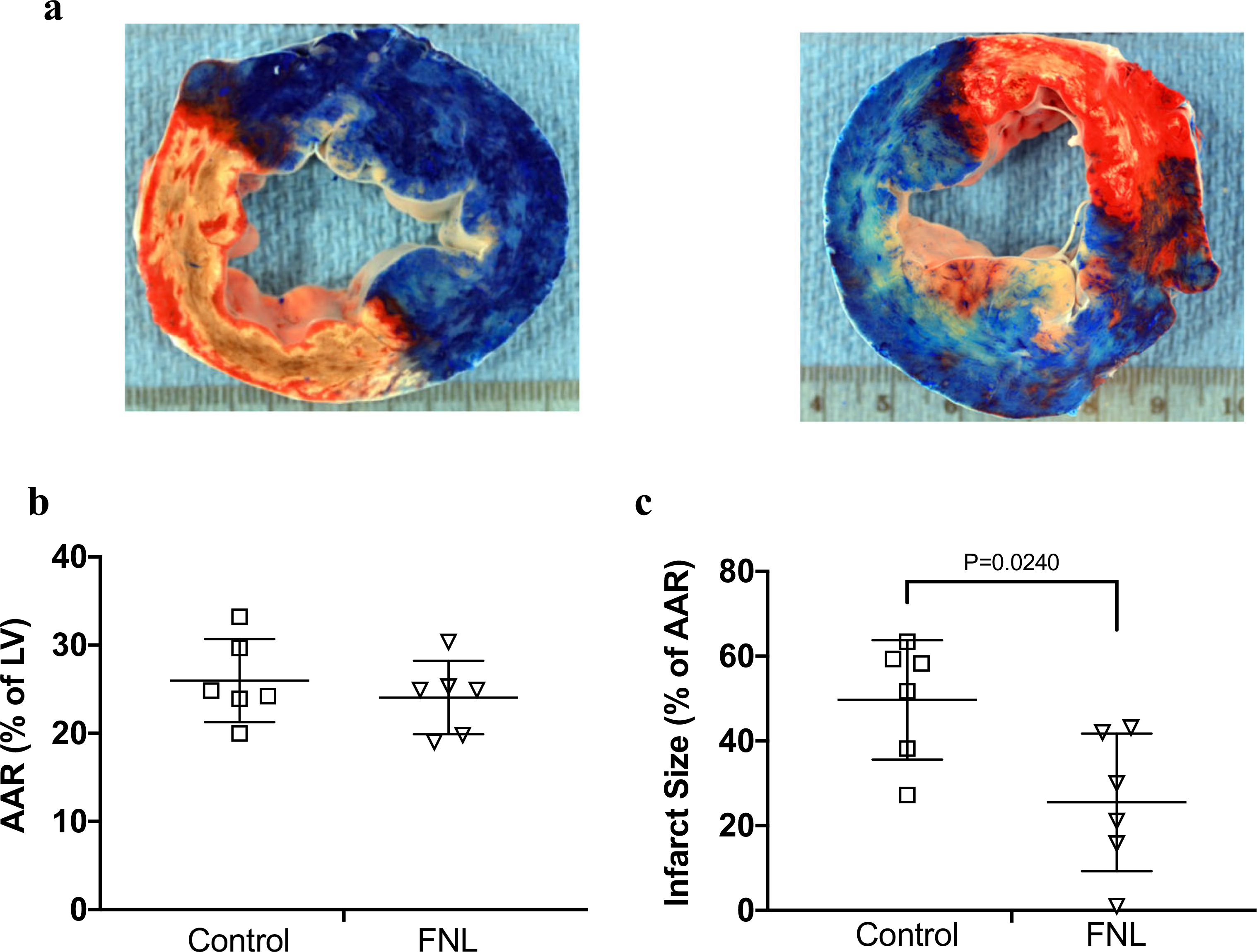
Methods: Using a myocardial ischemia/reperfusion swine model, the efficacy of FNL-MLT in reducing IS following a 60-min coronary artery occlusion was tested. Animals were randomized to receive IC Ringer's lactate solution with or without 10 mg/mL/min of FNLs for 10 min prior to reperfusion (n = 6 per group).
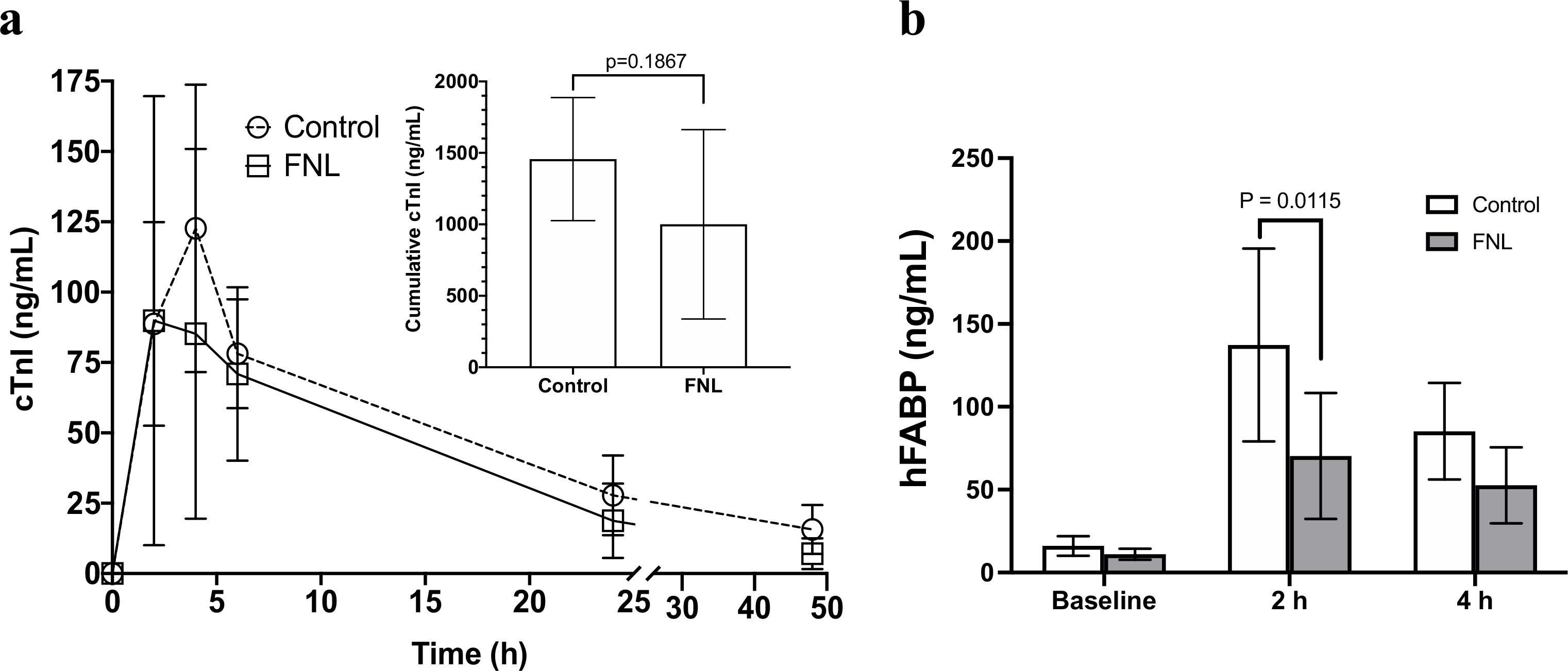
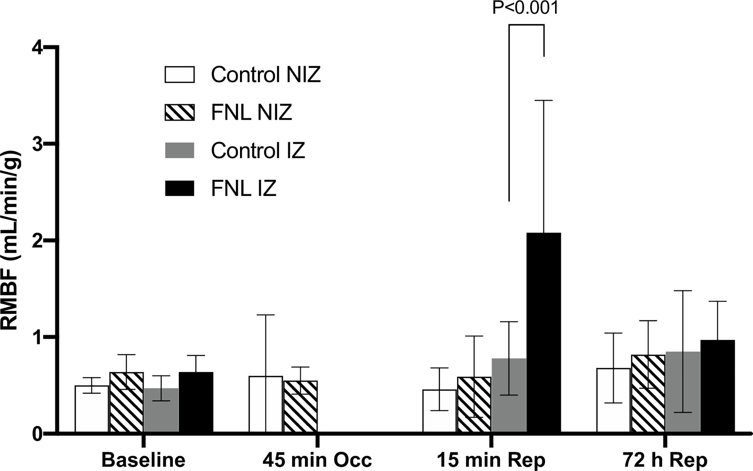
Results: The IC FNL-MLT reduced IS (25.45 ± 16.4% vs. 49.7 ± 14.1%, P < 0.02) and enhanced regional myocardial blood flow (RMBF) in the ischemic zone at 15 min of reperfusion (2.13 ± 1.48 mL/min/g vs. 0.70 ± 0.43 mL/min/g, P < 0.001). The total cumulative plasma levels of the cardiac injury biomarker cardiac troponin I (cTnI) were trending downward but were not significant (999.3 ± 38.7 ng/mL vs. 1456.5 ± 64.8 ng/mL, P = 0.1867). However, plasma levels of heart-specific fatty acid binding protein (hFABP), another injury biomarker, were reduced at 2 h of reperfusion (70.3 ± 38.0 ng/mL vs. 137.3 ± 58.2 ng/mL, P = 0.0115). CONCLUSION: The IC FNL-MLT reduced IS compared to vehicle in this swine model. The FNL-MLT maybe a promising adjuvant to PCI in the treatment of acute MI.
Keywords: Endothelium; Ischemia/reperfusion; Lipid rafts; Liposomes; Membrane lipid therapy; Myocardial infarction; Percutaneous coronary intervention.
Figures
Similar articles
-
Serial measurement of hFABP and high-sensitivity troponin I post-PCI in STEMI: how fast and accurate can myocardial infarct size and no-reflow be predicted?Am J Physiol Heart Circ Physiol. 2013 Oct 1;305(7):H1104-10. doi: 10.1152/ajpheart.00447.2013. Epub 2013 Jul 19. Am J Physiol Heart Circ Physiol. 2013. PMID: 23873799
-
HMG-CoA reductase inhibition prior reperfusion improves reparative fibrosis post-myocardial infarction in a preclinical experimental model.Int J Cardiol. 2014 Aug 20;175(3):528-38. doi: 10.1016/j.ijcard.2014.06.040. Epub 2014 Jul 2. Int J Cardiol. 2014. PMID: 25023790
-
Effects of intracoronary melatonin on ischemia-reperfusion injury in ST-elevation myocardial infarction.Heart Vessels. 2016 Jan;31(1):88-95. doi: 10.1007/s00380-014-0589-1. Epub 2014 Oct 16. Heart Vessels. 2016. PMID: 25319673
-
Pharmacological prevention of reperfusion injury in acute myocardial infarction. A potential role for adenosine as a therapeutic agent.Am J Cardiovasc Drugs. 2004;4(3):159-67. doi: 10.2165/00129784-200404030-00003. Am J Cardiovasc Drugs. 2004. PMID: 15134468 Review.
-
Sodium-hydrogen exchange inhibition: novel strategy to prevent myocardial injury following ischemia and reperfusion.Am J Cardiol. 1999 May 20;83(10A):19G-22G. doi: 10.1016/s0002-9149(99)00316-1. Am J Cardiol. 1999. PMID: 10482176 Review.
Cited by
-
Coronary Endothelium No-Reflow Injury Is Associated with ROS-Modified Mitochondrial Fission through the JNK-Drp1 Signaling Pathway.Oxid Med Cell Longev. 2021 Jan 30;2021:6699516. doi: 10.1155/2021/6699516. eCollection 2021. Oxid Med Cell Longev. 2021. PMID: 33613824 Free PMC article.
-
Challenges and future scope of exosomes in the treatment of cardiovascular diseases.J Physiol. 2023 Nov;601(22):4873-4893. doi: 10.1113/JP282053. Epub 2022 Dec 3. J Physiol. 2023. PMID: 36398654 Free PMC article. Review.
-
Unraveling the role of lactate-related genes in myocardial infarction.Heliyon. 2024 Sep 19;10(18):e38152. doi: 10.1016/j.heliyon.2024.e38152. eCollection 2024 Sep 30. Heliyon. 2024. PMID: 39347425 Free PMC article.
-
Therapeutic Applications of Extracellular Vesicles for Myocardial Repair.Front Cardiovasc Med. 2021 Dec 9;8:758050. doi: 10.3389/fcvm.2021.758050. eCollection 2021. Front Cardiovasc Med. 2021. PMID: 34957249 Free PMC article. Review.
-
Structural Basis of the Interaction of the G Proteins, Gαi1, Gβ1γ2 and Gαi1β1γ2, with Membrane Microdomains and Their Relationship to Cell Localization and Activity.Biomedicines. 2023 Feb 14;11(2):557. doi: 10.3390/biomedicines11020557. Biomedicines. 2023. PMID: 36831093 Free PMC article.
References
-
- Lingwood D, Simons K. Lipid rafts as a membrane-organizing principle. Science. 2010;327(5961):46–50. - PubMed
-
- Vogler O, Casas J, Capo D, Nagy T, Borchert G, Martorell G, et al.The Gbetagamma dimer drives the interaction of heterotrimeric Gi proteins with nonlamellar membrane structures. J Biol Chem. 2004;279(35):36540–5. - PubMed
-
- Epand RM. Proteins and cholesterol-rich domains. Biochim Biophys Acta. 2008;1778(7–8):1576–82. - PubMed
-
- Post JA, Ruigrok TJ, Verkleij AJ. Phospholipid reorganization and bilayer destabilization during myocardial ischemia and reperfusion: a hypothesis. J Mol Cell Cardiol. 1988;20(Suppl 2):107–11. - PubMed
Publication types
MeSH terms
Substances
Grants and funding
LinkOut - more resources
Full Text Sources
Research Materials
Miscellaneous
