. 2021 Mar;69(3):779-791.
doi: 10.1002/glia.23929. Epub 2020 Oct 20.

Microglial reduction of colony stimulating factor-1 receptor expression is sufficient to confer adult onset leukodystrophy

Affiliations

Microglial reduction of colony stimulating factor-1 receptor expression is sufficient to confer adult onset leukodystrophy

Fabrizio Biundo et al. Glia. 2021 Mar.

Abstract

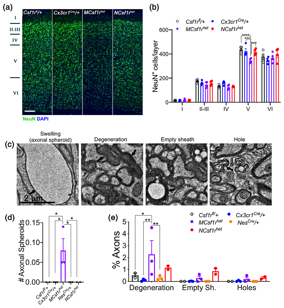
Adult onset leukoencephalopathy with axonal spheroids and pigmented glia (ALSP) is a dementia resulting from dominantly inherited CSF1R inactivating mutations. The Csf1r+/- mouse mimics ALSP symptoms and pathology. Csf1r is mainly expressed in microglia, but also in cortical layer V neurons that are gradually lost in Csf1r+/- mice with age. We therefore examined whether microglial or neuronal Csf1r loss caused neurodegeneration in Csf1r+/- mice. The behavioral deficits, pathologies and elevation of Csf2 expression contributing to disease, previously described in the Csf1r+/- ALSP mouse, were reproduced by microglial deletion (MCsf1rhet mice), but not by neural deletion. Furthermore, increased Csf2 expression by callosal astrocytes, oligodendrocytes, and microglia was observed in Csf1r+/- mice and, in MCsf1rhet mice, the densities of these three cell types were increased in supraventricular patches displaying activated microglia, an early site of disease pathology. These data confirm that ALSP is a primary microgliopathy and inform future therapeutic and experimental approaches.

Keywords: Csf2 expression; GM-CSF; adult onset leukoencephalopathy with axonal spheroids and pigmented glia (ALSP); axonal pathology; colony stimulating factor-1 receptor (CSF-1R); demyelination; microgliopathy.


Conflict of interest statement

CONFLICT OF INTEREST

The authors declare no conflict of interest.

Figures

FIGURE 1
FIGURE 1
Deletion of a single Csf1r allele in the microglial lineage reproduces the behavioral deficits of adult onset leukoencephalopathy with axonal spheroids and pigmented glia (ALSP) (Csf1r+/−) mice. The test performed, age and number of mice, and retention interval (RI) are indicated in each panel. (a–c) Cognition: (a) Left: Similar exploratory activity for all genotypes assessed by the number of total entries into the arms of the Y-maze (ANOVA, Dunnett’s). Right: Lack of exploratory preference for the novel arm by MCsf1rhet mice (one-way ANOVA, Bonferroni’s). (b) Morris water maze. Left: No visual or motivational deficit in any genotype assessed by the path length covered to reach the visible platform (two-way ANOVA, Dunnett’s). Center: Csf1r+/− mice exhibit deficits in learning the location of the hidden platform (two-way ANOVA, Dunnett’s). Right: Long-term deficits in Csf1r+/− mice revealed by the number of counter crossings into the platform location (one-way ANOVA, Fisher’s). (c) Fear conditioning: Deficitof long-term associative memory in Csf1r+/− and MCsf1rhet mice assessed by the percentage of freezing during the contextual memory test (one-way ANOVA, Holm-Sidak’s). (d,e) Anxiety: (d) Behavior spectrometer: Reduction of the track length in the central arena of the field reveals an overt anxious phenotype for MCsf1rhet mice (Kruskal–Wallis test, Dunn’s). (e) Elevated zero maze: Older MCsf1rhet mice spend less time in the open zone indicating the persistence of anxiety (Kruskal–Wallis test, Dunn’s). (f) Motor coordination: Increased number of slips on the balance beam in McCsf1rhet mice (Kruskal–Wallis test, Dunn’s). Data are presented as means ± SEM. Only significantly different changes are marked by asterisks. *, p < .05; **, p < .01; ***, p < .001 [Color figure can be viewed at wileyonlinelibrary.com]
FIGURE 2
FIGURE 2
Csf1r microglial heterozygosity reproduces the cerebral microgliosis, supraventricular microglial activation and oxidative stress of Csf1r+/− mice (a, b) Iba1+ cell densities (green) in different areas of brains of 18 to 21-month-old Cx3cr1Cre/+ and MCsf1rhet mice. (a) Gray Matter: OB, olfactory bulb; Cx, primary motor cortex; Hp, hippocampus; DCbNu, deep cerebellar nuclei; CbCx, cerebellar cortex; (b) white matter: Fornix; CC, corpus callosum; CbWM, cerebellar white matter; Scale bar, 100 μm applies to all panels. (c) Quantification of data in a,b (4 Cx3cr1Cre/+ and 5 MCsf1rhet mice). (d) Microglial patch located in the supraventricular region of the corpus callosum (arrow). Scale bar, 350 μm applies to both panels. (e) Quantification of the percentage of sections presenting callosal patches of dense microglia (5 mice per genotype). (f,g) Morphology (f) and quantification (g) of the Iba1+ cell ramifications in the supraventricular region of the corpus callosum (three mice per genotype). Scale bar, 50 μm applies to both panels. (h) Expression of Poly(ADP-Ribose) (PAR) in the CC of 18 to 21-month-old Cx3cr1Cre/+ and MCsf1rhet mice. Scale bar, 100 μm, applies to all panels. (i) Quantification of the percentage of PAR+ area in the supraventricular region of the corpus callosum (three mice per genotype). Data, presented as means ± SEM, were analyzed using the Unpaired t test. *, p < .05 [Color figure can be viewed at wileyonlinelibrary.com]
FIGURE 3
FIGURE 3
Csf1r microglial heterozygosity phenocopies the callosal demyelination of Csf1r+/− mice (a) Left, expression of Cystatin F in the CC of 18 to 21-month-old Cx3cr1Cre/+ and MCsf1rhet mice. Scale bar, 100 μm, applies to all panels. Right, quantification of the percentage of Cystatin F area in the supraventricular region of CC of Cx3cr1Cre/+ and MCsf1rhet mice (unpaired t test). (b) Fluoromyelin staining and fluorescence quantification in corpus callosum, fimbria and cerebellar white matter (WM) of 18 to 21-month-old Cx3cr1Cre/+ and MCsf1rhet mice. Scale bar, 100 μm, applies to all panels. Data are presented as means ± SEM (5 mice per genotype, unpaired t test). (c) Myelin and axonal ultrastructure in callosal cross sections from 12-month-old mice. Scale bar, 2 μm, applies to all panels. (d–g) Scatter plots of G-ratio distribution where trend line indicates an overt reduction in myelin thickness in MCsf1rhet compared with Cx3cr1Cre/+ mice. (h) Significant G-ratio increases in small and medium diameter neuronal fibers of MCsf1rhet mice (two to three mice per genotype, two-way ANOVA, Dunnett’s multiple comparisons test, p = .07 for the Cx3cr1Cre/+-MCsf1rhet comparison in the 1,001–1,700 nm fiber diameter range). (i,j) Analysis of age-induced myelin pathology in 12-month-old mice: (i) Representative images of abnormalities of myelin sheath structure. Scale bar, 2 μm, applies to all panels. (j) Quantification of myelin abnormalities reveals an increased percentage of axons exhibiting myelin degeneration. Data are presented as means ± SEM (two to three mice per genotype, two-way ANOVA, Dunnett’s multiple comparisons test, *, p < .05; **, p < .01) [Color figure can be viewed at wileyonlinelibrary.com]
FIGURE 4
FIGURE 4
Microglial deletion of a single allele of Csf1r phenocopies the neurodegenerative phenotype of Csf1r+/− mice. (a) Loss in cortical layer V neurons in the primary motor cortex of 18 to 21-month-old MCsf1rhet mice. Scale bar, 100 μm, applies to all panels. (b) Quantification of the number of NeuN+ neurons in the layers of the primary motor cortex (data from 5 mice/genotype, two-way ANOVA, Dunnett’s multiple comparisons test). (c) Representative images of age-induced axonal pathologies. Scale bar, 2 μm, applies to all panels. (d) Quantification of age-induced axonal pathologies in Csf1rfl/+, Cx3cr1Cre/+, MCsf1rhet, NesCre/+, NCsf1rhet mice (two to three mice/genotype, >2,000 neurons/genotype, two-way ANOVA, Dunnett’s multiple comparisons test, *, p < .05; **, p < .01) [Color figure can be viewed at wileyonlinelibrary.com]
FIGURE 5
FIGURE 5
Neuroglia are the major sources of increased Csf2 expression in Csf1r+/− mice (a,b) Single-molecule RNA in situ hybridization (RNAscope) analysis of Csf2 expression in glia and endothelial cells in the corpus callosum (a) and cerebral cortex (b) of 18-month-old mice, showing Csf2 expression in Gfap+ (astrocytes), Sox10+ (oligodendrocytes), Aif1+ (microglia), and Pecam1+ (endothelial) cells. Scale bar, 10 μm, applies to all panels. Nuclear fragmentation was caused by tissue processing. Quantitation: (a) four mice per genotype for all panels except for the upper and lower panels (three mice per genotype); (b) four mice per genotype for all panels except for the upper panels (three mice per genotype) and lower panels (3 Csf1r+/− mice). Data analyzed by unpaired Student’s t test. (c) Increased expression of Csf2 in 6-month-old MCsf1rhet mice (4 Cx3Cr1Cre/+ and eight MCsf1rhet mice, unpaired t test). (d) Increased densities of astrocytes and oligodendrocytes in supraventricular microglial patches of 18 to 21-month-old MCsf1rhet mice. Quantitation: Left panel, 4 Cx3Cr1Cre/+ and 5 MCsf1rhet mice; middle panel, 5 Cx3Cr1Cre/+ mice and 4 MCsf1rhet mice; right panel, 4 Cx3Cr1Cre/+ and 5 MCsf1rhet mice; unpaired t test. Scale bar, 100 μm, applies to all panels. Data are presented as means ± SEM. *p < .05, **p < .01 [Color figure can be viewed at wileyonlinelibrary.com]

References

    1. Biundo F, del Prete D, Zhang H, Arancio O, & D’Adamio L (2018). A role for tau in learning, memory and synaptic plasticity. Scientific Reports, 8(1), 3184. 10.1038/s41598-018-21596-3 - DOI - PMC - PubMed
    1. Biundo F, Ishiwari K, del Prete D, & D’Adamio L (2016). Deletion of the gamma-secretase subunits Aph1B/C impairs memory and worsens the deficits of knock-in mice modeling the Alzheimer-like familial Danish dementia. Oncotarget, 7(11), 11923–11944. 10.18632/oncotarget.7389 - DOI - PMC - PubMed
    1. Brodkin J, Frank D, Grippo R, Hausfater M, Gulinello M, Achterholt N, & Gutzen C (2014). Validation and implementation of a novel high-throughput behavioral phenotyping instrument for mice. Journal of Neuroscience Methods, 224, 48–57. 10.1016/j.jneumeth.2013.12.010 - DOI - PMC - PubMed
    1. Chitu V, Biundo F, Shlager GGL, Park ES, Wang P, Gulinello ME, … Stanley ER (2020). Microglial homeostasis requires balanced CSF-1/CSF-2 receptor signaling. Cell Reports, 30(9), 3004–3019 e3005. 10.1016/j.celrep.2020.02.028 - DOI - PMC - PubMed
    1. Chitu V, Gokhan S, Gulinello M, Branch CA, Patil M, Basu R, … Stanley ER (2015). Phenotypic characterization of a Csf1r haploinsufficient mouse model of adult-onset leukodystrophy with axonal spheroids and pigmented glia (ALSP). Neurobiology of Disease, 74, 219–228. 10.1016/j.nbd.2014.12.001 - DOI - PMC - PubMed

Publication types

Substances