. 2020 Nov;19(11):e13259.
doi: 10.1111/acel.13259. Epub 2020 Oct 20.

Ovarian stiffness increases with age in the mammalian ovary and depends on collagen and hyaluronan matrices

Affiliations

Ovarian stiffness increases with age in the mammalian ovary and depends on collagen and hyaluronan matrices

Farners Amargant et al. Aging Cell. 2020 Nov.

Abstract

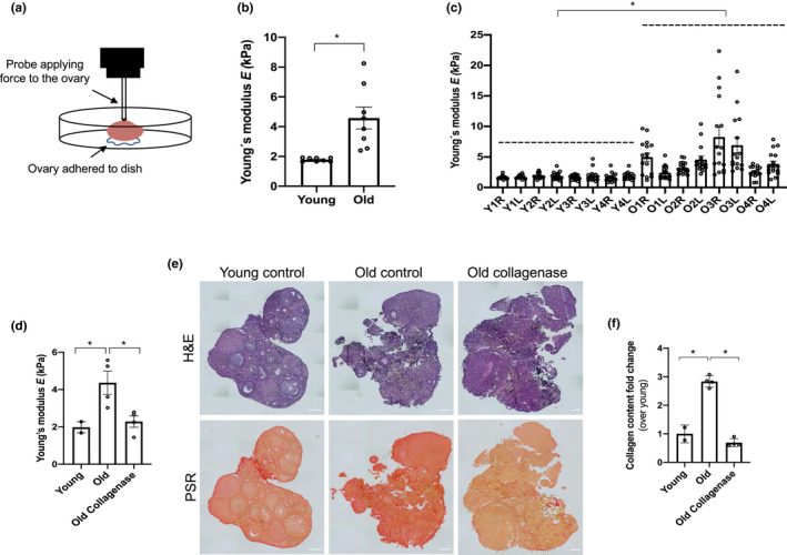
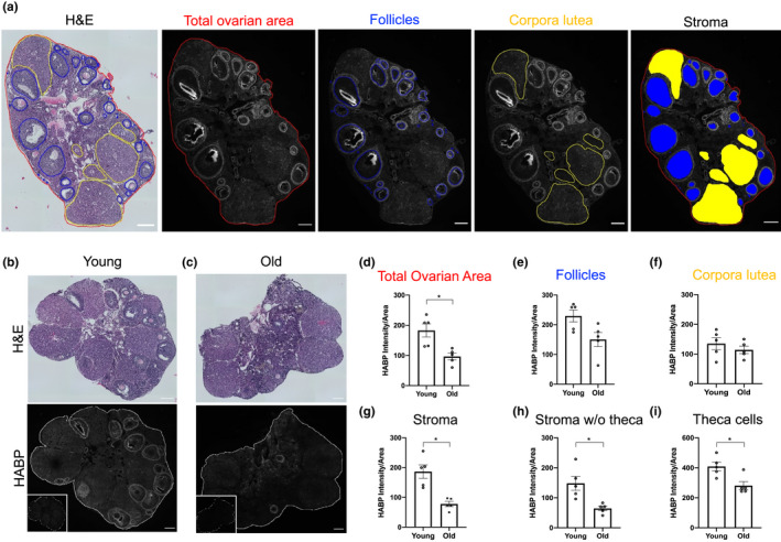
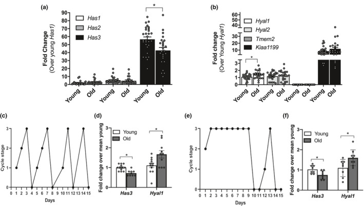
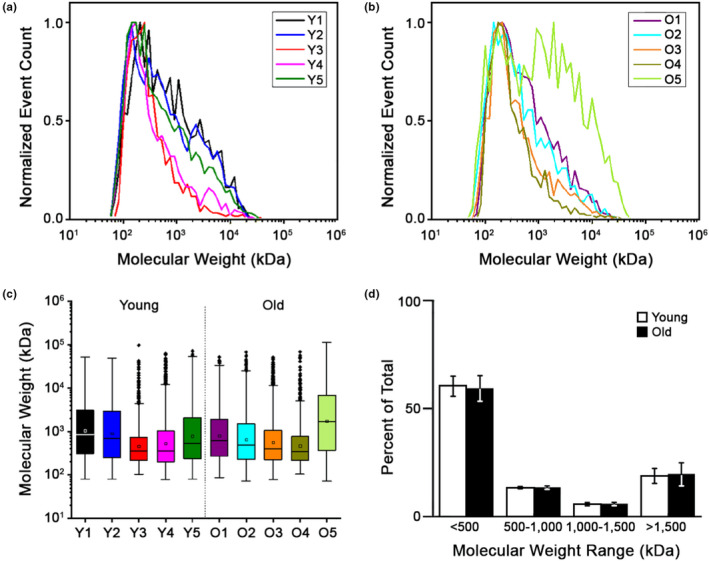
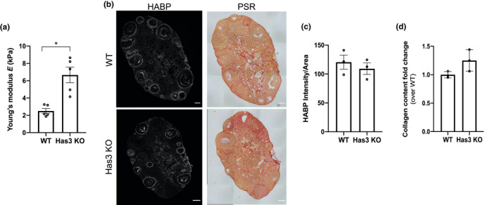
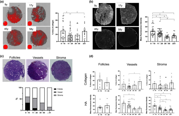
Fibrosis is a hallmark of aging tissues which often leads to altered architecture and function. The ovary is the first organ to show overt signs of aging, including increased fibrosis in the ovarian stroma. How this fibrosis affects ovarian biomechanics and the underlying mechanisms are unknown. Using instrumental indentation, we demonstrated a quantitative increase in ovarian stiffness, as evidenced by an increase in Young's modulus, when comparing ovaries from reproductively young (6-12 weeks) and old (14-17 months) mice. This ovarian stiffness was dependent on collagen because ex vivo enzyme-mediated collagen depletion in ovaries from reproductively old mice restored their collagen content and biomechanical properties to those of young controls. In addition to collagen, we also investigated the role of hyaluronan (HA) in regulating ovarian stiffness. HA is an extracellular matrix glycosaminoglycan that maintains tissue homeostasis, and its loss can change the biomechanical properties of tissues. The total HA content in the ovarian stroma decreased with age, and this was associated with increased hyaluronidase (Hyal1) and decreased hyaluronan synthase (Has3) expression. These gene expression differences were not accompanied by changes in ovarian HA molecular mass distribution. Furthermore, ovaries from mice deficient in HAS3 were stiffer compared to age-matched WT mice. Our results demonstrate that the ovary becomes stiffer with age and that both collagen and HA matrices are contributing mechanisms regulating ovarian biomechanics. Importantly, the age-associated increase in collagen and decrease in HA are conserved in the human ovary and may impact follicle development and oocyte quality.

Keywords: Biomechanics; extracellular matrix; fibrosis; hyaluronan synthase; hyaluronidase; reproduction.


Conflict of interest statement

The authors have no conflicts of interest to declare.

Figures

Figure 1
Figure 1
Ovarian stiffness increases with advanced reproductive age and depends in part on the collagen matrix. (a) Schematic of the indentation method. (b‐c) The graphs show ovarian stiffness (Young's modulus, E) in all the ovaries combined (b) or (c) in individual ovaries (Y: young, O: old, R: right, L: left). N = 8 ovaries from reproductively young mice, or N = 8 ovaries from reproductively old mice. (d) Graph showing the stiffness (Young's modulus, E) of reproductively young (N = 2), reproductively old (control; N = 4), and reproductively old ovaries treated with collagenase (N = 4). (e) Images of post‐indented ovaries fixed and stained with H&E and PSR. Scale bar=100 μm. (f) PSR‐positive area represented as fold change over young (in indented reproductively young and old control ovaries and collagenase treated reproductively old ovaries). Asterisks indicate significant differences (p < 0.05)
Figure 2
Figure 2
HA content decreases in the ovarian stroma and theca layer with advanced reproductive age. (a) Reproductively young mouse ovarian serial sections stained with H&E to identify ovarian structures and with the HABP assay (HA in white) to analyze total ovarian HA intensity in the follicles, in the CL, and in the stroma. (b,c) H&E and HABP assay images from ovarian tissue sections of reproductively young (b) and old (c) mice. Insets are sequential ovarian sections treated with hyaluronidase as negative controls. (d‐g) Quantification of HA intensity in the total ovarian area, follicles, CL, and stroma. Graphs showing HA intensity per area in the stroma without theca cells (h) and in the theca cell layer (i). N = 5 reproductively young and N = 5 reproductively old mice. Asterisks indicate significant differences (p < 0.05). Scale bar=100 μm.
Figure 3
Figure 3
Ovarian stromal Has3 and Hyal1 undergo age‐dependent transcriptional changes independent of estrous cyclicity. Relative gene expression levels of (a) hyaluronan synthases and (b) hyaluronidases in ovarian stroma from reproductively young and old mice. The data are shown as a fold change expression over Has1 (a) or Hyal1 (b) in reproductively young mice. (c) A representative plot of a mouse exhibiting normal estrous cyclicity over a 15‐day period. (d) Graph showing the relative expression of Has3 and Hyal1 in reproductively young and old mice with normal estrous cycles. (e) A representative plot of a mouse exhibiting abnormal estrous cyclicity over a 15‐day period. (f) Relative expression of Has3 and Hyal1 in reproductively young and old mice with abnormal estrous cycles. (c and e) 0‐3 represent the different cycle stages: diestrus (0), proestrus (1), estrus (2), and metestrus (3). N = 30 stromal samples from reproductively young mice; N = 29 stromal samples from reproductively old mice. Asterisks indicate significant differences (p < 0.05)
Figure 4
Figure 4
LMW‐HA fragments do not accumulate in the aging ovary. MW distributions of total HA isolated from reproductively young (a) and old (b) mouse ovaries. (c) Box plots showing the mean HA MW for each individual ovary studied. (d) Plot comparing the allocation of total ovarian HA in the MW ranges of <500, 500‐1000, 1000‐1500, and >1500 kDa, respectively, between reproductively young and old mice. N = 5 reproductively young, N = 5 reproductively old ovaries
Figure 5
Figure 5
HA dysregulation is associated with increased ovarian stiffness. (a) Ovarian stiffness of WT (N = 5) and Has3 KO (N = 5) mouse ovaries. (b) Representative images of WT and Has3 KO ovaries stained with HABP (HA in white) and PSR (collagen in red). Scale bar=100 μm. (c,d) HA and collagen quantification in WT (N = 3) and Has3 KO (N = 3) mouse ovaries (contralateral to the nanoindented ovaries). HA is represented as intensity per area and collagen as fold change over WT. The asterisk indicates statistical significance
Figure 6
Figure 6
Age‐associated collagen and HA changes are conserved in humans. (a) On the left, representative processed PSR‐stained and color thresholded images for selected participants ranging from youngest to oldest cohort are shown. Scale bar=100 μm. N = 120. Graph showing the percent fibrotic area for each cohort. (b) Representative images of HABP stained TMA samples for selected participants in each of the 4 age cohorts. Scale bar=100 μm. N = 120. On the right, the mean fluorescence intensity HA is represented for each age cohort. (c) Representative H&E‐stained images containing follicles, vessels, or stroma, and the percentage of category within each given cohort is represented below. Scale bar=100 μm. (d) Graphs showing collagen and HA quantification in follicle, vessel, and stroma compartments. Asterisks indicate significant differences (< 0.05).

References

    1. Achterberg, V. F. , Buscemi, L. , Diekmann, H. , Smith‐Clerc, J. , Schwengler, H. , Meister, J.‐J. , Wenck, H. , Gallinat, S. , & Hinz, B. (2014). The nano‐scale mechanical properties of the extracellular matrix regulate dermal fibroblast function. The Journal of Investigative Dermatology, 134(7), 1862‐1872. 10.1038/jid.2014.90 - DOI - PubMed
    1. Bai, K. J. , Spicer, A. P. , Mascarenhas, M. M. , Yu, L. , Ochoa, C. D. , Garg, H. G. , & Quinn, D. A. (2005). The role of hyaluronan synthase 3 in ventilator‐induced lung injury. American Journal of Respiratory and Critical Care Medicine, 172(1), 92‐98. 10.1164/rccm.200405-652OC - DOI - PMC - PubMed
    1. Biernacka, A. , & Frangogiannis, N. G. (2011). Aging and Cardiac Fibrosis. Aging Disease, 2(2), 158‐173. - PMC - PubMed
    1. Briley, S. M. , Jasti, S. , McCracken, J. M. , Hornick, J. E. , Fegley, B. , Pritchard, M. T. , & Duncan, F. E. (2016). Reproductive age‐associated fibrosis in the stroma of the mammalian ovary. Reproduction, 152(3), 245‐260. 10.1530/REP-16-0129 - DOI - PMC - PubMed
    1. Broekmans, F. J. , Soules, M. R. , & Fauser, B. C. (2009). Ovarian aging: mechanisms and clinical consequences. Endocrine Reviews, 30(5), 465‐493. 10.1210/er.2009-0006 - DOI - PubMed

Publication types

LinkOut - more resources