. 2020 Nov 9;12(1):1-17.
doi: 10.1080/19490976.2020.1829962.

Dietary cellulose induces anti-inflammatory immunity and transcriptional programs via maturation of the intestinal microbiota

Affiliations

Dietary cellulose induces anti-inflammatory immunity and transcriptional programs via maturation of the intestinal microbiota

Florence Fischer et al. Gut Microbes. .

Abstract

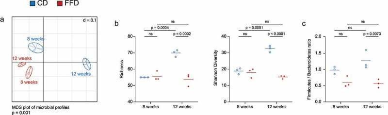
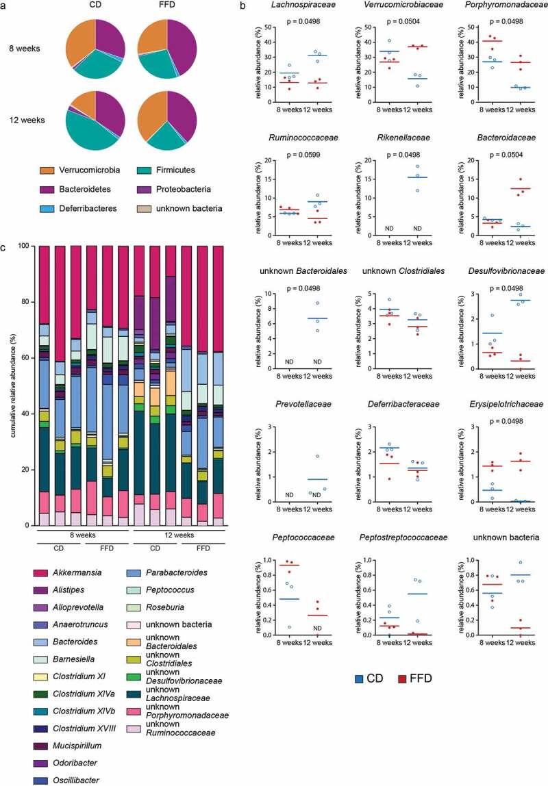
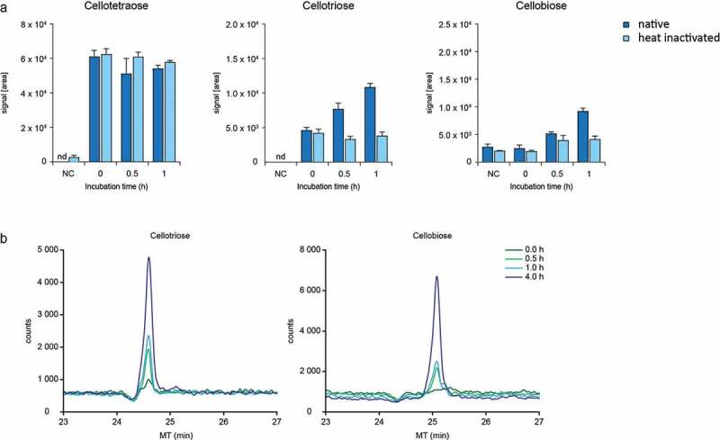
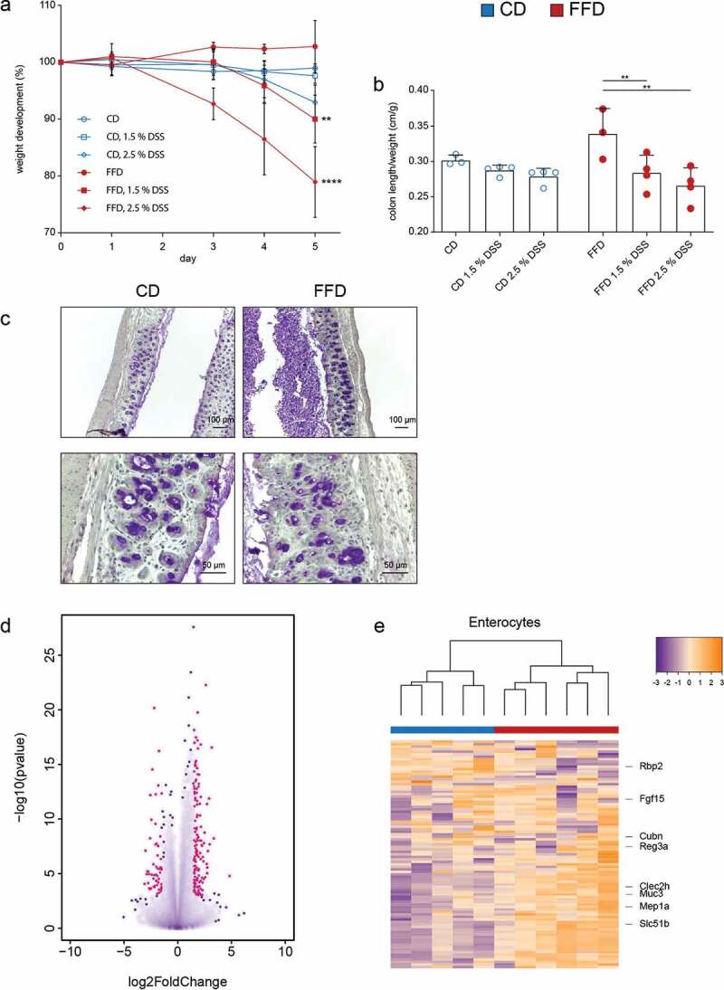
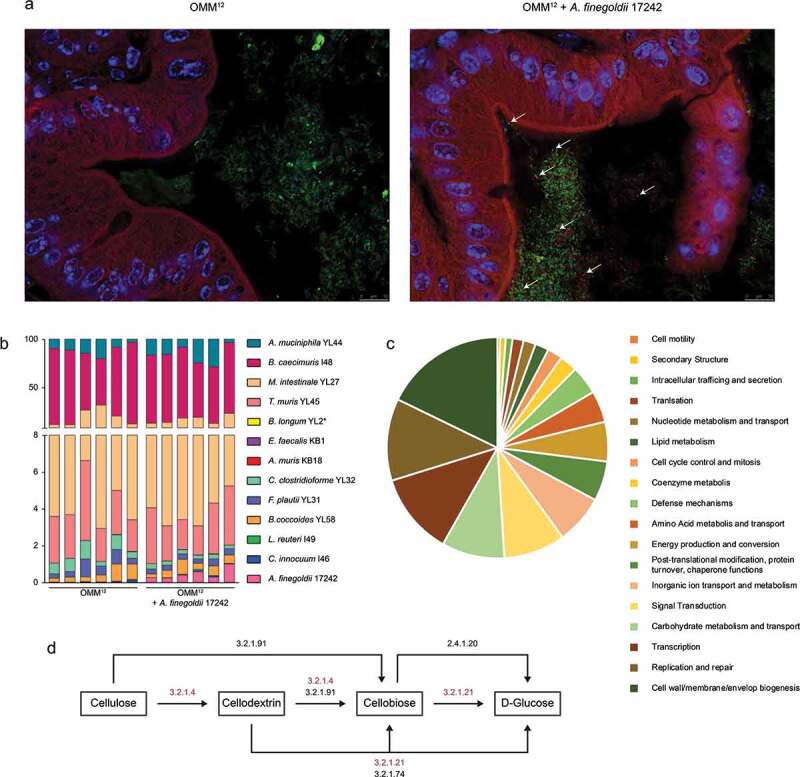
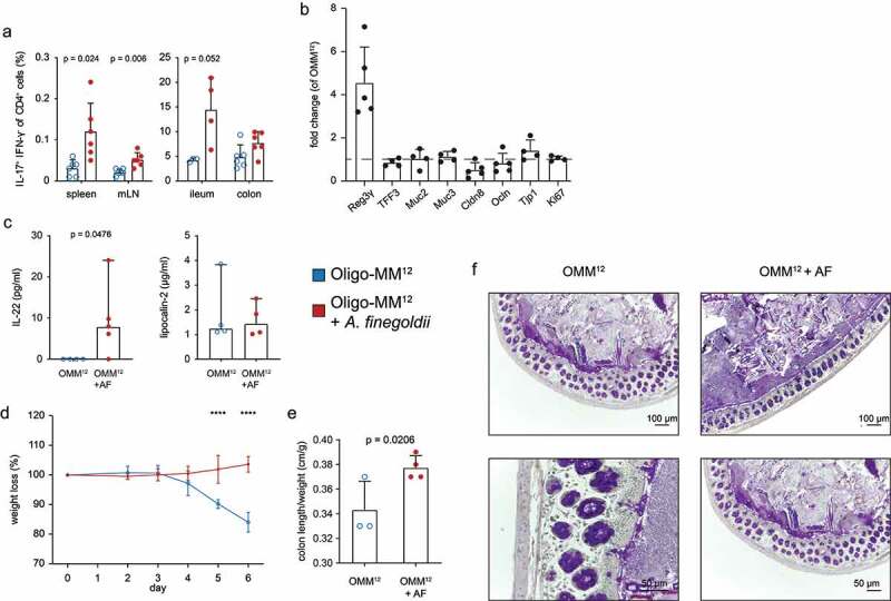
Although it is generally accepted that dietary fiber is health promoting, the underlying immunological and molecular mechanisms are not well defined, especially with respect to cellulose, the most ubiquitous dietary fiber. Here, the impact of dietary cellulose on intestinal microbiota, immune responses and gene expression in health and disease was examined. Lack of dietary cellulose disrupted the age-related diversification of the intestinal microbiota, which subsequently remained in an immature state. Interestingly, one of the most affected microbial genera was Alistipes which is equipped with enzymes to degrade cellulose. Absence of cellulose changed the microbial metabolome, skewed intestinal immune responses toward inflammation, altered the gene expression of intestinal epithelial cells and mice showed increased sensitivity to colitis induction. In contrast, mice with a defined microbiota including A. finegoldii showed enhanced colonic expression of intestinal IL-22 and Reg3γ restoring intestinal barrier function. This study supports the epidemiological observations and adds a causal explanation for the health promoting effects of the most common biopolymer on earth.

Keywords: Alistipes; Cellulose; IL-22; Reg3γ; bile acids; inflammation; insoluble fiber; microbial diversity; microbiota maturation; mucosal homeostasis.


Conflict of interest statement

No potential conflict of interest was reported by the authors

Figures

Figure 1.
Figure 1.
The impact of cellulose on the diversification of the intestinal microbiota. The diversity of the intestinal microbiota of eight and twelve-week-old B6 mice was analyzed by 16S rRNA gene amplicon analysis. (a) Diversity shown as multi-dimensional scaling (MDS) plot based on generalized UniFrac dissimilarities (n = 3). (b) Alpha diversity shown as richness and Shannon diversity and (c) the Firmicutes/Bacteroidetes ratio (n = 3). Statistical analysis: (a) non-parametric multivariate analysis of variance (Rhea), (b, c) one-way ANOVA. Data are shown as individual mice and means and are representative of two independent experiments
Figure 2.
Figure 2.
Alterations of intestinal microbiota at taxonomic levels. Taxonomic composition of the intestinal microbiota of CD or FFD mice. (a) Phyla, (b) family and (c) genera at the age of eight and twelve weeks was measured via 16S rRNA gene amplicon analysis (n = 3). Statistical analysis: (b) Kruskal-Wallis Rank Sum Test for all groups (corrected for multiple comparison by Benjamini-Hochberg method). Data are shown as individual mice and means and are representative of two independent experiments
Figure 3.
Figure 3.
Degradation of cellotetraose by enzymes of the cecal microbiota. (a) Signals intensity (AUC) measured by HPLC-MS and (b) CE-MS electropherogram of degradation products after incubation of cellotetraose with native and heat-inactivated cecal enzymes of CD mice (n = 1). Data are shown as means ± SD and are representative of two independent experiments
Figure 4.
Figure 4.
Influence of dietary cellulose on the intestinal immune and epithelial cells. T-lymphocytes isolated from spleen, mLN and intestinal lamina propria of CD and FFD mice were analyzed by FACS. (a) Cytokine secretion in indicated organs and (b) representative plots for ileal CD4+ T cells (n = 4). (c) Expression of transcription factors in indicated organs and (d) representative plots for transcription factor expression in ileal CD4+ T cells (n = 4). (e) RT-PCR of indicated genes from colonic tissue of FFD mice normalized to CD animals (n = 4). Statistical analysis: (a, c) Welch’s test. Data are shown as individual mice and means ± SD and are representative of two independent experiments
Figure 5.
Figure 5.
Influence of dietary cellulose on development of colitis and transcriptional profiles of gut epithelial cells. (a) Weight loss and (b) colon length/weight ratio in CD or FFD mice after treatment with DSS (1.5% and 2.5%) in the drinking water. (c) Periodic acid-Schiff histology (PAS) of the colon (2.5% DSS) (n – 3-4). (d) Differentially expressed genes of colonic epithelial cells of CD or FFD RAG KO mice after treatment with DSS (1.5%) for five days; red points represent genes statistically differentially expressed with p = .005 and log fold change > 1.5 (FFD vs CD). (e) Unsupervised clustering of genes related to distinct intestinal enterocytes differentially expressed in CD and FFD RAG KO mice (n = 5–6). Statistical analysis: (a) two-way ANOVA (* are related to weight loss at day five in comparison to untreated mice), (b, d) multiple t test corrected by Benjamini-Hochberg-method. *p < .03, **p < .002, ***p < .0002, and ****p < .0001. Data are shown as individual mice and means ± SD and are representative of two independent experiments
Figure 6.
Figure 6.
A. finegoldii enriches the Oligo-MM12 with cellulolytic potential. Oligo-MM12 mice were colonized with A. finegoldii 17242 and (a) viable A. finegoldii (white arrows) was visualized by FISH in the cecum (green, all bacteria; red, A. finegoldii; blue, DAPI). (b) Relative abundance of A. finegoldii within the Oligo-MM12 consortium was quantified by RT-PCR (n = 6). (c) Singletons were assigned to cluster of orthologous genes (EDGAR). (d) Scheme of the cellulose degrading pathways. Genes encoding cellulose degrading enzymes marked in red were exclusively found in A. finegoldii but not Oligo-MM12
Figure 7.
Figure 7.
A. finegoldii enhances the intestinal barrier and protects from colitis. Oligo-MM12 mice were colonized with A. finegoldii three weeks prior to analysis. (a) IL-17 and IFN-γ secretion in CD4+ T cells in indicated organs was measured via FACS (n = 4–6). (b) RT-PCR of indicated genes from colon normalized to Oligo-MM12 mice without A. finegoldii. (c) Cytokines from colon cultures measured by LEGENDplexTM and ELISA. Impact of A. finegoldii on (d) weight loss and (e) colon length/weight ratio of Oligo-MM12 after DSS treatment (3.5%) (n = 3–4). (f) Periodic acid-Schiff histology (PAS) of the colon from Oligo-MM12 and Oligo-MM12 + A. finegoldii mice (n = 2). Statistical analysis: (a) Welch’s test, (c) Mann-Whitney Test, (d) two-way ANOVA and (e) Student’s t test. *p < .03, **p < .002, ***p < .0002, and ****p < .0001. Data are shown as individual mice and (a, b, d, e) means ± SD or (c) median ± CI and are representative of two independent experiments

References

    1. Cordain L, Eaton SB, Sebastian A, Mann N, Lindeberg S, Watkins BA, O’Keefe JH, Brand-Miller J.. Origins and evolution of the Western diet: health implications for the 21st century. Am J Clin Nutr. 2005;81(2):341–354. doi:10.1093/ajcn.81.2.341. - DOI - PubMed
    1. Burkitt D, Trowell HC. Refined carbohydrate foods and disease: some implications of dietary fibre. New York: Academic Press Inc; 1975.
    1. Cummings JH, Engineer A. Denis Burkitt and the origins of the dietary fibre hypothesis. Nutr. Res. Rev. 2018;31(1):1–15. doi:10.1017/S0954422417000117. - DOI - PubMed
    1. Stephen AM, Champ MM-J, Cloran SJ, Fleith M, van Lieshout L, Mejborn H, Burley VJ. Dietary fibre in Europe: current state of knowledge on definitions, sources, recommendations, intakes and relationships to health. Nutr Res Rev. 2017;30(2):149–190. doi:10.1017/S095442241700004X. - DOI - PubMed
    1. Jones JM. CODEX-aligned dietary fiber definitions help to bridge the ‘fiber gap’. Nutr J. 2014;13(1):34. doi:10.1186/1475-2891-13-34. - DOI - PMC - PubMed

Publication types

MeSH terms

Supplementary concepts