. 2021 Jan 4;131(1):e137377.
doi: 10.1172/JCI137377.

Lung megakaryocytes are immune modulatory cells

Affiliations

Lung megakaryocytes are immune modulatory cells

Daphne N Pariser et al. J Clin Invest. .

Abstract

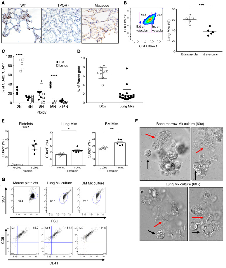
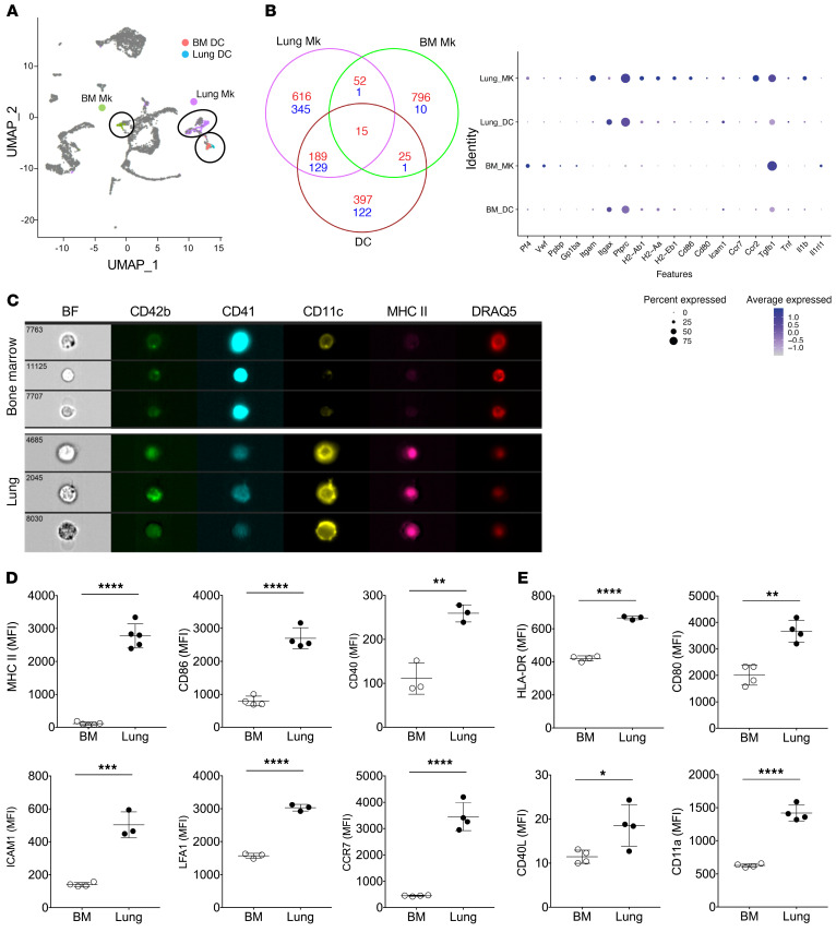
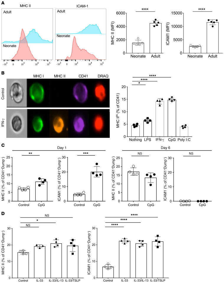
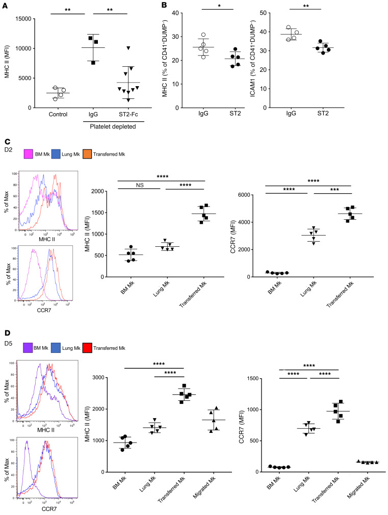
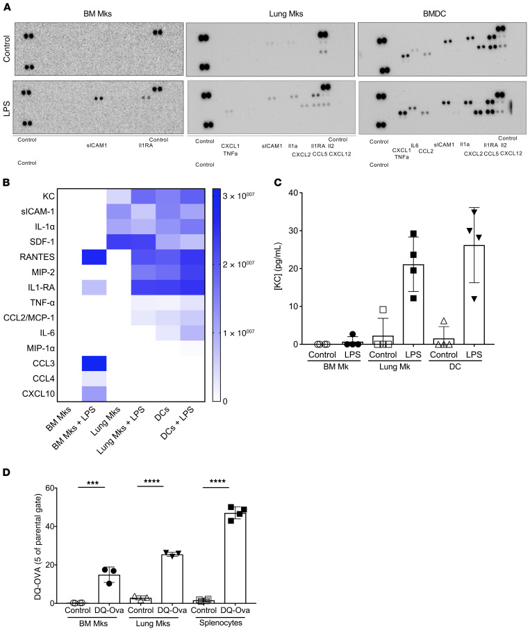
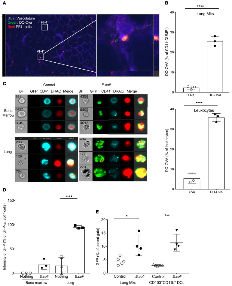
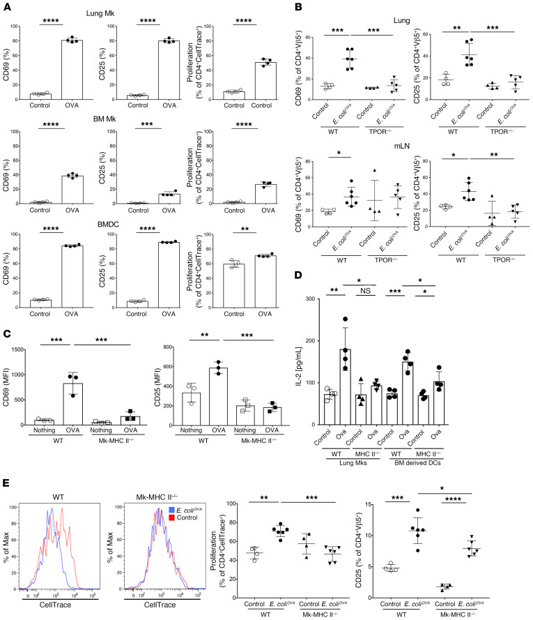
Although platelets are the cellular mediators of thrombosis, they are also immune cells. Platelets interact both directly and indirectly with immune cells, impacting their activation and differentiation, as well as all phases of the immune response. Megakaryocytes (Mks) are the cell source of circulating platelets, and until recently Mks were typically only considered bone marrow-resident (BM-resident) cells. However, platelet-producing Mks also reside in the lung, and lung Mks express greater levels of immune molecules compared with BM Mks. We therefore sought to define the immune functions of lung Mks. Using single-cell RNA sequencing of BM and lung myeloid-enriched cells, we found that lung Mks, which we term MkL, had gene expression patterns that are similar to antigen-presenting cells. This was confirmed using imaging and conventional flow cytometry. The immune phenotype of Mks was plastic and driven by the tissue immune environment, as evidenced by BM Mks having an MkL-like phenotype under the influence of pathogen receptor challenge and lung-associated immune molecules, such as IL-33. Our in vitro and in vivo assays demonstrated that MkL internalized and processed both antigenic proteins and bacterial pathogens. Furthermore, MkL induced CD4+ T cell activation in an MHC II-dependent manner both in vitro and in vivo. These data indicated that MkL had key immune regulatory roles dictated in part by the tissue environment.

Keywords: Hematology; Inflammation; Platelets.


Conflict of interest statement

Conflict of interest: The authors have declared that no conflict of interest exists.

Figures

Figure 1
Figure 1. Lung and BM Mks are phenotypically distinct.
(A) Mks are present in the lung. Lung sections from WT mice, TPOR–/– mice, and macaques were immunostained with anti-CD42c antibody (mouse tissue) or anti-CD41 (macaque). Mks were noted in WT mouse and primate lungs (representative images). Original magnification, ×40. (B) Mks are both intravascular and extravascular in the lung. Tissue and vascular discrimination between the lung Mks using anti-CD41 BV421 and BV786 by flow cytometry (unpaired t test). (C) BM Mk ploidy is greater than that of lung Mks. BM and lung Mk ploidy determined by flow cytometry (2-way ANOVA with Šidák’s multiple-comparison test). (D) The percentage of Mks relative to DCs in digested and single-cell resuspended lung tissue. Approximately 2% are Mks compared with 7% resident lung DCs (CD103+CD11b+). (E) Lung and BM Mks upregulate P-selectin (CD62P) in response to thrombin. Isolated lung and BM Mks were stimulated with 1 U/mL thrombin and CD62P surface expression determined by flow cytometry as a marker of degranulation (unpaired t test). (F) Lung Mks produce platelets in vitro. FRET imaging was used to obtain images of proplatelet production from both BM and lung Mks in culture. The red arrows indicate proplatelet formation, while the black arrows indicate an Mk. (G) Flow cytometry confirmation of platelet production in the Mk cultures. Freshly isolated mouse platelets were used as a control. *P = 0.01 to 0.05; **P = 0.001 to 0.01; ***P = 0.0001 to 0.001; ****P < 0.0001.
Figure 2
Figure 2. Lung and Mk immune molecule expression.
(A) Integration of BM and lung scRNA-seq data. BM and lung Mks had distinct mRNA expression. (B) Venn diagram and dot plot from scRNA-seq. Wilcoxon’s rank sum test (Seurat FindMarkers function) was performed and genes differentially expressed in the clusters of interest (Mks or DCs) against all other clusters were identified (adjusted P value < 1 × 10⁻³). Positive markers in red, and negative markers in blue. Dot plot indicates the average expression and proportion of cells expressing genes of interest. (C) Mk characterization using imaging flow cytometry. Lung Mks expressed more CD11c and MHC II compared with BM Mks. BF, bright field. (D) Comparison of mouse lung and BM Mk APC-related molecule expression by flow cytometry. Lung Mks express more APC-associated molecules (unpaired t test). (E) Comparison of primate lung and BM APC-related molecule expression. Lung Mks expressed more APC-associated molecules (unpaired t test). *P = 0.01 to 0.05, **P = 0.001 to 0.01, ***P = 0.0001 to 0.001, ****P < 0.0001.
Figure 3
Figure 3. Lung Mk immune phenotype is environmentally regulated.
(A) MHC II and ICAM1 expression on Mks from P0 and adult mice. Neonatal lung Mks had reduced MHC II and ICAM1 compared with adult lung Mks (unpaired t test). (B) BM Mks increased MHC II expression in response to immune stimuli. BM Mks were incubated with immune stimuli for 48 hours and MHC II expression determined. LPS, INF-γ, and CpG increased MHC II (1-way ANOVA with Tukey’s multiple-comparison test). (C) BM Mks respond to CpG within 24 hours and the expression of immune molecules is similar to that of control BM Mks at day 6 (unpaired t test). (D) Lung-derived immune modulatory cytokines induced BM Mk immune differentiation. BM Mks were incubated with IL-33 or IL-33 in combination with other common lung cytokines. Forty-eight hours later immune differentiation was determined (1-way ANOVA with Tukey’s multiple-comparison test). *P = 0.01 to 0.05; **P = 0.001 to 0.01; ***P = 0.0001 to 0.001; ****P < 0.0001.
Figure 4
Figure 4. In vivo regulators of lung Mk phenotype.
(A) IL-33 promoted lung Mk immune differentiation in vivo. Mice were treated with Mk-depleting antibody or control IgG. Mice were then treated with either ST2-Fc as an IL-33 blocking agent or control IgG. Recovering Mks had increased MHC II that was greatly attenuated by IL-33 blocking (unpaired t test). (B) P0 mice were treated with IgG or ST2-Fc and on P7 the lung Mk immune phenotype determined. IL-33 blocking reduced postnatal lung Mk immune differentiation (unpaired t test). (C and D) BM Mk immune phenotype plasticity in vivo. BM Mks were isolated, labeled with CFSE, and o.p. delivered to control mice. (C) Two days and (D) 5 days later transferred BM Mk MHC II and CCR7 levels were determined. BM-transferred BM Mks had increased immune molecule expression in the lung environment. After 5 days (D), BM Mk cells transferred to the lung were also identified in the BM and had reduced immune molecule expression compared with those transferred and in the lung (1-way ANOVA with Tukey’s multiple-comparison test). *P = 0.01 to 0.05; **P = 0.001 to 0.01; ***P = 0.0001 to 0.001; ****P < 0.0001.
Figure 5
Figure 5. Lung Mks process and present antigen.
(A and B) Lung Mks had greater LPS-induced inflammatory molecule production compared with BM Mks. Mks were incubated for 24 hours with control buffer or LPS. Inflammatory molecules were identified by a (A) cytokine array and (B) relative intensities compared with control plotted as a heatmap. (C) KC in the supernatant measured by ELISA. (D) Antigen processing in vitro. Mks were incubated with DQ-Ova for 30 minutes and fluorescence was determined by flow cytometry (unpaired t test). ***P = 0.0001 to 0.001, ****P < 0.0001.
Figure 6
Figure 6. Lung Mks process and present antigen in vivo.
(A) Mice were treated with control buffer or DQ-Ova via the o.p. route. Eighty minutes later real-time in vivo lung imaging was performed (representative image). (B) Twenty-four hours later DQ-Ova lungs were also isolated to quantify fluorescent Mks. Lung Mks internalized antigen (unpaired t test). (C and D) Lung Mks are more phagocytic than BM Mks. BM and lung Mks were incubated with control buffer or GFP E. coli and 30 minutes later fluorescence was determined by (C) ImageStream (representative images) and (D) bacteria internalization was quantified (unpaired t test). BF, bright field. (E) Lung Mks take up E. coli in vivo. E. coli was delivered via the o.p. route and 3 hours later E. coli–positive lung Mks and DCs were quantified by flow cytometry (unpaired t test). *P = 0.01 to 0.05; ***P = 0.0001 to 0.001; ****P < 0.0001.
Figure 7
Figure 7. Lung Mks present antigen.
(A) Lung Mks activated OTII T cells in vitro. T cells were cocultured with lung Mks or splenocytes and on day 3 T cell activation was determined (unpaired t test). (B) Mice lacking Mks had reduced antigen-specific T cell responses in vivo. WT and TPOR–/– mice were given OTII T cells and 24 hours later mice were o.p. treated with E. coliOVA. OTII T cell activation was determined on day 3 (unpaired t test). (C and D) Lung Mks present antigen in the context of MHC II in vitro. WT and MHC II–/– lung Mks were incubated with OTII T cells and Ova/LPS. On day 3 T cell activation was determined by flow cytometry (unpaired t test). (C) WT lung Mks induced more T cell activation than did MHC II–/– lung Mks and had (D) more IL-2 production on day 8. (E) Lung Mks present antigen in the context of MHC II in vivo. WT and Mk-specific MHC II–/– mice were given OTII T cells and E. coliOVA via the o.p. route. On day 3, OTII T cell activation was determined. WT mice had more CD25-positive OTII cells and OTII T cell proliferation compared with Mk-MHC II–/– mice (upaired t test). *P = 0.01 to 0.05, **P = 0.001 to 0.01, ***P = 0.0001 to 0.001, ****P < 0.0001.

Comment in

References

    1. Morrell CN, Pariser DN, Hilt ZT, Vega Ocasio D. The platelet Napoleon complex — small cells, but big immune regulatory functions. Annu Rev Immunol. 2019;37:125–144. doi: 10.1146/annurev-immunol-042718-041607. - DOI - PubMed
    1. Lefrançais E, et al. The lung is a site of platelet biogenesis and a reservoir for haematopoietic progenitors. Nature. 2017;544(7648):105–109. doi: 10.1038/nature21706. - DOI - PMC - PubMed
    1. Megakaryocytes in the lungs Br Med J. 1966;1(5500):1377–1378. doi: 10.1136/bmj.1.5500.1377-a. - DOI - PMC - PubMed
    1. Aabo K, Hansen KB. Megakaryocytes in pulmonary blood vessels. I. Incidence at autopsy, clinicopathological relations especially to disseminated intravascular coagulation. Acta Pathol Microbiol Scand A. 1978;86(4):285–291. - PubMed
    1. Sharnoff JG, Kim ES. Pulmonary megakaryocyte studies in rabbits. AMA Arch Pathol. 1958;66(3):340–343. - PubMed

Publication types

Substances