. 2020 Oct 1;61(12):19.
doi: 10.1167/iovs.61.12.19.

Neuroretinal-Derived Caveolin-1 Promotes Endotoxin-Induced Inflammation in the Murine Retina

Affiliations

Neuroretinal-Derived Caveolin-1 Promotes Endotoxin-Induced Inflammation in the Murine Retina

Jami M Gurley et al. Invest Ophthalmol Vis Sci. .

Abstract

Purpose: The immune-privileged environment and complex organization of retinal tissue support the retina's essential role in visual function, yet confound inquiries into cell-specific inflammatory effects that lead to dysfunction and degeneration. Caveolin-1 (Cav1) is an integral membrane protein expressed in several retinal cell types and is implicated in immune regulation. However, whether Cav1 promotes or inhibits inflammatory processes in the retina (as well as in other tissues) remains unclear. Previously, we showed that global-Cav1 depletion resulted in reduced retinal inflammatory cytokine production but paradoxically elevated retinal immune cell infiltration. We hypothesized that these disparate responses are the result of differential cell-specific Cav1 functions in the retina.

Methods: We used Cre/lox technology to deplete Cav1 specifically in the neural retinal (NR) compartment to clarify the role NR-specific Cav1 (NR-Cav1) in the retinal immune response to intravitreal inflammatory challenge induced by activation of Toll-like receptor-4 (TLR4). We used multiplex protein suspension array and flow cytometry to evaluate innate immune activation. Additionally, we used bioinformatics assessment of differentially expressed membrane-associated proteins to infer relationships between NR-Cav1 and immune response pathways.

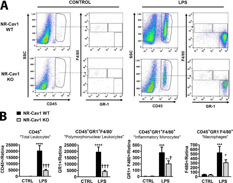
Results: NR-Cav1 depletion, which primarily affects Müller glia Cav1 expression, significantly altered immune response pathway regulators, decreased retinal inflammatory cytokine production, and reduced retinal immune cell infiltration in response to LPS-stimulated inflammatory induction.

Conclusions: Cav1 expression in the NR compartment promotes the innate TLR4-mediated retinal tissue immune response. Additionally, we have identified novel potential immune modulators differentially expressed with NR-Cav1 depletion. This study further clarifies the role of NR-Cav1 in retinal inflammation.


Conflict of interest statement

Disclosure: J.M. Gurley, None; G.B. Gmyrek, None; M.E. McClellan, None; E.A. Hargis, None; S.M. Hauck, None; M.G. Dozmorov, None; J.D. Wren, None; D.J.J. Carr, None; M.H. Elliott, None

Figures

Figure 1.
Figure 1.
Effect of neuroretinal-specific deletion on whole retinal tissue CAV1 protein expression (Chx10-Cre/Cav1-flox model). (A) Schematic diagram illustrating the strategy for neuroretinal-specific Cav1 deletion via Chx10-promoter driven Cre-mediated recombination of the floxed Cav1 gene. (B) Representative Western blots of whole cell lysates from various tissues showing retina-specific CAV1 protein reduction following Chx10-Cre expression in the floxed Cav1 model. Protein loads ranged from 5 µg to 35 µg, depending on tissue, to detect protein within the linear range of the assay. (C) Histogram of Western blot protein densitometry data quantification shows retinal tissue specific CAV1 protein reduction after NR-Cav1-depletion in Chx10-Cre/Cav1+/+ mice compared to Chx10-Cre/Cav−/− controls. β-actin was used as a loading control and for normalization. Data are mean ± SEM and were analyzed via Student's t-test (** P < 0.01; N = 7). ACT = β-actin.
Figure 2.
Figure 2.
Neuroretinal-specific Cav1 depletion targets retinal Müller glial cells. Immunohistochemical staining of mouse retinal sections stained with anti-CAV1 and costained with Müller cell-specific marker GS shows high Müller glial CAV1 protein expression and colocalization in cell regions associating with blood vessels, within Müller somata, and in apical processes near the external limiting membrane (white arrows). Blood vessel cross-sections representing the three layers of retinal vasculature also exhibit high levels of CAV1 protein expression, but are not targeted by the NR-Cav1 KO model (white arrowheads; s, i, d = superficial, intermediate, and deep retinal blood vessels, respectively). Sparse mosaic Cre-mediated recombination results in occasional residual Müller glial CAV1 expression (black arrows). WT, NR-Cav1 WT; KO, NR-Cav1 KO; INL, inner nuclear layer.
Figure 3.
Figure 3.
Effect of neuroretinal-specific depletion on membrane-associated protein composition. Mass spectrometry-based proteomics heat map analysis (pheatmap R) of membrane raft fractions from whole retinal tissue. Each replicate sample contained pooled retinas from eight mice. Blue = downregulated; red = upregulated. Heatmap represents peptide abundance quantification for the top 70 differentially expressed peptides as a result of NR-Cav1 depletion. Data are mean ± SEM and were analyzed via Student's t-test (* P < 0.05; *** P < 0.001).
Figure 4.
Figure 4.
Neuroretinal-specific Cav1 depletion blunts endotoxin-mediated retinal proinflammatory cytokine production. Multiplex protein suspension array analysis showing endotoxin-mediated inflammatory activation in retinas from Chx10-Cav1 WT and KO animals 24 hours after intravitreal LPS injection. Histograms represent measured concentrations of proinflammatory cytokines/chemokines (A) IL-6, (B) CXCL1/KC, (C) CCL2/MCP-1, and (D) IL-1β. Data are mean ± SEM and were analyzed using two-way analysis of variance, with Fisher's LSD post-hoc. Treatment effect: *P < 0.05, ***P < 0.001, ****P < 0.0001; Genotype effect: ††P < 0.01, †††P < 0.001. CXCL1/KC, C-X-C motif chemokine ligand 1; CCL2/MCP-1, monocyte chemoattractant protein 1; IL-1β, interleukin 1 beta.
Figure 5.
Figure 5.
Neuroretinal-specific Cav1 depletion reduces immune cell recruitment to retinal tissue. (A) Flow cytometry dot plots and (B) histogram quantifications representing immune cell infiltration into retinal tissue 24 hours after intraocular LPS challenge. Animals were perfused before collection of whole retinal tissue from Chx10-Cav1 WT and Chx10-Cav1 KO animals. Gating strategy represents the following cell populations: “total leukocytes” (CD45+), “PMNs” (CD45+F4/80/GR1+), “inflammatory monocytes” (CD45+F4/80+/Gr1+), and “macrophages/MΦ” (CD45+F4/80+/GR1). (B) Histogram quantification of flow cytometry data from control or LPS-treated Chx10-Cav1 WT and Chx10-Cav1 KO animals. Data are mean ± SEM and were analyzed using two-way analysis of variance, with Fisher's LSD post hoc. Treatment effect: *P < 0.05, **P < 0.01, ***P < 0.001, ****P < 0.0001; genotype effect: †P < 0.05, †††P < 0.001.
Figure 6.
Figure 6.
Validation of neuroretinal-specific depletion effects on retinal tissue immune cell recruitment using six-color flow cytometry. Upper panel: Representative dot plots from an LPS-treated NR-Cav1 WT retina and illustrates the gating strategy used for six-color flow cytometry. These data were obtained and analyzed with LPS-treated NR-Cav1 KO and PBS-treated NR-Cav1 WT and NR-Cav1 KO samples (representative dot plot for PBS samples is shown in Supplementary Figure S7). Lower panel: Corresponding histogram quantifications represent defined immune cell populations (AF). Animals were perfused before collection of whole retinal tissue. Data represent all PBS-treated and LPS-treated NR-Cav1 WT and NR-Cav1 KO samples and are mean ± SEM. Data were analyzed via two-way analysis of variance with Tukey's post-hoc. Treatment effect: *P < 0.05, **P < 0.01, ***P < 0.001, ****P < 0.0001; Genotype effect: ††P < 0.01.
Figure 7.
Figure 7.
Simplified model summarizing retinal immune response following LPS challenge. A simple diagram summarizes the sequence of events after intravitreal immune challenge with LPS, which highlights events affected by NR-Cav1 depletion versus global-Cav1 depletion.

Similar articles

Cited by

References

    1. Whitcup SM, Nussenblatt RB, Lightman SL, Hollander DA. Inflammation in retinal disease. Int J Inflam. 2013; 2013: 724648. - PMC - PubMed
    1. Murakami Y, Ishikawa K, Nakao S, Sonoda KH. Innate immune response in retinal homeostasis and inflammatory disorders. Prog Retin Eye Res. 2019; 74: 100778. - PubMed
    1. Streilein JW. Ocular immune privilege: the eye takes a dim but practical view of immunity and inflammation. J Leukoc Biol. 2003; 74: 179–185. - PubMed
    1. Zhou R, Caspi RR. Ocular immune privilege. F1000 Biol Rep. 2010; 2: 3. - PMC - PubMed
    1. Chidlow JH Jr., Sessa WC. Caveolae, caveolins, and cavins: complex control of cellular signalling and inflammation. Cardiovasc Res. 2010; 86: 219–225. - PMC - PubMed

Publication types

MeSH terms