

Preparation of Coralline Hydroxyapatite Implant with Recombinant Human Bone Morphogenetic Protein-2-Loaded Chitosan Nanospheres and Its Osteogenic Efficacy
- PMID: 33080108
- PMCID: PMC7767670
- DOI: 10.1111/os.12752
Preparation of Coralline Hydroxyapatite Implant with Recombinant Human Bone Morphogenetic Protein-2-Loaded Chitosan Nanospheres and Its Osteogenic Efficacy
Abstract
Objective: Spinal fusion is one of the most common surgical interventions for spine reconstruction. Despite the efforts to promote osteogenesis after spinal fusion, osteogenesis after spinal fusion remains a clinical challenge and new methods are still needed. The bone morphogenetic protein-2 (BMP-2) is a widely reported factor that can facilitate the osteogenesis in spinal fusion. In previous research, we found that the delivery of chitosan nanospheres could promote the effects of BMP-2 on osteogenic activity. The coralline hydroxyapatite (CHA) is one of the most frequently used implants in bone fusion. However, up to now no study has focused on the osteogenic efficacy of the CHA composite with recombinant human BMP-2 (rhBMP-2)-loaded chitosan nanospheres. This study aimed to investigate the effects of the CHA implant with rhBMP-2-loaded chitosan nanospheres on osteogenesis in spinal fusion.
Methods: The rhBMP-2-loaded microspheres and CHA composite (rhBMP-2 microspheres/CHA) were prepared and were used for implantation of the rats. All SD rats were divided into four groups: the rhBMP-2 microspheres/CHA composite group (containing 0.5 mg rhBMP-2), the rhBMP-2-loaded CHA (rhBMP-2/CHA) composite group (containing 0.5 mg rhBMP-2), the blank CHA group, and the negative control group. The microsphere morphology was scanned and analyzed using a scanning electron microscope. Micro-computed tomography examination and three-dimensional reconstruction were performed 4 weeks after the surgery. Hematoxylin and eosin staining was conducted for histological analysis. Both alkaline phosphatase (ALP) and calcium content were measured.
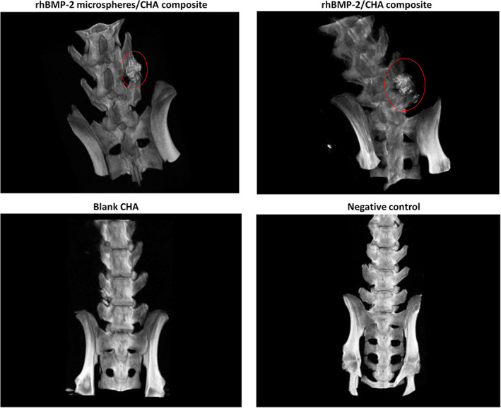
Results: The rhBMP-2-loaded CHA (rhBMP-2/CHA) composite was successfully prepared. Spherical regularity and a smooth and unwrinkled surface of the spheres were observed in all chitosan (CS)/rhBMP-2 microspheres. No side effects, infections, or abnormal behaviors were found in the animals. After 4 weeks of surgery, obvious new bone formation and bone fusion could be observed around the implant in both the rhBMP-2 microspheres/CHA composite group and the rhBMP-2/CHA composite group. No ectopic osteogenesis was found in the vertebral canal or other muscle tissues. After 4 weeks of implantation, in both the rhBMP-2 microspheres/CHA composite group and the rhBMP-2/CHA composite group, osteoid tissues could be found, and bone cells, bone marrow, and trabecular bone turned into mature sclerotin, obvious bone tissue formation could be also seen. Both ALP activity and calcium content in the rhBMP-2 microspheres/CHA composite group (6.52 ± 0.50 kat/g and 17.54 ± 2.49 μg/mg) were significantly higher than in all other groups.
Conclusion: The composite with rhBMP-2-loaded CS nanospheres could enhance osteogenic efficacy and increase the ALP activity and calcium content. These results might provide a novel method for osteogenesis in spinal fusion and offer new insight into the role of BMP-2 in osteogenesis.
Keywords: Chitosan nanospheres; Coralline hydroxyapatite implant; Osteogenesis; Spinal fusion; rhBMP-2.
© 2020 The Authors. Orthopaedic Surgery published by Chinese Orthopaedic Association and John Wiley & Sons Australia, Ltd.
Figures


Similar articles
-
[Radiological evaluation of dextran sulfate/recombinant human bone morphogenetic protein 2/chitosan composite microspheres combined with coral hydroxyapatite artificial bone in repairing large segmental bone defects].Zhongguo Xiu Fu Chong Jian Wai Ke Za Zhi. 2017 Nov 15;31(11):1384-1389. doi: 10.7507/1002-1892.201703094. Zhongguo Xiu Fu Chong Jian Wai Ke Za Zhi. 2017. PMID: 29798596 Free PMC article. Chinese.
-
[ECTOPIC OSTEOGENESIS EVALUATION OF RECOMBINANT HUMAN BONE MORPHOGENETIC PROTEIN 2 LOADED CHITOSAN/DEXTRAN SULFATE BY MICRO-CT].Zhongguo Xiu Fu Chong Jian Wai Ke Za Zhi. 2016 Mar;30(3):286-91. Zhongguo Xiu Fu Chong Jian Wai Ke Za Zhi. 2016. PMID: 27281871 Chinese.
-
The mechanism research of non-Smad dependent TAK1 signaling pathway in the treatment of bone defects by recombination BMP-2-loaded hollow hydroxyapatite microspheres/chitosan composite.J Mater Sci Mater Med. 2019 Nov 27;30(12):130. doi: 10.1007/s10856-019-6340-9. J Mater Sci Mater Med. 2019. PMID: 31776786
-
A Narrative Review on Recombinant Human Bone Morphogenetic Protein 2: Where Are We Now?Cureus. 2024 Aug 26;16(8):e67785. doi: 10.7759/cureus.67785. eCollection 2024 Aug. Cureus. 2024. PMID: 39188335 Free PMC article. Review.
-
Bone Morphogenetic Protein-2 in Development and Bone Homeostasis.J Dev Biol. 2020 Sep 13;8(3):19. doi: 10.3390/jdb8030019. J Dev Biol. 2020. PMID: 32933207 Free PMC article. Review.
Cited by
-
Bone Regeneration by Multichannel Cylindrical Granular Bone Substitute for Regeneration of Bone in Cases of Tumor, Fracture, and Arthroplasty.Int J Environ Res Public Health. 2022 Jul 6;19(14):8228. doi: 10.3390/ijerph19148228. Int J Environ Res Public Health. 2022. PMID: 35886080 Free PMC article.
References
-
- Group PVLS . Risk factors associated with ischemic optic neuropathy after spinal fusion surgery. Anesthesiology, 2016, 116: 15. - PubMed
-
- Lu VM, Kerezoudis P, Gilder HE, McCutcheon BA, Phan K, Bydon M. Minimally invasive surgery versus open surgery spinal fusion for Spondylolisthesis. A systematic review and meta‐analysis. Spine (Phila Pa 1976), 2016, 42: E177. - PubMed
-
- Shamsul BS, Tan KK, Chen HC, Aminuddin BS, Ruszymah BH. Posterolateral spinal fusion with osteogenesis induced BMSC seeded TCP/HA in a sheep model. Tissue Cell, 2014, 46: 152–158. - PubMed
-
- Lin S, Liao C, Chen G, et al PLGA/β‐TCP composite scaffold incorporating salvianolic acid B promotes bone fusion by angiogenesis and osteogenesis in a rat spinal fusion model. Biomaterials, 2018, 196: 109–121. - PubMed
MeSH terms
Substances
Grants and funding
LinkOut - more resources
Full Text Sources
