Review
. 2021 Mar 16;22(6):949-960.
doi: 10.1002/cbic.202000661. Epub 2020 Nov 17.

Hydrogen Sulfide Oxidation by Sulfide Quinone Oxidoreductase

Affiliations
Review

Hydrogen Sulfide Oxidation by Sulfide Quinone Oxidoreductase

Aaron P Landry et al. Chembiochem. .

Abstract

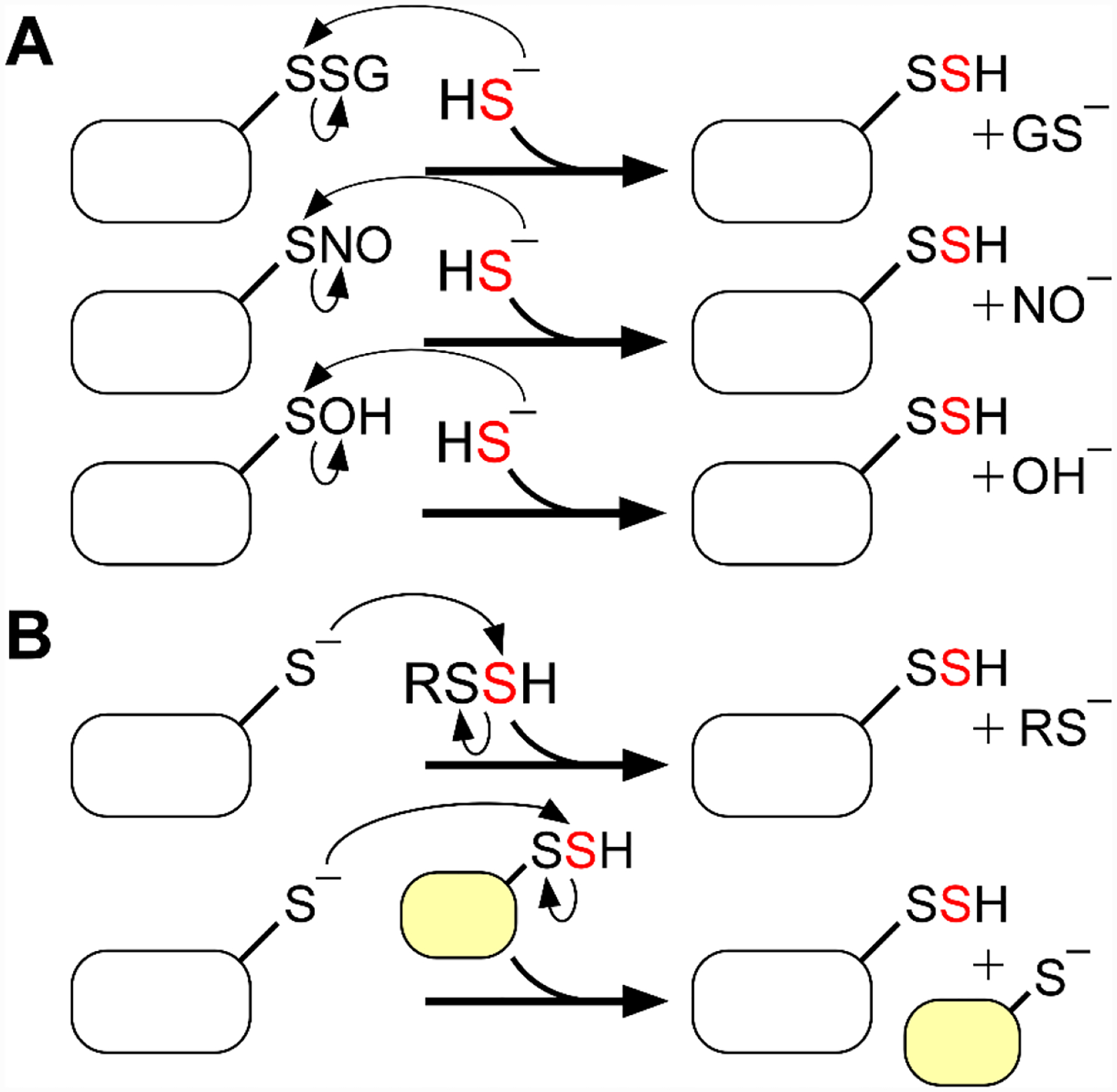
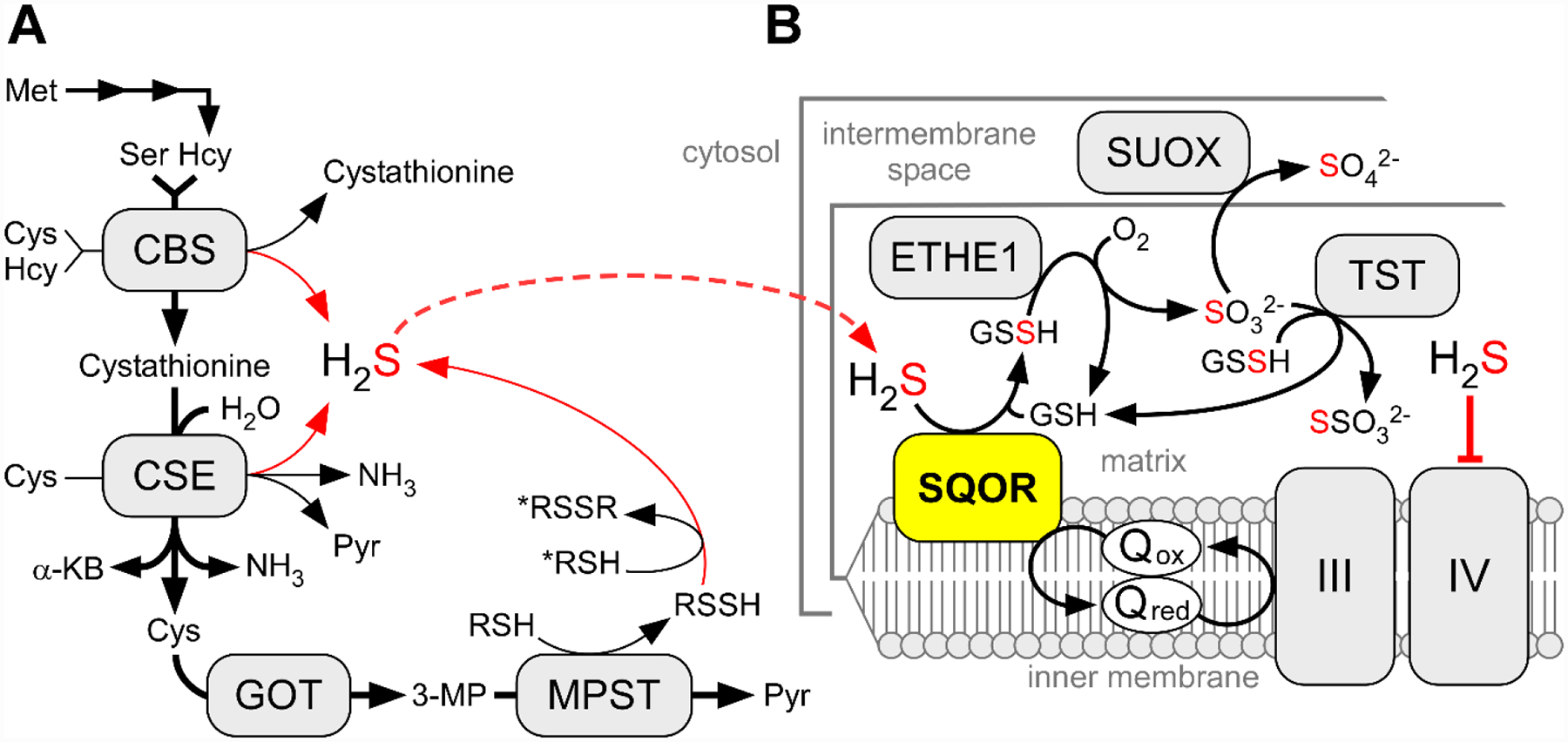
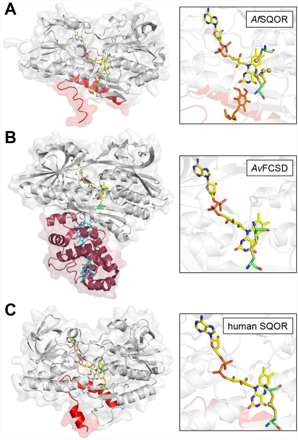
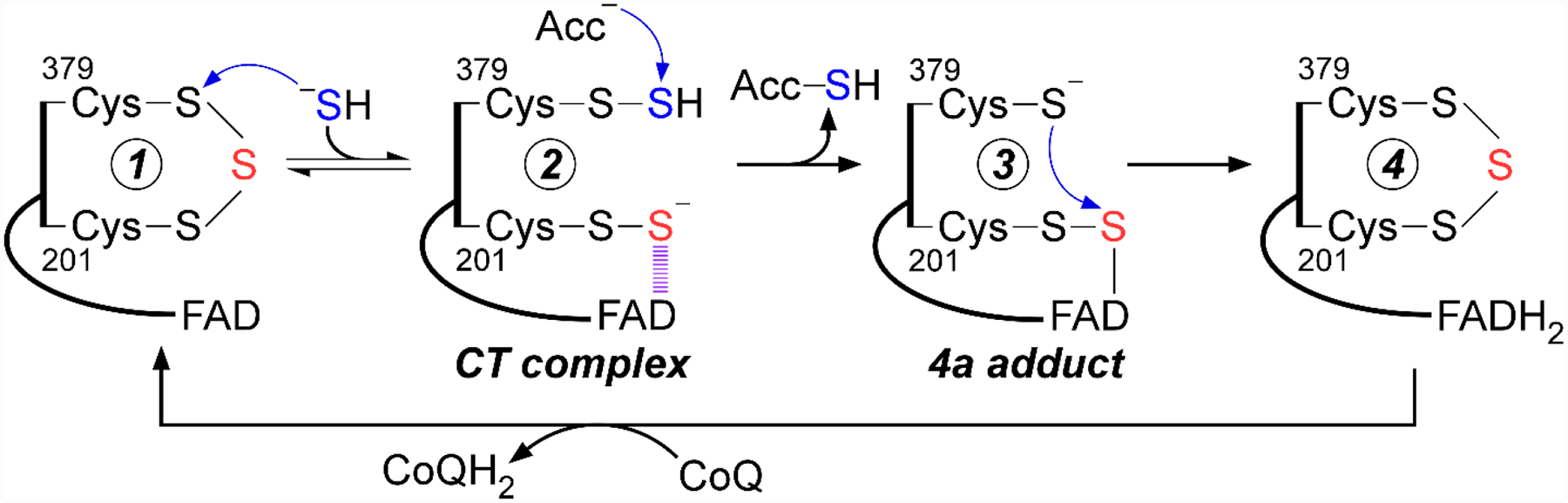
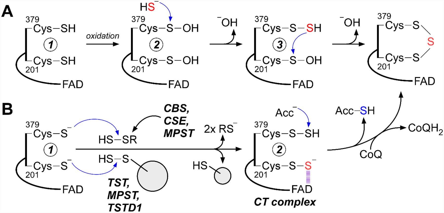
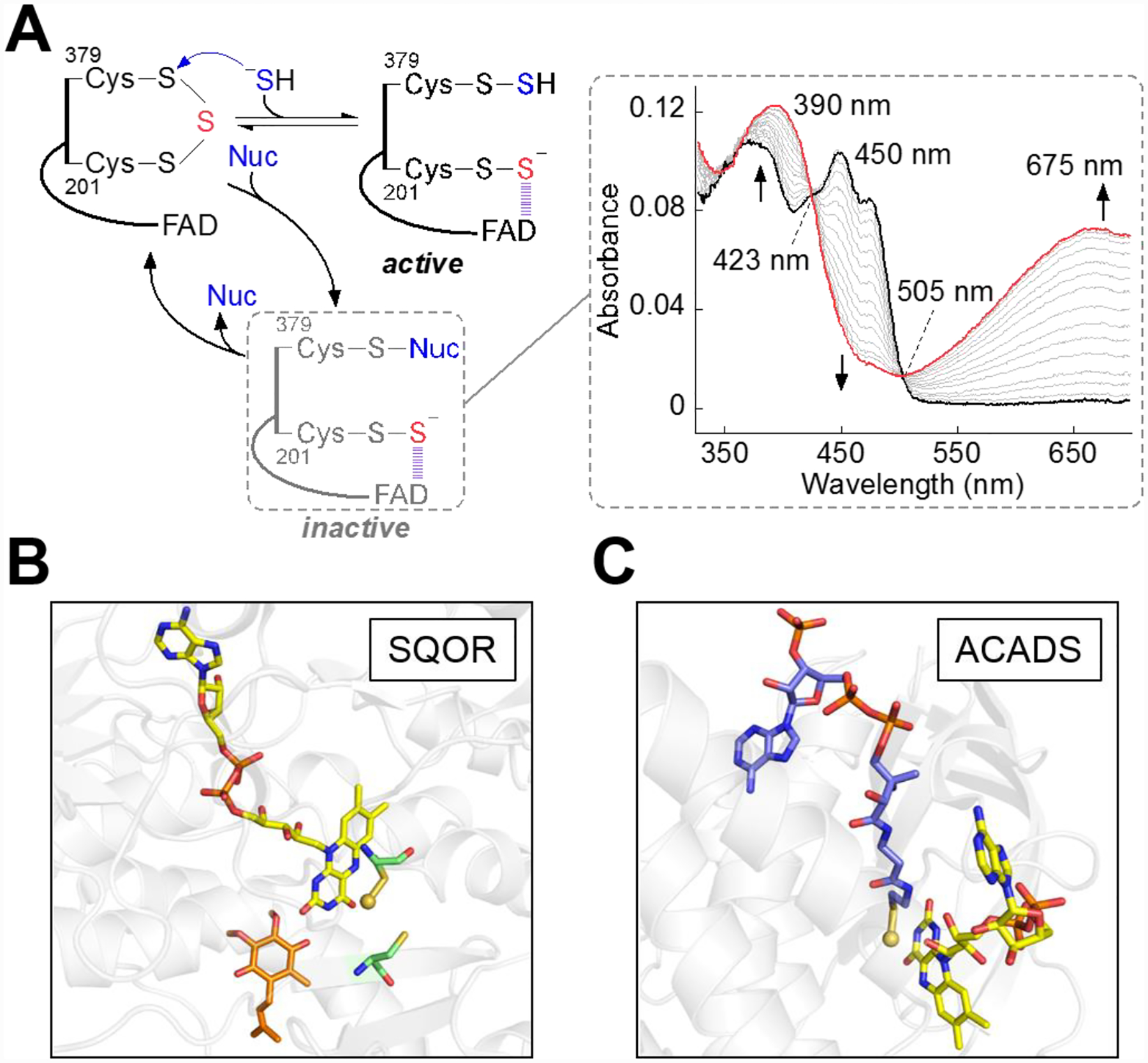
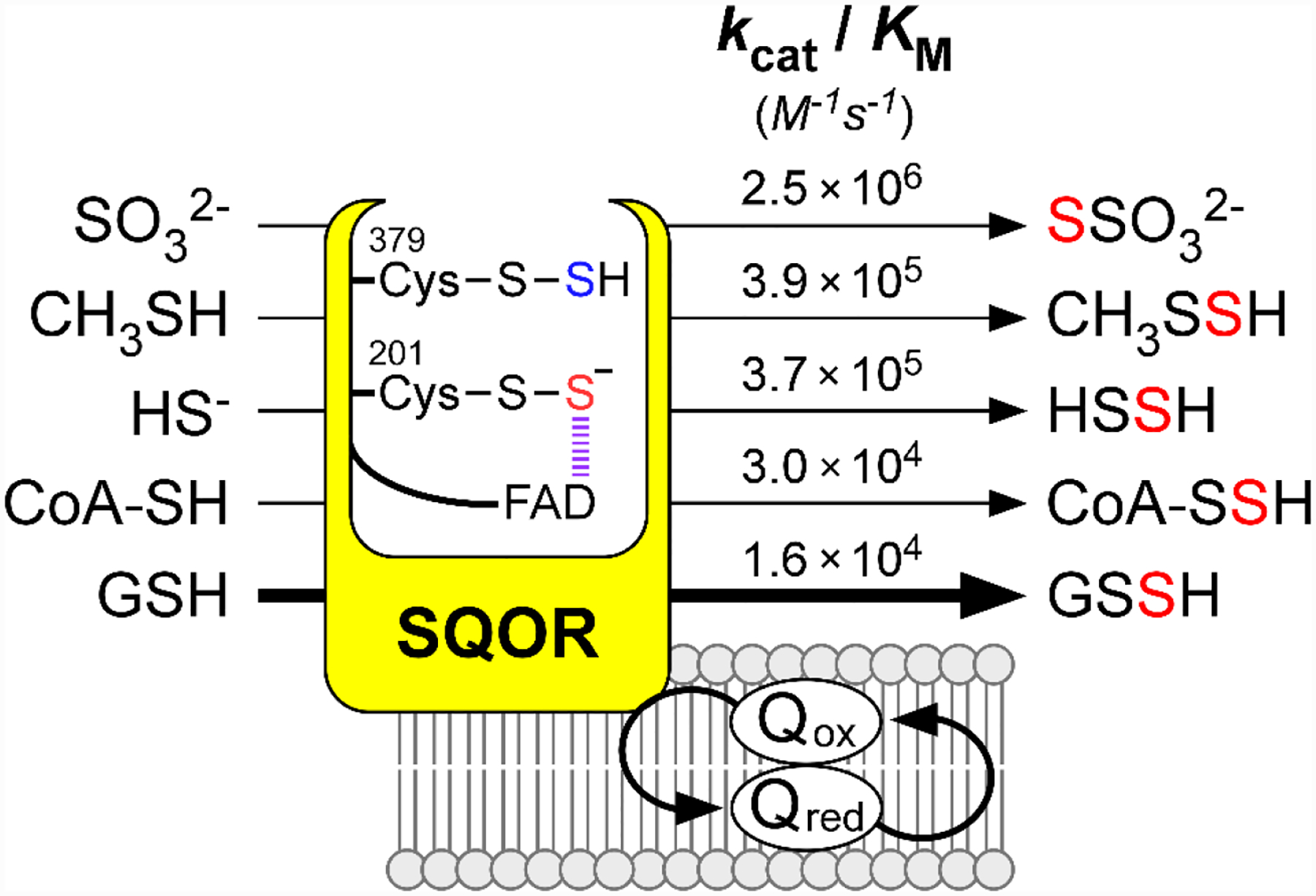
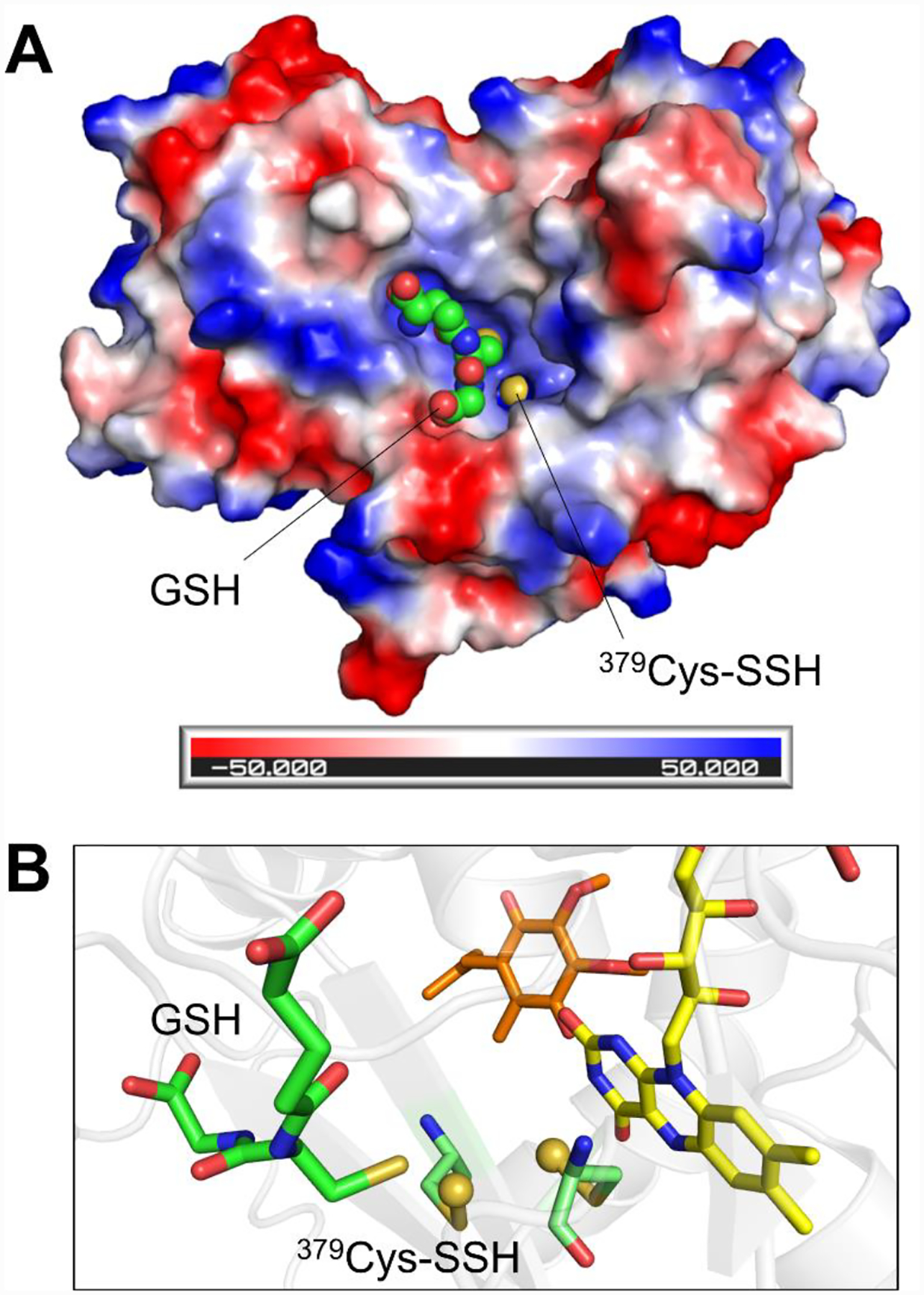
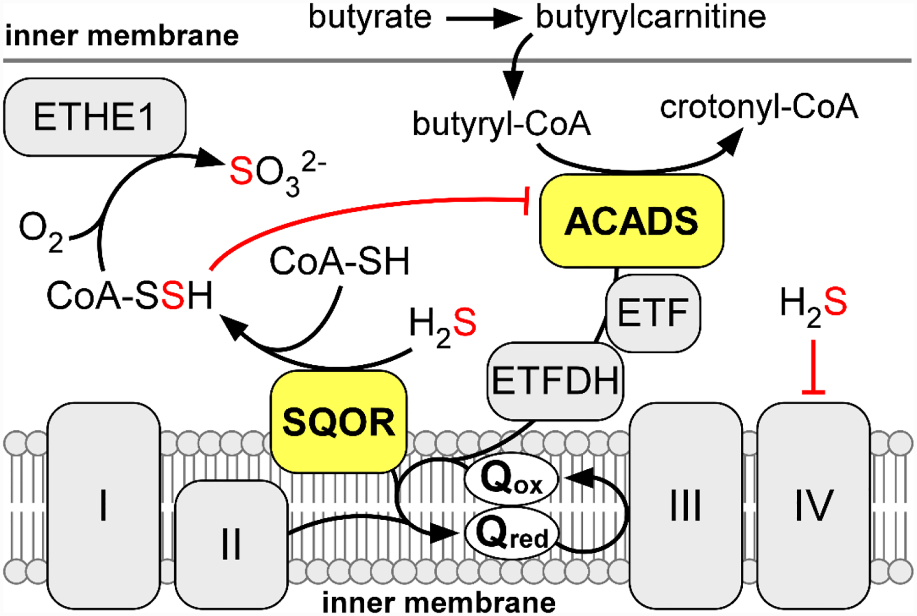
Hydrogen sulfide (H2 S) is an environmental toxin and a heritage of ancient microbial metabolism that has stimulated new interest following its discovery as a neuromodulator. While many physiological responses have been attributed to low H2 S levels, higher levels inhibit complex IV in the electron transport chain. To prevent respiratory poisoning, a dedicated set of enzymes that make up the mitochondrial sulfide oxidation pathway exists to clear H2 S. The committed step in this pathway is catalyzed by sulfide quinone oxidoreductase (SQOR), which couples sulfide oxidation to coenzyme Q10 reduction in the electron transport chain. The SQOR reaction prevents H2 S accumulation and generates highly reactive persulfide species as products; these can be further oxidized or can modify cysteine residues in proteins by persulfidation. Here, we review the kinetic and structural characteristics of human SQOR, and how its unconventional redox cofactor configuration and substrate promiscuity lead to sulfide clearance and potentially expand the signaling potential of H2 S. This dual role of SQOR makes it a promising target for H2 S-based therapeutics.

Keywords: flavins; metabolism; protein structure; redox chemistry; sulfides.


Conflict of interest statement

Conflict of Interest

The authors declare no conflict of interest.

Figures

Figure 1.
Figure 1.. Mechanisms of protein persulfidation.
A, Persulfidation of oxidized cysteines on proteins. B, Persulfidation of reduced cysteines on proteins via reaction with a low molecular weight persulfide (RSSH) or a sulfurtransferase.
Figure 2.
Figure 2.. H2S biosynthesis and oxidation pathways.
A, The canonical reactions catalyzed by CBS and CSE and MPST are depicted by bold black arrows while the H2S-synthesizing reactions are shown by thin black and red arrows. α-KB and Pyr denote α-ketobutyrate and pyruvate, respectively. GOT and 3-MP denote cysteine aminotransferase and its product, 3-mercaptopyruvate, respectively. To note, MPST has both cytoplasmic and mitochondrial isoforms. B, The mitochondrial sulfide oxidation pathway. TST, SUOX, III and IV denote rhodanese, sulfite oxidase, and respiratory complexes III and IV, respectively.
Figure 3.
Figure 3.. Structures of bacterial SQOR, FCSD, and human SQOR.
Cartoon and transparent surface overlay representations of A, Acidithiobacillus ferrooxidans SQOR (AfSQOR, PDB ID: 3T31); B, Allochromatium vinosum FCSD (AvFCSD, PDB ID: 1FCD); and C, human SQOR (PDB ID: 6OI5). The amphipathic helices of membrane-anchored AfSQOR and human SQOR are depicted in red, and the diheme cytochrome subunit of cytosolic AvFCSD is depicted in burgundy. Yellow sticks represent the FAD cofactor in each structure. The CoQ analog decylubiquinone in AfSQOR is shown in orange stick display and the dual heme cofactors in the cytochrome subunit of AvFCSD in cyan. An enlarged view of the active sites is shown in each panel on the right, with the redox active cysteines displayed in green sticks. Sulfane sulfur atoms in AfSQOR and bridging the active site cysteines in human SQOR, are shown as yellow spheres.
Figure 4.
Figure 4.. Proposed catalytic mechanism for human SQOR.
The bridging sulfur within the active site cysteine trisulfide is shown in red. The sulfur that undergoes oxidation and transfer to a thiophilic acceptor (Acc) is shown in blue.
Figure 5.
Figure 5.. Possible mechanisms for in vivo building of the SQOR cysteine trisulfide.
A, Oxidation of active site cysteines in newly synthesized SQOR (e.g., formation of cysteine sulfenic acid) (2) can promote cysteine persulfide formation (3) via nucleophilic attack of sulfide. Attack on the sulfenic acid by the persulfide generates the trisulfide. B, Persulfidation of SQOR at each cysteine by small molecule persulfides generated by the H2S synthesizing enzymes (CBS: cystathionine β-synthase; CSE: cystathionine γ-lyase; MPST: 3-mercaptopyruvate sulfurtransferase), or by an enzyme-catalyzed sulfur transfer via a sulfurtransferase (TST: rhodanese; MPST, or TSTD1), generates the CT complex (2). The latter leads to cysteine trisulfide formation following electron transfer to FAD and subsequent oxidation by CoQ.
Figure 6.
Figure 6.. Promiscuous nucleophilic addition and active site persulfide formation.
A, Alternative to sulfide, nucleophiles (Nuc) including GSH, sulfite, and methanethiol are capable of adding into the cysteine trisulfide to form a slowly decaying dead-end complex. The right panel (adapted from Ref. Supplemental Figure 2) shows SQOR embedded in nanodiscs (20 μM, black trace) rapidly mixed with sulfite (500 μM) and monitored over a period of 14 s for the formation of an alternative CT complex (red trace), with an absorbance maximum at 675 nm. B, Active site of human SQOR co-crystallized with CoQ (orange sticks) and soaked with sulfite (PDB: 6OIC), which contained a stable 201Cys persulfide-to-FAD CT complex in crystallo. C, Active site of ACADS crystallized with bound CoA persulfide (PDB: 2VIG), shown as blue sticks. In panels B and C, the sulfane sulfur in 201Cys-SSH and in CoA-SSH persulfide are shown as yellow spheres.
Figure 7.
Figure 7.. Catalytic promiscuity of the SQOR reaction.
The catalytic efficiencies denoted for small thiophilic acceptors are derived from the most recent kinetic characterizations using SQOR embedded in nanodiscs (Refs. 54, 73, 93). In the CT complex intermediate, the sulfur derived from the active site cysteine trisulfide is shown in red, and the sulfur derived from H2S is shown in blue.
Figure 8.
Figure 8.. Structural basis for catalytic promiscuity in SQOR.
A, Electrostatic surface potential map of the SQOR monomer, revealing a large electropositive cavity containing the exposed 379Cys-SSH persulfide, shown as a yellow sphere. GSH is docked in the cavity and shown as green spheres. B, Orientation of the docked GSH, shown as green sticks, relative to the active site cysteine persulfides, shown as pale green sticks with the persulfide sulfurs shown as yellow spheres. The thiol moiety of GSH is oriented proximal to 379Cys-SSH, which would facilitate sulfur transfer (PDB: 6OIB).
Figure 9.
Figure 9.. Interplay of sulfide and butyrate oxidation.
The activities of SQOR and ACADS both drive electrons into the mitochondrial Q pool, which restricts the capacity of sulfide oxidation during acute H2S exposures. As a countermeasure, SQOR can catalyze the formation of CoA-SSH, a tight-binding inhibitor of ACADs. Inhibition of ACADS by CoA-SSH relieves competition for the Q pool to prioritize sulfide oxidation.

Similar articles

Cited by

References

    1. Yant WP, Am. J. Public Health 1930, 20, 598–608 - PMC - PubMed
    1. Evans CL, Exp QJ. Physiol. Cogn. Med. Sci 1967, 52, 231–248 - PubMed
    1. Bouillaud F, Blachier F, Antioxid. Redox Signal 2011, 15, 379–391 - PubMed
    1. Beauchamp RO Jr., Bus JS, Popp JA, Boreiko CJ, Andjelkovich DA, Crit. Rev. Toxicol 1984, 13, 25–97 - PubMed
    1. Abe K, Kimura H, J. Neurosci 1996, 16, 1066–1071 - PMC - PubMed

Publication types

MeSH terms