Review
. 2020 May;38(2-03):108-118.
doi: 10.1055/s-0040-1718922. Epub 2020 Oct 20.

Pathology and Pathogenesis of Adenomyosis

Affiliations
Review

Pathology and Pathogenesis of Adenomyosis

Maria Facadio Antero et al. Semin Reprod Med. 2020 May.

Abstract

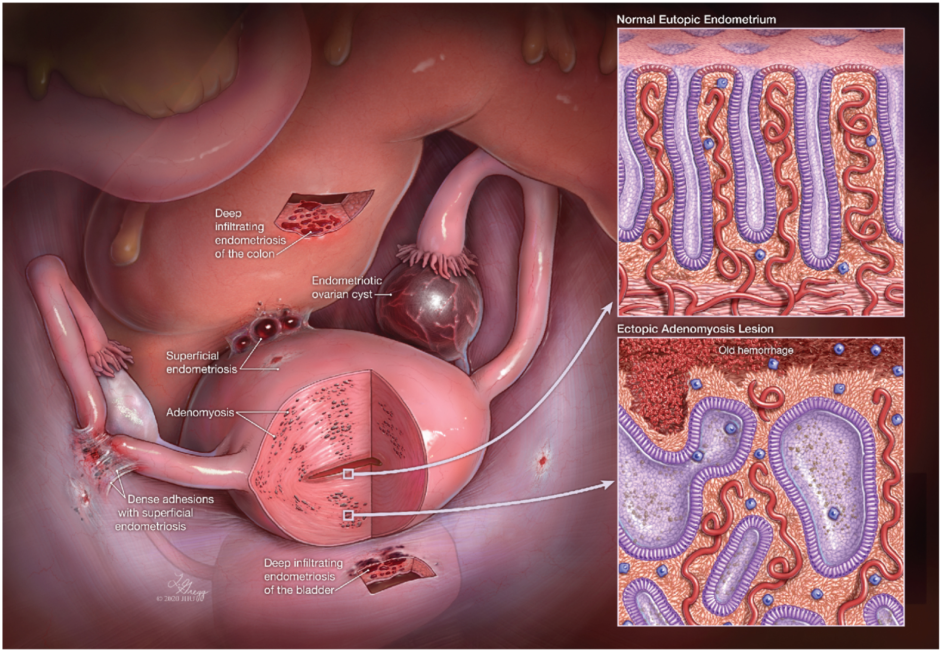
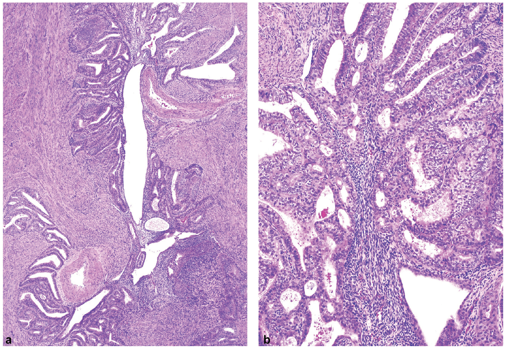
Adenomyosis represents a unique pathophysiological condition in which normal-appearing endometrial mucosa resides within myometrium and is thus protected from menstrual shedding. The resulting ectopic presence of endometrial tissue composed of glands and stroma is thought to affect normal contractile function and peristalsis of uterine smooth muscle, causing menometrorrhagia, infertility, and adverse obstetric outcomes. Since the first description of adenomyosis more than 150 years ago, pathologists have studied this lesion by examining tissue specimens, and have proposed multiple explanations to account for its pathogenesis. However, as compared with endometriosis, progress of adenomyosis research has been, at best, incremental mainly due to the lack of standardized protocols in sampling tissue and a lack of consensus diagnostic criteria in pathology practice. Despite these limitations, recent advances in revealing the detailed anatomy and biology of eutopic endometrium offer an unprecedented opportunity to study this common but relatively understudied disorder. Here, we briefly summarize the pathological aspects of adenomyosis from an historical background, and discuss conventional morphology and recent tissue-based molecular studies with a special emphasis on elucidating its tissue of origin from a pathologist's perspective. We also discuss unmet needs in pathology studies that would be important for advancing adenomyosis research.


Conflict of interest statement

None declared.

Figures

Fig. 1
Fig. 1
The ectopic endometrium and eutopic endometrium. The diseases related to ectopic endometrium include adenomyosis, deep infiltrating endometriosis, peritoneal endometriosis, and ovarian endometriotic cyst (endometrioma). The insets illustrate the similar histology of eutopic endometrium (top) and ectopic lesions (bottom), containing both glandular epithelium and stroma. (Modified from Wang et al; [© IM Shih, Johns Hopkins University].)
Fig. 2
Fig. 2
Gross and microscopic features of adenomyosis. (a) The involved uterus appears slightly enlarged and globular. (b) A bivalved surface from a hysterectomy specimen shows multiple foci of adenomyosis with petechia-like areas (yellow arrows) in myometrium. The smooth muscle appears hypertrophied and disarrayed. (c, d) Comparison of gross appearance of adenomyosis (AD) and leiomyoma (LM). Formalin fixed cross-section in C and hematoxylin and eosin (H&E) stained section in D. (e) Microscopic features showing presence of glandular epithelium and stroma surrounded by hypertrophic smooth muscle cells. (H&E stain ×20 magnification). (f) Higher magnification view to show the resemblance of epithelial and stromal components of adenomyosis to eutopic endometrium (H&E stain ×40 magnification). (g) Ectopic endometrial tissue showing foci of hemorrhage with histiocytes and hemosiderin-laden macrophages (red arrow) (H&E stain ×20 magnification). (h) Marked decidualization with small inactive glands in adenomyosis seen after exogenous progestin therapy. (H&E stain ×20 magnification).
Fig. 3
Fig. 3
Atypical hyperplasia and low-grade endometrioid carcinoma arising from adenomyosis. (a) Foci of atypical hyperplasia within a preexisting adenomyosis lesion. (b) An incipient low-grade endometrioid carcinoma arising from adenomyosis with cancerous tissue coexisting with ectopic benign glandular tissue (hematoxylin and eosin stain ×20 magnification).
Fig. 4
Fig. 4
Adenomyosis originating from the basal layer of endometrium. (a) A schematic representation of the development of adenomyosis with the focal extension of a single clonally distinct endometrial gland from the endometrial basalis into the myometrium. In the myometrium, the incipient adenomyosis continues to clonally expand to form intricate and interconnected glandular foci which are similar to the endometrial basalis layer. Together with the “invading” gland, stromal cells also migrate and clonally expand. Different color lines (glands) and dots (stroma) represent clonally distinct subpopulations. (b) Immunostaining of cytokeratin suggesting the process of early adenomyosis development. The adenomyotic tissue appears to descend from the basal endometrium through the endometrial myometrial interface (dashed red line) with the vertical “invasion” of ectopic glandular and stromal tissue into myometrium. (×20 magnification).

References

    1. Wang Y, Nicholes K, Shih IM. The origin and pathogenesis of endometriosis. Annu Rev Pathol 2020;15:71–95 - PMC - PubMed
    1. Bruun MR, Arendt LH, Forman A, Ramlau-Hansen CH. Endometriosis and adenomyosis are associated with increased risk of preterm delivery and a small-for-gestational-age child: a systematic review and meta-analysis. Acta Obstet Gynecol Scand 2018; 97(09):1073–1090 - PubMed
    1. Harada T, Taniguchi F, Amano H Japan Environment and Children’s Study Group., et al.;. Adverse obstetrical outcomes for women with endometriosis and adenomyosis: a large cohort of the Japan Environment and Children’s Study. PLoS One 2019;14(08):e0220256. - PMC - PubMed
    1. Orazov M, Nosenko EN, Radzinsky VE, Khamoshina MB, Lebedeva MG, Sounov MA. Proangiogenic features in chronic pelvic pain caused by adenomyosis. Gynecol Endocrinol 2016;32 (Suppl 2):7–10 - PubMed
    1. Seidman JD, Kjerulff KH. Pathologic findings from the Maryland Women’s Health Study: practice patterns in the diagnosis of adenomyosis. Int J Gynecol Pathol 1996;15(03):217–221 - PubMed