

Adenomyoepithelioma with a human epidermal growth factor receptor 2-fluorescence in situ hybridization-confirmed ductal carcinoma in situ component: A case report and review of the literature
- PMID: 33080708
- PMCID: PMC7572011
- DOI: 10.1097/MD.0000000000022665
Adenomyoepithelioma with a human epidermal growth factor receptor 2-fluorescence in situ hybridization-confirmed ductal carcinoma in situ component: A case report and review of the literature
Abstract
Introduction: Breast adenomyoepithelioma (AME) is a rare tumor composed of myoepithelial cells and ductal or luminal cells. Most cases of AME are benign, but rare cases in which either or both cell types exhibited malignant features have been reported. Due to its rarity, no diagnostic criteria for malignancy have been established for AME.
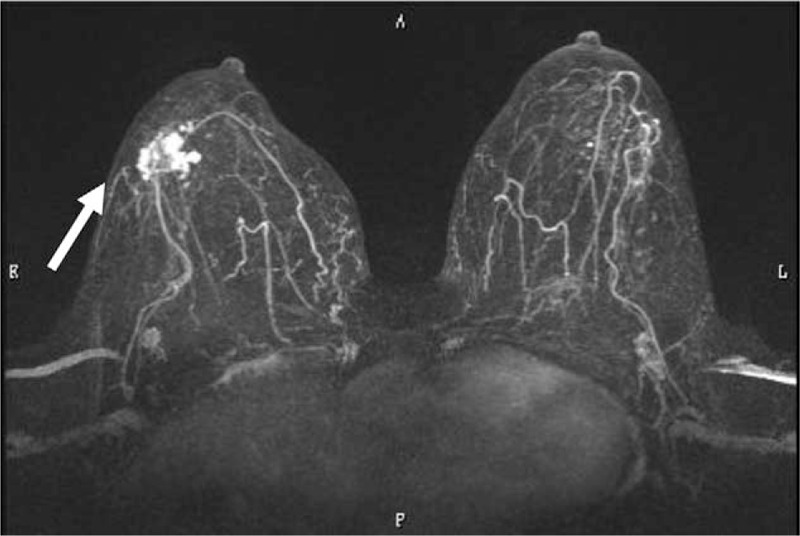
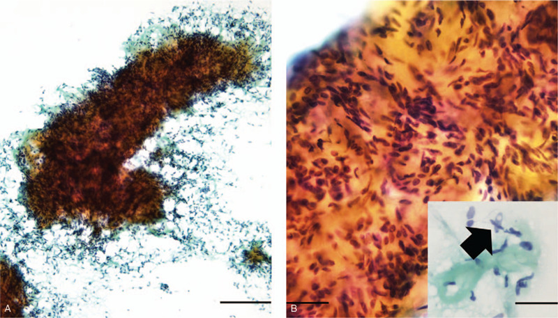
Patient concerns: A 64-year-old woman presented with a mass in her right breast. Fine-needle aspiration cytology and biopsy examinations revealed lesions composed of spindle-shaped cells and round epithelial cells. AME was suspected, and partial mastectomy was performed.
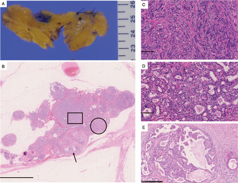
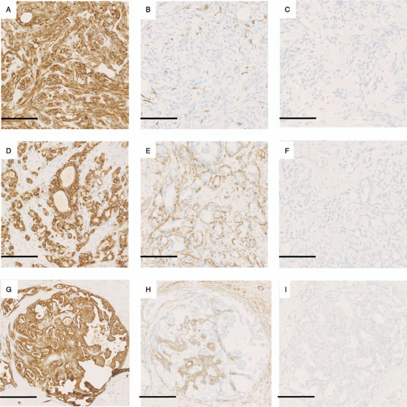
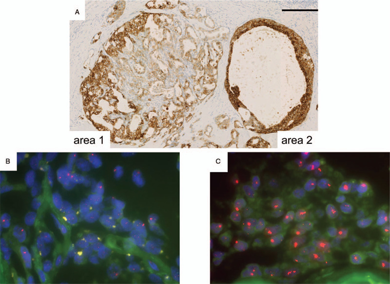
Diagnosis: The tumor specimen showed AME, which mainly consisted of spindle-shaped myoepithelial cells with slight atypia, admixed with tubular luminal cells and small areas of atypical intraductal proliferative lesions. No apparent features of malignancy, such as necrosis or invasion, were seen in the myoepithelial cells or the luminal or intraductal component. However, the atypical intraductal component exhibited focal nuclear atypia, a cribriform pattern, and moderate to strong membranous human epidermal growth factor receptor 2 (HER2) immunoreactivity. HER2 amplification was detected in focal regions of the atypical intraductal component by fluorescence in situ hybridization (FISH), which resulted in a diagnosis of AME with ductal carcinoma in situ.
Outcomes: The patient did not receive further therapy and was free from tumor recurrence at 23 months after the operation.
Conclusion: HER2 FISH might be useful for evaluating suspected AME tumors for malignancy when an atypical ductal lesion that lacks definitive features of malignancy is encountered.
Conflict of interest statement
The authors have no conflicts of interest to disclose.
Figures
Similar articles
-
Adenomyoepithelioma of the breast: a proposal for classification.Histopathology. 2021 Oct;79(4):465-479. doi: 10.1111/his.14380. Epub 2021 Jun 13. Histopathology. 2021. PMID: 33829532 Review.
-
Breast adenomyoepithelioma and adenomyoepithelioma with carcinoma (malignant adenomyoepithelioma) with associated breast malignancies: A case series emphasizing histologic, radiologic, and clinical correlation.Breast. 2016 Oct;29:132-9. doi: 10.1016/j.breast.2016.07.018. Epub 2016 Aug 3. Breast. 2016. PMID: 27494340
-
Spindle cell ductal carcinoma in situ. An unusual variant of ductal intra-epithelial neoplasia that simulates ductal hyperplasia or a myoepithelial proliferation.Virchows Arch. 2001 Jul;439(1):70-7. doi: 10.1007/s004280100446. Virchows Arch. 2001. PMID: 11499843
-
Malignant adenomyoepithelioma of the breast: A case report.Medicine (Baltimore). 2021 Feb 5;100(5):e24461. doi: 10.1097/MD.0000000000024461. Medicine (Baltimore). 2021. PMID: 33592899 Free PMC article.
-
Adenomyoepithelioma with carcinoma of the breast: A report of two cases and a review of the literature.Pathol Res Pract. 2016 Feb;212(2):130-4. doi: 10.1016/j.prp.2015.09.008. Epub 2015 Sep 8. Pathol Res Pract. 2016. PMID: 26596263 Review.
Cited by
-
Concurrence of Adenomyoepithelioma of the Breast and Gastrointestinal Stromal Tumor of the Stomach: A Case Report and Review of the Literature.Med Sci (Basel). 2023 Sep 10;11(3):57. doi: 10.3390/medsci11030057. Med Sci (Basel). 2023. PMID: 37755162 Free PMC article.
References
-
- Hamperl H. The myothelia (myoepithelial cells). Normal state; regressive changes; hyperplasia; tumors. Curr Top Pathol 1970;53:161–220. - PubMed
-
- Hayes MM. Adenomyoepithelioma of the breast: a review stressing its propensity for malignant transformation. J Clin Pathol 2011;64:477–84. - PubMed
-
- Moritz AW, Wiedenhoefer JF, Profit AP, et al. Breast adenomyoepithelioma and adenomyoepithelioma with carcinoma (malignant adenomyoepithelioma) with associated breast malignancies: a case series emphasizing histologic, radiologic, and clinical correlation. Breast 2016;29:132–9. - PubMed
-
- Loose JH, Patchefsky AS, Hollander IJ, et al. Adenomyoepithelioma of the breast. A spectrum of biologic behavior. Am J Surg Pathol 1992;16:868–76. - PubMed
-
- Xu J, Tang X, Iida Y, et al. Adenomyoepithelioma with carcinoma of the breast: a report of two cases and a review of the literature. Pathol Res Pract 2016;212:130–4. - PubMed
Publication types
MeSH terms
Substances
LinkOut - more resources
Full Text Sources
Medical
Research Materials
Miscellaneous
