Review
. 2020 Oct 17;12(10):3022.
doi: 10.3390/cancers12103022.

Carbon Ion Radiobiology

Affiliations
Review

Carbon Ion Radiobiology

Walter Tinganelli et al. Cancers (Basel). .

Abstract

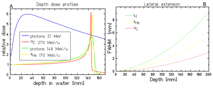
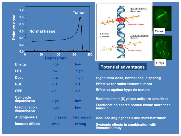
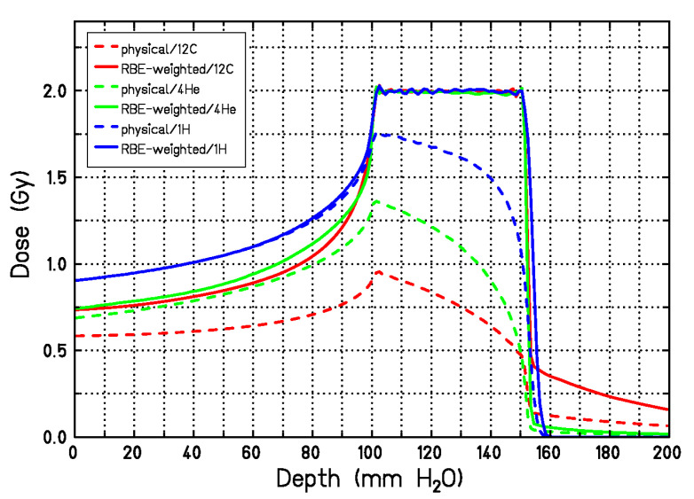
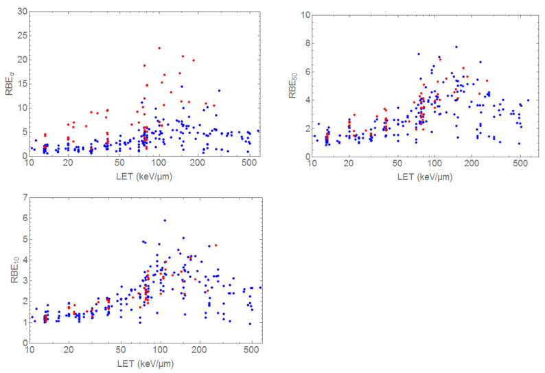
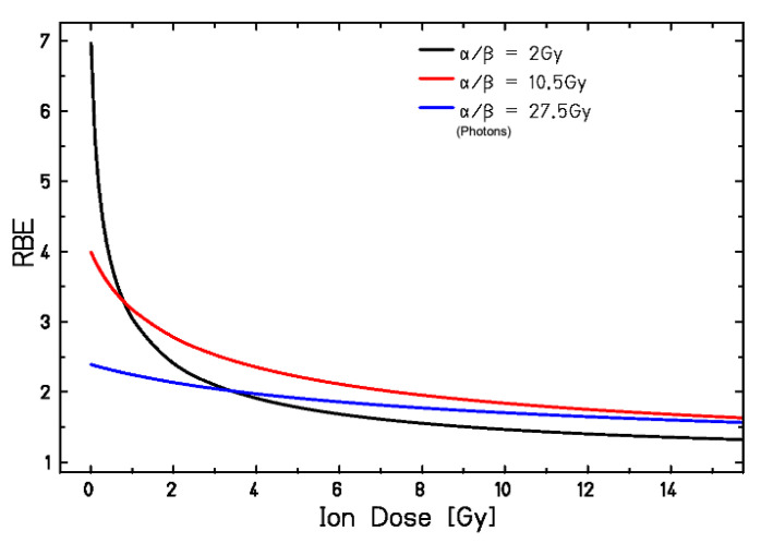
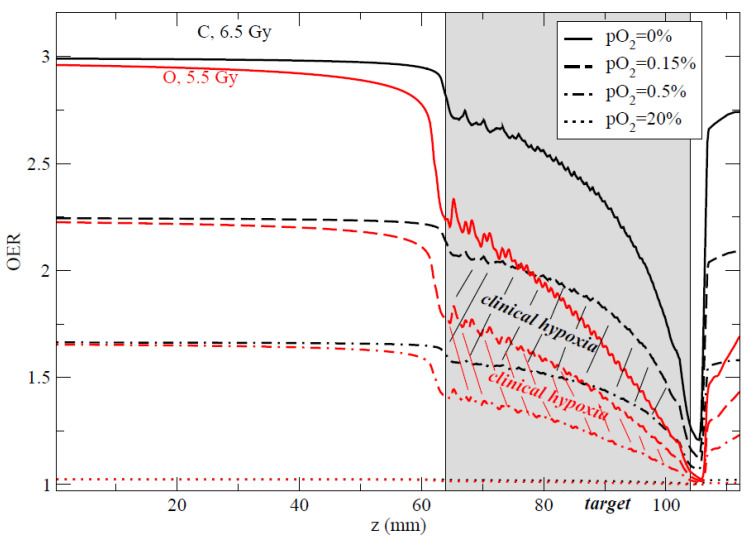
Radiotherapy using accelerated charged particles is rapidly growing worldwide. About 85% of the cancer patients receiving particle therapy are irradiated with protons, which have physical advantages compared to X-rays but a similar biological response. In addition to the ballistic advantages, heavy ions present specific radiobiological features that can make them attractive for treating radioresistant, hypoxic tumors. An ideal heavy ion should have lower toxicity in the entrance channel (normal tissue) and be exquisitely effective in the target region (tumor). Carbon ions have been chosen because they represent the best combination in this direction. Normal tissue toxicities and second cancer risk are similar to those observed in conventional radiotherapy. In the target region, they have increased relative biological effectiveness and a reduced oxygen enhancement ratio compared to X-rays. Some radiobiological properties of densely ionizing carbon ions are so distinct from X-rays and protons that they can be considered as a different "drug" in oncology, and may elicit favorable responses such as an increased immune response and reduced angiogenesis and metastatic potential. The radiobiological properties of carbon ions should guide patient selection and treatment protocols to achieve optimal clinical results.

Keywords: RBE; carbon ions; hypoxia; immunotherapy; metastasis; particle therapy; radiobiology; radiotherapy.


Conflict of interest statement

The authors declare no conflict of interest.

Figures

Figure 1
Figure 1
Physical properties of carbon ions in comparison to X-rays, protons, and helium ions. (A). Depth-dose distributions showing the Bragg peak for all ions at the same range, and the reduced straggling of heavier ions. (B). Lateral scattering is reduced by increasing the ion mass.
Figure 2
Figure 2
Summary of the physical and radiobiological properties of heavy ions along the Bragg curve. The figures on top right show a sketch of the quality of DNA damage and the corresponding γH2AX foci distribution with carbon ions and X-rays. Adapted from [12].
Figure 3
Figure 3
LET versus depth in tissue for a single SOBP of p, He, C, and O providing a uniform physical dose (2 Gy). The grey area represents the tumor region, a 2.5 × 2.5 × 2.5 cm3 volume centered at 8 cm in water. The yellow and orange lines are 100 and 20 keV/μm level, respectively. Figure from [15], distributed under Creative Commons CC-BY.
Figure 4
Figure 4
DNA DSB repair kinetics of human fibroblasts exposed in the entrance channel (EC) or SOBP (2.4 cm width at a water-equivalent depth of 16 cm, two opposite fields, 2 Gy dose). Figure from [47], distributed under Creative Commons CC-BY.
Figure 5
Figure 5
Karyotypes of peripheral blood lymphocytes from two esophageal cancer patients treated with either C-ions (RBE-weighted dose 36 Gy in 10 fractions, radiation field 61 cm2) or X-rays (41.4 Gy in 23 fractions, radiation field 141 cm2 ) at NIRS-QST, Japan. The C-ion (left) karyotype is 46, XY, t(6;18). The X-ray karyotype (right) is 46, XY, t (4;12), t(8;9). Samples were obtained as described in [74].
Figure 6
Figure 6
Survival of CHO cells irradiated in a phantom with two opposite beams of protons or C-ions. Proton and carbon ion irradiations were performed at the HIT facility using an active pencil beam scanning technique with energies of 90–120 MeV/u and 175–230 MeV/u for protons and carbon ions, respectively. The dose levels were 1.5 Gy in the entrance channel for both protons and carbon ions; in the center of target region, doses of 5.3 Gy and 3.9 Gy were applied with protons and carbon ions, respectively. Lines are predictions from the LEM-IV model. The model use the X-rays survival dose of CHO cells with α/β = 4 Gy and a threshold dose Dt = 20 Gy. Adapted from [75], reproduced with permission from Elsevier.
Figure 7
Figure 7
Physical (dashed lines) and RBE-weighted dose (solid lines) for a single 5-cm SOBP using protons, He- or C-ions. The physical dose shape is calculated with LEM-IV to achieve the same RBE-weighted dose in the target region for all ions. Figure modified from [78].
Figure 8
Figure 8
RBE of C-ions from the PIDE database. The 3 panels represent the RBEα, RBE50, and RBE10 for radioresistant (red; α/β < 4 Gy) and radiosensitive (blu; α/β > 4 Gy) cells. Plot courtesy of Dr. Thomas Friedrich, GSI.
Figure 9
Figure 9
RBE of C-ions as a function of the dose per fraction calculated with the liner quadratic model for different α/β values. Figure from reference [96], reproduced with permission by Elsevier.
Figure 10
Figure 10
OER vs. LET of CHO cells at different oxygen concentration levels. Figure from reference [158], distributed under Creative Commons CC-BY.
Figure 11
Figure 11
Comparison of the computed OER along an SOBP for carbon (black curves) and oxygen (red curves) at different pO2 levels. The hatched areas represent the clinical interesting regions for hypoxia (0.15% < pO2 < 0.5%). Doses indicated are prescribed RBE-weighted doses in the target to achieve iso-survival. Plans for using 16O in the clinics are currently under way at HIT (Heidelberg). Plot from [159], © Institute of Physics and Engineering in Medicine. Reproduced by permission of IOP Publishing. All rights reserved.
Figure 12
Figure 12
Typical protocol used in various laboratories to compare C-ions and X-rays in combination to immunotherapy. Reproduced from ref. [295], distributed under Creative Commons CC-BY.

Similar articles

Cited by

References

    1. Wilson R.R. Radiological use of fast protons. Radiology. 1946;47:487–491. doi: 10.1148/47.5.487. - DOI - PubMed
    1. Tobias C.A. Failla Memorial lecture. The future of heavy-ion science in biology and medicine. Radiat. Res. 1985;103:1–33. doi: 10.2307/3576668. - DOI - PubMed
    1. Durante M., Paganetti H. Nuclear physics in particle therapy: A review. Rep. Prog. Phys. 2016;79:096702. doi: 10.1088/0034-4885/79/9/096702. - DOI - PubMed
    1. Schardt D., Elsässer T., Schulz-Ertner D. Heavy-ion tumor therapy: Physical and radiobiological benefits. Rev. Mod. Phys. 2010;82:383–425. doi: 10.1103/RevModPhys.82.383. - DOI
    1. Durante M., Cucinotta F.A. Physical basis of radiation protection in space travel. Rev. Mod. Phys. 2011;83:1245–1281. doi: 10.1103/RevModPhys.83.1245. - DOI

LinkOut - more resources