Review
. 2020 Oct 18;9(10):2314.
doi: 10.3390/cells9102314.

Pathways for Sensing and Responding to Hydrogen Peroxide at the Endoplasmic Reticulum

Affiliations
Review

Pathways for Sensing and Responding to Hydrogen Peroxide at the Endoplasmic Reticulum

Jennifer M Roscoe et al. Cells. .

Abstract

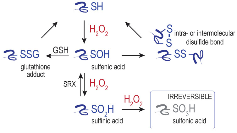
The endoplasmic reticulum (ER) has emerged as a source of hydrogen peroxide (H2O2) and a hub for peroxide-based signaling events. Here we outline cellular sources of ER-localized peroxide, including sources within and near the ER. Focusing on three ER-localized proteins-the molecular chaperone BiP, the transmembrane stress-sensor IRE1, and the calcium pump SERCA2-we discuss how post-translational modification of protein cysteines by H2O2 can alter ER activities. We review how changed activities for these three proteins upon oxidation can modulate signaling events, and also how cysteine oxidation can serve to limit the cellular damage that is most often associated with elevated peroxide levels.

Keywords: BiP; IRE1; SERCA2; cysteine oxidation; endoplasmic reticulum (ER); hydrogen peroxide; reactive oxygen species (ROS); redox signaling; unfolded protein response (UPR).


Conflict of interest statement

The authors declare no conflict of interest.

Figures

Figure 1
Figure 1
Reactive oxygen species. Scheme shows several of the types of ROS generated through the successive reduction molecular oxygen (O2).
Figure 2
Figure 2
Cysteine modifications initiated by H2O2. Hydrogen peroxide (H2O2) reacts with target protein cysteine thiol (–SH), producing a sulfenic acid adduct. Subsequent reactions with glutathione (GSH), protein thiols, or H2O2 can generate additional modifications. Glutathione adducts, disulfides, and sulfinylated proteins can be reduced to the free thiol state through the action of proteins and low-molecular weight thiols [4]. The only established route for reduction of sulfinylated cysteines is by sulfiredoxin (SRX). Sulfonylated cysteines are considered irreversible oxidation products. This figure depicts the further modification of sulfenic acid by thiols (GSH, proteins) or H2O2. However, sulfenic acids can react also with a backbone amide (forming sulfenylamide), condense with another sulfenic acid (forming a thiosulfinate), or undergo further modification by hydrogen sulfide; these modifications are not shown here and are further discussed in [4,25,26].
Figure 3
Figure 3
Sources of H2O2 at the endoplasmic reticulum (ER). Several direct and indirect enzymatic sources of H2O2 are depicted and colored in dark orange (refer to text for more details). Two examples of peroxiporins that facilitate bi-directional peroxide transport across membranes are noted in blue.
Figure 4
Figure 4
The Saccharomyces cerevisiae Hsp70 BiP is reversibly modified by H2O2. A conserved cysteine (red circle) in the nucleotide-binding domain (NDB, orange) of yeast BiP can be oxidized in the presence of elevated H2O2. Upon oxidation, BiP ATPase activity is inhibited yet the peptide-binding domain (grey) maintains an ability to hold polypeptides (blue). Oxidation of BiP can be reversed by Sil1, which restores the canonical Hsp70 chaperone cycle.
Figure 5
Figure 5
Oxidation of the Caenorhabditis elegans IRE-1 kinase domain inhibits canonical unfolded protein response (UPR) signaling and promotes an antioxidant response. High levels of hydrogen peroxide (H2O2) triggers oxidation of a conserved cysteine (red circle) in the activation loop in the kinase domain, which inhibits kinase activity and prevents activation of the RNase domain, preventing canonical UPR signaling. Dissociation of BiP from IRE-1 is normally associated with UPR induction, and BiP remains associated with oxidized IRE-1 [114]. Oxidation results in the recruitment of the scaffold TRF-1 and kinase NSY-1, which mediate initiation of an antioxidant transcriptional response through the transcription factor SKN-1.
Figure 6
Figure 6
Mammalian SERCA2 oxidation at the cytosolic Cys674 modulates Ca2+ uptake into the ER in response to nitric oxide (NO). Cys674 (red circle) is located within the SERCA2 P domain, between the nucleotide binding domain (N) and the transmembrane domains (TMDs). The presence of NO in combination with cellular ROS and reduced glutathione results in SERCA2 glutathionylation (–SSG) and increased pumping of Ca2+ from the cytosol into the ER lumen. An accumulation of sulfonylated SERCA2 (–SO3H) is associated with a lack of responsiveness to NO and various disease pathologies, including artherosclerosis. A, actuator domain.

References

    1. Sies H. Oxidative stress: A concept in redox biology and medicine. Redox Biol. 2015;4:180–183. doi: 10.1016/j.redox.2015.01.002. - DOI - PMC - PubMed
    1. Reczek C.R., Chandel N.S. ROS-dependent signal transduction. Curr. Opin. Cell Biol. 2015;33:8–13. doi: 10.1016/j.ceb.2014.09.010. - DOI - PMC - PubMed
    1. Sies H., Berndt C., Jones D.P. Oxidative Stress. Annu. Rev. Biochem. 2017;86:715–748. doi: 10.1146/annurev-biochem-061516-045037. - DOI - PubMed
    1. Sies H., Jones D.P. Reactive oxygen species (ROS) as pleiotropic physiological signalling agents. Nat. Rev. Mol. Cell Biol. 2020;21:363–383. doi: 10.1038/s41580-020-0230-3. - DOI - PubMed
    1. Winterbourn C.C. Reconciling the chemistry and biology of reactive oxygen species. Nat. Chem. Biol. 2008;4:278–286. doi: 10.1038/nchembio.85. - DOI - PubMed

Publication types

LinkOut - more resources