

Clinical Relevance of Unexpected Findings of Post-Mortem Computed Tomography in Hospitalized Patients: An Observational Study
- PMID: 33081003
- PMCID: PMC7589901
- DOI: 10.3390/ijerph17207572
Clinical Relevance of Unexpected Findings of Post-Mortem Computed Tomography in Hospitalized Patients: An Observational Study
Abstract
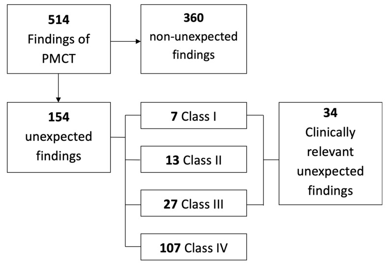
Background and objective: The current literature describing the use of minimally invasive autopsy in clinical care is mainly focused on the cause of death. However, the identification of unexpected findings is equally important for the evaluation and improvement of daily clinical care. The purpose of this study was to analyze unexpected post-mortem computed tomography (PMCT) findings of hospitalized patients and assess their clinical relevance. Materials and methods: This observational study included patients admitted to the internal medicine ward. Consent for PMCT and autopsy was requested from the next of kin. Decedents were included when consent for at least PMCT was obtained. Consent for autopsy was not obtained for all decedents. All findings reported by PMCT were coded with an International Classification of Diseases (ICD) code. Unexpected findings were identified and subsequently categorized for their clinical relevance by the Goldman classification. Goldman class I and III were considered clinically relevant. Additionally, correlation with autopsy results and ante-mortem imaging was performed. Results: In total, 120 decedents were included and evaluated for unexpected findings on PMCT. Of them, 57 decedents also underwent an autopsy. A total of 1020 findings were identified; 111 correlated with the cause of death (10.9%), 508 were previously reported (49.8%), 99 were interpreted as post-mortem changes (9.7%), and 302 were classified as unexpected findings (29.6%). After correlation with autopsy (in 57 decedents), 24 clinically relevant unexpected findings remained. These findings were reported in 18 of 57 decedents (32%). Interestingly, 25% of all unexpected findings were not reported by autopsy. Conclusion: Many unexpected findings are reported by PMCT in hospitalized patients, a substantial portion of which is clinically relevant. Additionally, PMCT is able to identify pathology and injuries not reported by conventional autopsy. A combination of PMCT and autopsy can thus be considered a more comprehensive and complete post-mortem examination.
Keywords: autopsy; post-mortem computed tomography; radiology; unexpected findings.
Conflict of interest statement
The authors declare no conflict of interest.
Figures


References
-
- Rutty G.N., Morgan B., Robinson C., Raj V., Pakkal M., Amoroso J., Visser T., Saunders S., Biggs M., Hollingbury F., et al. Diagnostic accuracy of post-mortem CT with targeted coronary angiography versus autopsy for coroner-requested post-mortem investigations: A prospective, masked, comparison study. Lancet. 2017;390:145–154. doi: 10.1016/S0140-6736(17)30333-1. - DOI - PMC - PubMed
Publication types
MeSH terms
LinkOut - more resources
Full Text Sources
Medical
