Thioredoxin Interacting Protein (TXNIP) Is Differentially Expressed in Human Tumor Samples but Is Absent in Human Tumor Cell Line Xenografts: Implications for Its Use as an Immunosurveillance Marker
- PMID: 33081035
- PMCID: PMC7603212
- DOI: 10.3390/cancers12103028
Thioredoxin Interacting Protein (TXNIP) Is Differentially Expressed in Human Tumor Samples but Is Absent in Human Tumor Cell Line Xenografts: Implications for Its Use as an Immunosurveillance Marker
Abstract
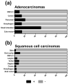
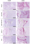
Thioredoxin interacting protein (TXNIP) is a metabolic protein critically involved in redox homeostasis and has been proposed as a tumor suppressor gene in a variety of malignancies. Accordingly, TXNIP is downregulated in breast, bladder, and gastric cancer and in tumor transplant models TXNIP overexpression inhibits growth and metastasis. As TXNIP protein expression has only been investigated in few malignancies, we employed immunohistochemical detection in a large multi-tumor tissue microarray consisting of 2,824 samples from 94 different tumor entities. In general, TXNIP protein was present only in a small proportion of primary tumor samples and in these cases was differently expressed depending on tumor stage and subtype (e.g., renal cell carcinoma, thyroid cancer, breast cancer, and ductal pancreatic cancer). Further, TXNIP protein expression was determined in primary mouse xenograft tumors derived from human cancer cell lines and was immunohistochemically absent in all xenograft tumors investigated. Intriguingly, TXNIP expression became gradually lower in the proximity of the primary tumor tissue and was absent in leukocytes directly adjacent to tumor tissue. In conclusion, these findings suggest that TXNIP downregulation is as a common feature in human tumor xenograft models and that intra-tumoral leukocytes down-regulate TXNIP. Hence TXNIP expression might be used to monitor the functional state of tumor-infiltrating leukocytes in tissue sections.
Keywords: cancer; immunotherapy; reactive oxygen species; thioredoxin; thioredoxin interacting protein; tissue array; tumorigenesis; xenograft.
Conflict of interest statement
The authors declare no conflict of interest.
Figures


Similar articles
-
Thioredoxin interacting protein (TXNIP) is a novel tumor suppressor in thyroid cancer.Mol Cancer. 2014 Mar 19;13:62. doi: 10.1186/1476-4598-13-62. Mol Cancer. 2014. PMID: 24645981 Free PMC article.
-
Identification of thioredoxin-interacting protein 1 as a hypoxia-inducible factor 1alpha-induced gene in pancreatic cancer.Pancreas. 2008 Mar;36(2):178-86. doi: 10.1097/MPA.0b013e31815929fe. Pancreas. 2008. PMID: 18376310
-
YKL-40 protein expression in human tumor samples and human tumor cell line xenografts: implications for its use in tumor models.Cell Oncol (Dordr). 2021 Oct;44(5):1183-1195. doi: 10.1007/s13402-021-00630-z. Epub 2021 Aug 25. Cell Oncol (Dordr). 2021. PMID: 34432260 Free PMC article.
-
Research Progress of TXNIP as a Tumor Suppressor Gene Participating in the Metabolic Reprogramming and Oxidative Stress of Cancer Cells in Various Cancers.Front Oncol. 2020 Oct 21;10:568574. doi: 10.3389/fonc.2020.568574. eCollection 2020. Front Oncol. 2020. PMID: 33194655 Free PMC article. Review.
-
Roles of thioredoxin binding protein (TXNIP) in oxidative stress, apoptosis and cancer.Mitochondrion. 2013 May;13(3):163-9. doi: 10.1016/j.mito.2012.06.004. Epub 2012 Jun 27. Mitochondrion. 2013. PMID: 22750447 Review.
Cited by
-
Immunomodulatory Role of Thioredoxin Interacting Protein in Cancer's Impediments: Current Understanding and Therapeutic Implications.Vaccines (Basel). 2022 Nov 10;10(11):1902. doi: 10.3390/vaccines10111902. Vaccines (Basel). 2022. PMID: 36366411 Free PMC article. Review.
-
Thioredoxin System Protein Expression in Carcinomas of the Pancreas, Distal Bile Duct, and Ampulla in the United Kingdom.Diseases. 2024 Sep 24;12(10):227. doi: 10.3390/diseases12100227. Diseases. 2024. PMID: 39452470 Free PMC article.
-
Contribution of HSP90 Cleavage to the Cytotoxic Effect of Suberoylanilide Hydroxamic Acid In Vivo and the Involvement of TXNIP in HSP90 Cleavage.Biomol Ther (Seoul). 2024 Jan 1;32(1):115-122. doi: 10.4062/biomolther.2023.104. Biomol Ther (Seoul). 2024. PMID: 38148557 Free PMC article.
-
Identification and Validation of a Pyroptosis-Related Prognostic Model for Gastric Cancer.Front Genet. 2022 Feb 25;12:699503. doi: 10.3389/fgene.2021.699503. eCollection 2021. Front Genet. 2022. PMID: 35280928 Free PMC article.
-
Identification of a key ceRNA network associated with ferroptosis in gastric cancer.Sci Rep. 2022 Nov 22;12(1):20088. doi: 10.1038/s41598-022-24402-3. Sci Rep. 2022. PMID: 36418919 Free PMC article.
References
LinkOut - more resources
Full Text Sources
Research Materials

