. 2020 Oct 18;21(20):7709.
doi: 10.3390/ijms21207709.

Human Isogenic Cell Line Models for Neutrophils and Myeloid-Derived Suppressor Cells

Affiliations

Human Isogenic Cell Line Models for Neutrophils and Myeloid-Derived Suppressor Cells

Yuting Zhang et al. Int J Mol Sci. .

Abstract

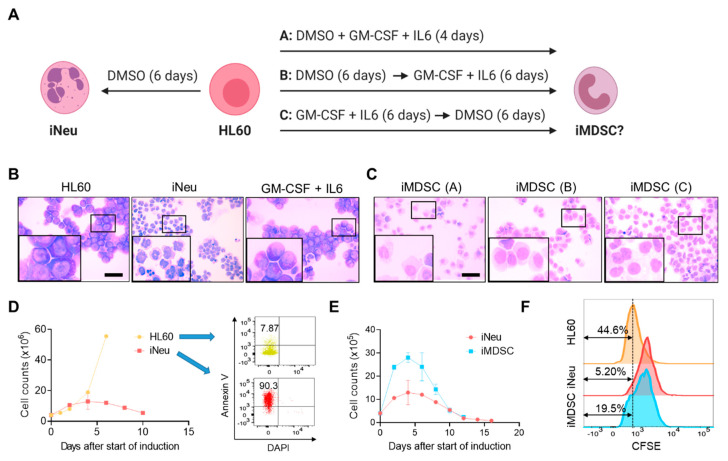
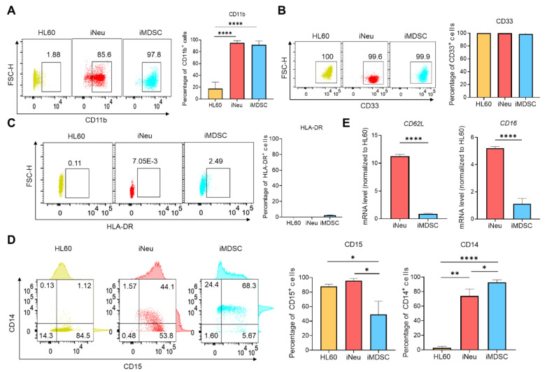
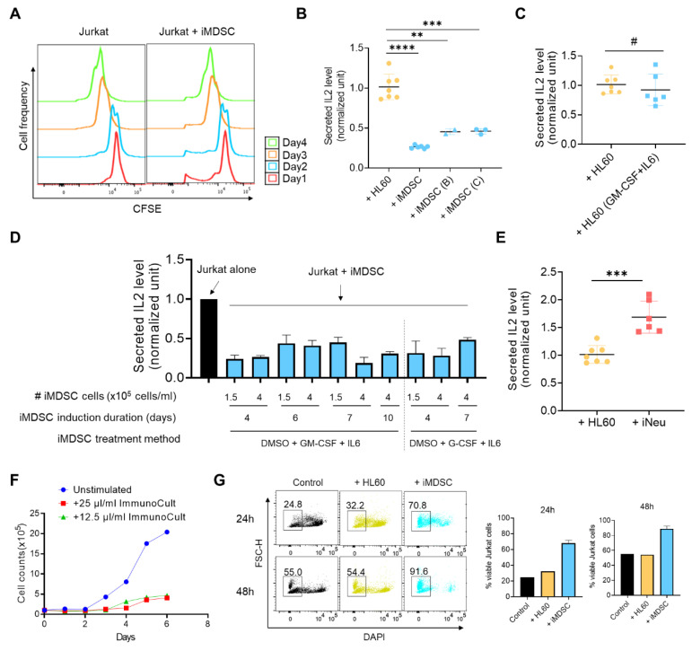
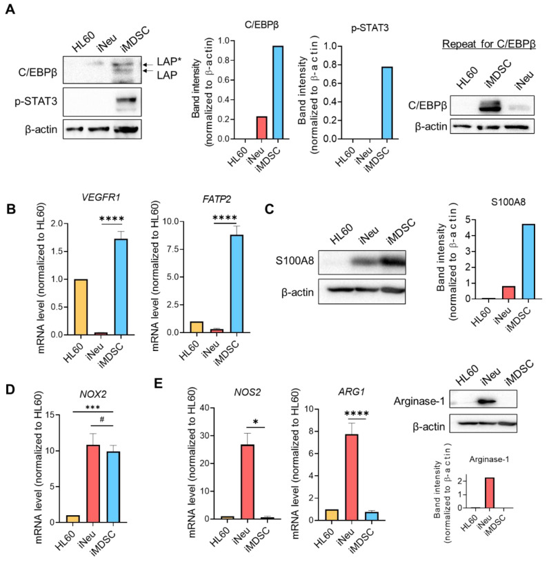
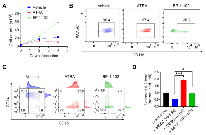
Neutrophils with immunosuppressive activity are polymorphonuclear myeloid-derived suppressor cells (MDSCs) and may contribute to the resistance to cancer immunotherapy. A major gap for understanding and targeting these cells is the paucity of cell line models with cardinal features of human immunosuppressive neutrophils and their normal counterparts, especially in an isogenic manner. To address this issue, we employ the human promyelocytic cell line HL60 and use DMSO and cytokines (granulocyte macrophage-colony stimulating factor (GM-CSF) and interleukin 6 (IL6)) to induce the formation of either neutrophils or MDSCs. The induced MDSCs are CD11b+ CD33+ HLA-DR-/low and are heterogeneous for CD15 and CD14 expression. The induced MDSCs abrogate IL2 production and activation-induced cell death of the human T cell line Jurkat stimulated by CD3/CD28 antibodies, whereas the induced neutrophils enhance IL2 production from Jurkat cells. The induced MDSCs upregulate the expression of C/EBPβ, STAT3, VEGFR1, FATP2 and S100A8. Lastly, the immunosuppressive activity of the induced MDSCs is inhibited by all-trans retinoic acid and STAT3 inhibitor BP-1-102 through cellular differentiation and dedifferentiation mechanisms, respectively. Together, our study establishes a human isogenic cell line system for neutrophils and MDSCs and this system is expected to facilitate future studies on the biology and therapeutics of human immunosuppressive neutrophils.

Keywords: GM-CSF; HL60; IL6; Jurkat; PMN-MDSC; STAT3 inhibitor; all-trans retinoic acid; myeloid-derived suppressor cell; neutrophil.


Conflict of interest statement

The authors declare no conflict of interest.

Figures

Figure 1
Figure 1
Induction of neutrophils and myeloid-derived suppressor cells (MDSCs) from HL60. (A) Experimental scheme for induced neutrophil (iNeu) and induced myeloid-derived suppressor cell (iMDSC) induction. Created with biorender.com. (B) Wright–Giemsa staining of HL60, iNeu, and HL60 treated with granulocyte macrophage-colony stimulating factor (GM-CSF) + interleukin 6 (IL6) for 6 days. Scale bar 50 μm. (C) Wright–Giemsa staining of iMDSC induced by the three conditions shown in (A). Scale bar 50 μm. (D) Proliferation curves and apoptosis analysis of HL60 and iNeu. Apoptotic cells were identified as Annexin V+ DAPI cells. (E) Proliferation curves of iNeu and iMDSC. (F) Representative carboxyfluorescein succinimidyl ester (CFSE) flow cytometer histograms showing the differentiation dilution of the CFSE labeling of HL60, iNeu and iMDSC on Day 4 after the start of induction.
Figure 2
Figure 2
MDSC surface maker analysis for HL60, iNeu and iMDSC. (AD) Flow cytometry analysis of CD11b, CD33, HLA-DR, CD14 and CD15 expression in HL60, iNeu and iMDSC. Both representative plots and average frequencies were shown. (E) Expression of CD62L and CD16 at the mRNA level in HL60, iNeu and iMDSC, measured by qRT-PCR. Relative mRNA was normalized to GAPDH (internal control) and HL60 (the reference baseline of 1.0). * p < 0.05, ** p < 0.01, **** p < 0.0001, two-tailed Student’s t-test. For (A,D), one-way ANOVA followed by multiple comparisons returned equivalent significance levels.
Figure 3
Figure 3
iMDSC suppress IL2 secretion and activation-induced cell death in stimulated Jurkat cells. (A) Representative CFSE flow cytometer histograms showing the proliferation-caused CFSE dilution of Jurkat cells alone or Jurkat cells co-cultured with iMDSCs during a 4-day incubation. (B) Relative IL2 level (measured by ELISA) in the medium from stimulated Jurkat cells co-cultured with HL60 or iMDSC (induced by conditions A, B and C as shown in Figure 1A). (C) Relative IL2 level (measured by ELISA) in the medium from stimulated Jurkat cells co-cultured with untreated HL60 or HL60 pre-treated with 10ng/mL GM-CSF + IL6 for 6 days. (D) Relative IL2 level (measured by ELISA) in the medium from stimulated Jurkat cells alone or co-cultured with iMDSC induced under indicated conditions. (E) Relative IL2 level (measured by ELISA) in the medium from stimulated Jurkat cells co-cultured with HL60 or iNeu. The results in (BE) were normalized to the IL2 concentration secreted from ImmunoCult CD3/CD28-stimulated Jurkat alone. (F) Proliferation curves of unstimulated Jurkat or Jurkat stimulated by 25 μL/mL or 12.5 μL/mL ImmunoCult CD3/CD28 T Cell Activator. (G) Cell viability measurement with DAPI in flow cytometry of stimulated Jurkat alone, stimulated Jurkat co-cultured with HL60, or stimulated Jurkat co-cultured with iMDSC for 24h or 48h, respectively. Jurkat were stimulated by 25 μL/mL ImmunoCult CD3/CD28 T Cell Activator. In (B,C,E), # p > 0.05, ** p < 0.01, *** p < 0.001, **** p < 0.0001, two-tailed Student’s t-test. For (B), one-way ANOVA followed by multiple comparisons returned equivalent significance levels.
Figure 4
Figure 4
iMDSC selectively upregulate the expression of MDSC functional players. (A) Protein levels of C/EBPβ and phospho-STAT3 (Tyr705) in HL60, iNeu and iMDSC, detected by Western blot. The normalized band intensity was shown. Shown on the right is the repeat experiment for C/EBPβ with independently extracted cell lysates. (B) Expression of VEGFR1 and FATP2 (measured by qRT-PCR) in HL60, iNeu and iMDSC. Relative mRNA was normalized to GAPDH (internal control) and HL60 (the reference baseline of 1.0). (C) Protein level of S100A8 in HL60, iNeu and iMDSC, detected by Western blot. The normalized band intensity was shown. (D) Expression of NOX2 (measured by qRT-PCR) in HL60, iNeu and iMDSC. Relative mRNA was normalized to GAPDH (internal control) and HL60 (the reference baseline of 1.0). (E) Expression of NOS2 and ARG1 at the mRNA level measured by qRT-PCR, and Arginase-1 at the protein level detected by Western blot in HL60, iNeu and iMDSC. In (B,D,E), # p > 0.05, * p < 0.05, *** p < 0.001, **** p < 0.0001, two-tailed Student’s t-test. For (D), one-way ANOVA followed by multiple comparisons returned equivalent significance levels.
Figure 5
Figure 5
All-trans retinoic acid (ATRA) and STAT3 inhibitor BP-1-102 direct iMDSC to distinct non-suppressive states. (A) Proliferation curves of iMDSC cells treated with vehicle, ATRA and BP-1-102 throughout the 4-day induction course. (B,C) Flow cytometry analysis of CD11b, CD14 and CD15 expression on iMDSC treated with vehicle, ATRA and BP-1-102 at the end of the 4-day induction course. (D) Relative IL2 level (measured by ELISA) in the medium from stimulated Jurkat alone or co-cultured with iMDSC pre-treated with vehicle, ATRA and BP-1-102. * p < 0.05, *** p < 0.001, two-tailed Student’s t-test. For (D), one-way ANOVA followed by multiple comparisons returned equivalent significance levels.

Similar articles

Cited by

References

    1. Gabrilovich D.I., Ostrand-Rosenberg S., Bronte V. Coordinated regulation of myeloid cells by tumours. Nat. Rev. Immunol. 2012;12:253–268. doi: 10.1038/nri3175. - DOI - PMC - PubMed
    1. Veglia F., Perego M., Gabrilovich D. Myeloid-derived suppressor cells coming of age. Nat. Immunol. 2018;19:108–119. doi: 10.1038/s41590-017-0022-x. - DOI - PMC - PubMed
    1. Gabrilovich D.I., Nagaraj S. Myeloid-derived suppressor cells as regulators of the immune system. Nat. Rev. Immunol. 2009;9:162–174. doi: 10.1038/nri2506. - DOI - PMC - PubMed
    1. Condamine T., Gabrilovich D.I. Molecular mechanisms regulating myeloid-derived suppressor cell differentiation and function. Trends Immunol. 2011;32:19–25. doi: 10.1016/j.it.2010.10.002. - DOI - PMC - PubMed
    1. Filipazzi P., Huber V., Rivoltini L. Phenotype, function and clinical implications of myeloid-derived suppressor cells in cancer patients. Cancer Immunol. Immunother. 2012;61:255–263. doi: 10.1007/s00262-011-1161-9. - DOI - PMC - PubMed

MeSH terms