. 2020 Oct 16;9(10):1003.
doi: 10.3390/antiox9101003.

Allium subhirsutum L. as a Potential Source of Antioxidant and Anticancer Bioactive Molecules: HR-LCMS Phytochemical Profiling, In Vitro and In Vivo Pharmacological Study

Affiliations

Allium subhirsutum L. as a Potential Source of Antioxidant and Anticancer Bioactive Molecules: HR-LCMS Phytochemical Profiling, In Vitro and In Vivo Pharmacological Study

Riadh Badraoui et al. Antioxidants (Basel). .

Abstract

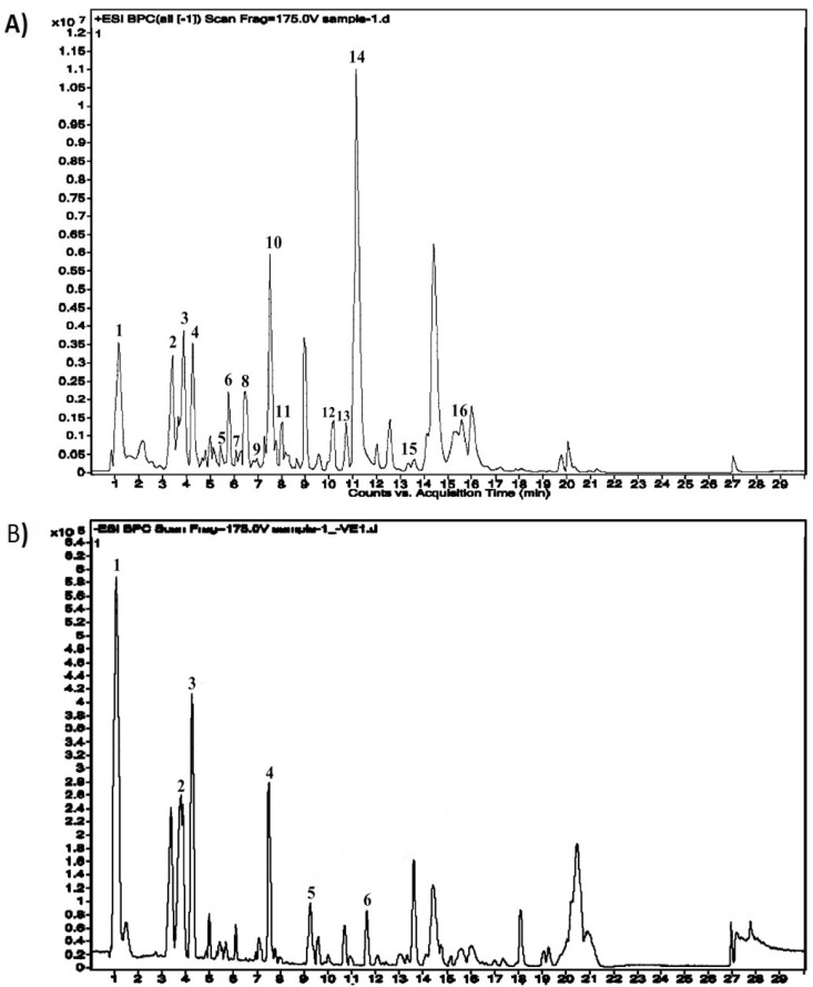
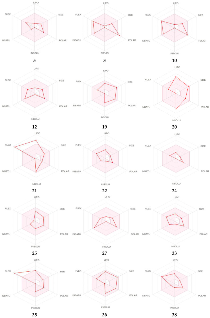
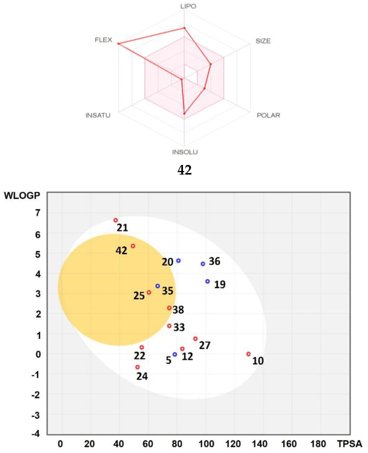
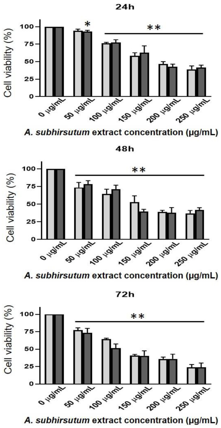
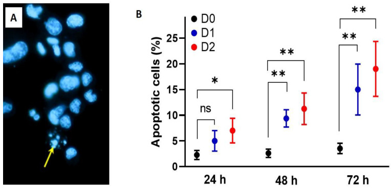
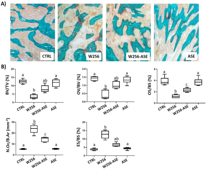
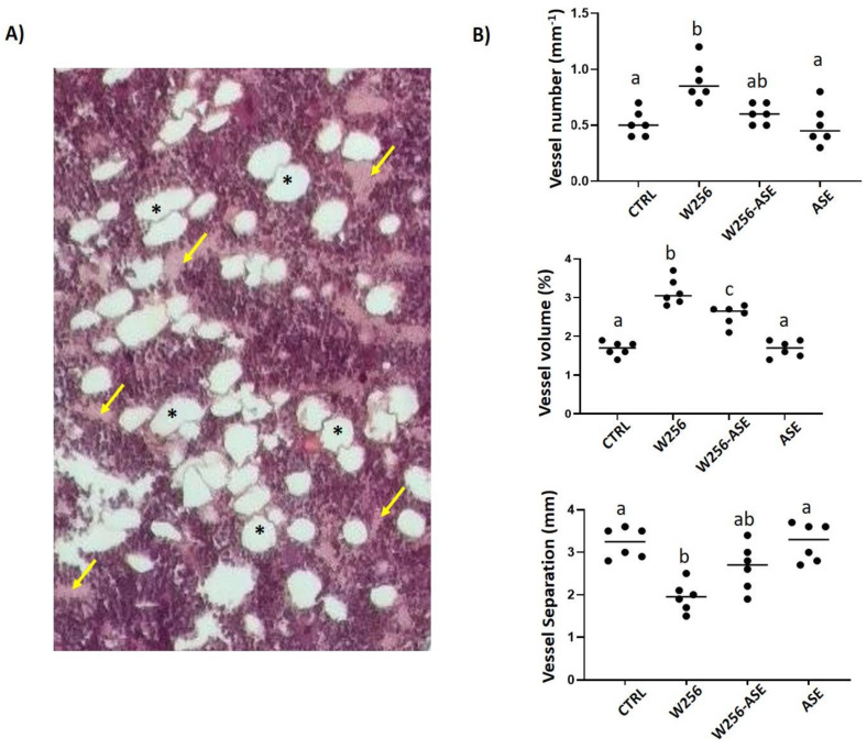
This study investigated Allium subhirsutum L. (AS) anticancer and antioxidant effects and inhibition of tumor angiogenesis in a murine model of skeletal metastases due to inoculation of Walker 256/B cells. Phytochemical composition of AS extract (ASE) was studied by High Resolution-Liquid Chromatography Mass Spectroscopy (HR-LCMS). Total phenolic and flavonoid contents (TPC and TFC) were determined. In vitro, the antioxidant properties were evaluated by reducing power and antiradical activity against DPPH. Cancer cells' proliferation, apoptosis, metastatic development and angiogenesis were evaluated using Walker 256/B and MatLyLu cells. The p-coumaric acid was the major phenolic acid (1700 µg/g extract). ASE showed high levels of TPC and TFC and proved potent antioxidant effects. ASE inhibited Walker 256/B and MatLyLu cells' proliferation (Half-maximal inhibitory concentration: IC50 ≃ 150 µg/mL) and induced apoptosis. In silico and in vivo assays confirmed these findings. ASE effectively acts as a chemo-preventive compound, induces apoptosis and attenuates angiogenesis and osteolytic metastases due to Walker 256/B malignant cells.

Keywords: Allium subhirsutum; angiogenesis; anticancer; antioxidant; apoptosis; bone metastases; cell proliferation; phytochemistry.


Conflict of interest statement

The authors declare no conflict of interest. The funder had no role in the design of the study; in the collection, analyses, or interpretation of data; in the writing of the manuscript, or in the decision to publish the results.

Figures

Figure 1
Figure 1
Phytochemical composition profile of Allium subhirsutum methanolic extract using the High Resolution-Liquid Chromatography Mass Spectroscopy (HR-LCMS) technique. (A) Positive peak (1). Pro Leu, (2). Cys Tyr Trp, (3). Glu Thr, (4). Asp Arg Tyr, (5). 2-methylene-5-(2,5dioxotetrahydrofuran-3-yl)-6-oxo--10,10-dimethylbicyclo [7:2:0] undecane, (6). (22S)-1alpha,22,25-trihydroxy-26,27-dimethyl-23,23,24,24-tetradehydro-24ahomovitaminD3/(22S)-1al, (7). L-4-Hydroxy-3-methoxy-amethylphenylalanine, (8). 1-nonadecanoyl-2-(5Z,8Z,11Z,14Z,17Zeicosapentaenoyl)-sn-glycerol, (9). TG(16:1(9Z)/17:2(9Z,12Z)/20: 5(5Z,8Z,11Z,14Z,17Z))[iso6], (10). 11alpha-acetoxykhivorin, (11) Methyl gamboginate, (12). C16 Sphinganine, (13). 4-Oxomytiloxanthin, (14). Sebacic acid, (15). Linolenoyl lysolecithin, (16). 3beta, 7alpha, 12alpha-Trihydroxy-5alpha-cholestan- 26-oic acid, (17). N-(2-hydroxyethyl) stearamide. (B) Negative peak (1). Asn Asn Asn, (2). His Asp, (3). Cepharanthine, (4). Asn Gln Ala, (5). 6 alpha-Hydroxy Castasterone, (6). 6-Deoxocastasterone. Abbreviations: Ala: Alanine, Arg: Arginine, Asn: Asparagine, Asp: Aspartic acid, Cys: Cysteine, Gln: Glutamine, Glu: Glutamic acid, His: Histidine, Leu: Leucine, Pro: Proline, Thr: Threonine, Tyr: Tyrosine.
Figure 2
Figure 2
Bioavailability radar of identified compounds based on physicochemical indices ideal for oral bioavailability. LIPO, Lipophilicity: −0.7 < XLOGP3 < þ 5; SIZE, Molecular size: 150 g/mol < mol. wt. < 500 g/mol; POLAR, Polarity: 20 Å2 < TPSA < 130 Å2; INSOLU, Insolubility: 0 < Log S (ESOL) < 6; INSATU, Insaturation: 0.25 < Fraction Csp3 < 1; FLEX, Flexibility: 0 < Number of rotatable bonds < 9. The colored zone is the suitable physicochemical space for oral bioavailability (Top). Boiled-egg model of identified compounds (Bottom). ESOL: Estimating Aqueous Solubility from Molecular Structure, TPSA: topological polar surface area.
Figure 2
Figure 2
Bioavailability radar of identified compounds based on physicochemical indices ideal for oral bioavailability. LIPO, Lipophilicity: −0.7 < XLOGP3 < þ 5; SIZE, Molecular size: 150 g/mol < mol. wt. < 500 g/mol; POLAR, Polarity: 20 Å2 < TPSA < 130 Å2; INSOLU, Insolubility: 0 < Log S (ESOL) < 6; INSATU, Insaturation: 0.25 < Fraction Csp3 < 1; FLEX, Flexibility: 0 < Number of rotatable bonds < 9. The colored zone is the suitable physicochemical space for oral bioavailability (Top). Boiled-egg model of identified compounds (Bottom). ESOL: Estimating Aqueous Solubility from Molecular Structure, TPSA: topological polar surface area.
Figure 3
Figure 3
Time course and dose-response effects of Allium subhirsutum methanolic extract at different doses: 0, 50 and 100 µg/mL, on Walker 256/B and MatLyLu cells’ vitality. The cells were incubated for 24, 48 and 72 h. Cell viability was assessed using the MTT assay. Pure (100%) EthO was used as a positive control. Note that the effect is dose- and time-dependent. The most prominent effect was noticed at 72 h with the highest dose (250 µg/mL). Values represent mean ± standard deviation (SD) of n = 3. Statistical difference between the groups * p < 0.05, ** p < 0.01 following one-way analysis of variance (ANOVA) and Newman–Keuls post hoc tests. The experiment was run in triplicate.
Figure 4
Figure 4
Time course and dose-response effects of Allium subhirsutum methanolic extract at different doses: 0, 50 and 100 µg/mL (D0, D1 and D2, respectively), on Walker 256/B cell apoptosis. The cells were incubated for 24, 48 and 72 h. Cell apoptosis was assessed using the Hoechst 33,342 assay (A). Note that D1 and D2 of AS extract were found to induce statistically high significant (* p < 0.001) increases after 48 h and the effect was more prominent at 72 h (B). Values represent mean ± SD of n = 3. Groups with different letters exhibit significant statistical difference at least at ** p < 0.05 following one-way ANOVA and Newman–Keuls post hoc tests. The experiment was run in triplicate.
Figure 5
Figure 5
(A) Bone histological sections of the distal operated femurs from CTRL, W256, W256-ASE and ASE groups 20 days after surgery. The sections were stained with Goldner Modified Trichrome. W256/B slide exhibits cancellous and trabecular osteolysis once compared with CTRL and ASE. The inter-trabecular spaces are occupied with both hematopoietic cells together with tumor foci due to Walker 256/B cells’ inoculation. Note the alleviated trabecular network and bone quality in the W256-ASE histological section. Original magnification ×200. (B) Bone histomorphometric data for the distal operated femurs from CTRL, W256, W256-ASE and ASE. BV/TV: trabecular bone volume, OV/BV and OS/BS: osteoid volume and surface respectively, N.Oc/B.Ar: osteoclasts number per bone area, ES/BS: eroded surface. Values represent mean ± SD. Groups with different letters exhibit significant statistical difference at least p < 0.05 following one-way ANOVA and Newman–Keuls post hoc tests.
Figure 6
Figure 6
(A) Bone histological section of the distal operated femur from W256 rat 20 days after surgery. The cross-section was realized close to the tumoral foci, just below the growth plate. The section exhibits a high number of blood vessels (arrows) and adipocytes (asterisks) surrounded by a polymorph aspect of hematopoietic lineage and Walker 256/B cells. Original magnification ×200. (B) Angio-architecture of the distal operated femurs from CTRL, W256, W256-ASE and ASE 20 days after surgery. The angioarchitecture was assessed by two-dimensional (2D) histomorphometric analysis. Values represent mean ± SD. Groups with different letters exhibit significant statistical difference at least * p < 0.05 following one-way ANOVA and Newman–Keuls post hoc tests.

Similar articles

Cited by

References

    1. Lozano R., Naghavi M., Foreman K., Lim S., Shibuya K., Aboyans V., Abraham J., Adair T., Aggarwal R., Ahn S.Y., et al. Global and regional mortality from 235 causes of death for 20 age groups in 1990 and 2010: A systematic analysis for the global burden of disease study 2010. Lancet. 2012;380:2095–2128. doi: 10.1016/S0140-6736(12)61728-0. - DOI - PMC - PubMed
    1. Zambrowski J.J. Costs of cancer care. Rev. Prat. 2016;66:489–493. - PubMed
    1. Bharti A.C., Aggarwal B.B. Nuclear factor-kappa B and cancer: Its role in prevention and therapy. Biochem. Pharmacol. 2002;101:1053–1062. doi: 10.1016/S0006-2952(02)01154-1. - DOI - PubMed
    1. Badraoui R., Rebai T. Effect of malignant ascites on antioxidative potency of two tumoral cells-induced bone metastases: Walker 256/B and MatLyLu. Exp. Toxicol. Pathol. 2012;64:65–68. doi: 10.1016/j.etp.2010.06.007. - DOI - PubMed
    1. Blouin S., Baslé M.F., Chappard D. Rat models of bone metastases. Clin. Exp. Metastasis. 2005;22:605–614. doi: 10.1007/s10585-006-9002-5. - DOI - PubMed