Review
. 2020 Oct 16;9(4):55.
doi: 10.3390/antib9040055.

IgE Antibodies against Cancer: Efficacy and Safety

Affiliations
Review

IgE Antibodies against Cancer: Efficacy and Safety

Jitesh Chauhan et al. Antibodies (Basel). .

Abstract

Immunoglobulin E (IgE) antibodies are well known for their role in allergic diseases and for contributions to antiparasitic immune responses. Properties of this antibody class that mediate powerful effector functions may be redirected for the treatment of solid tumours. This has led to the rise of a new class of therapeutic antibodies to complement the armamentarium of approved tumour targeting antibodies, which to date are all IgG class. The perceived risk of type I hypersensitivity reactions following administration of IgE has necessitated particular consideration in the development of these therapeutic agents. Here, we bring together the properties of IgE antibodies pivotal to the hypothesis for superior antitumour activity compared to IgG, observations of in vitro and in vivo efficacy and mechanisms of action, and a focus on the safety considerations for this novel class of therapeutic agent. These include in vitro studies of potential hypersensitivity, selection of and observations from appropriate in vivo animal models and possible implications of the high degree of glycosylation of IgE. We also discuss the use of ex vivo predictive and monitoring clinical tools, as well as the risk mitigation steps employed in, and the preliminary outcomes from, the first-in-human clinical trial of a candidate anticancer IgE therapeutic.

Keywords: AllergoOncology; IgE; anaphylaxis; antibodies; basophil activation test (BAT); cancer; immunotherapy; in vivo models; safety; type I hypersensitivity.


Conflict of interest statement

V.F.C., J.F.S. and S.N.K. are founders and shareholders of Epsilogen Ltd.; H.J.B. and H.S.S. are employed through a fund provided by Epsilogen Ltd. All other authors declare no conflicts of interest. The authors are solely responsible for decision to publish, and preparation of the manuscript. The views expressed are those of the author(s) and not necessarily those of the NHS, the NIHR or the Department of Health.

Figures
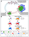
Figure 1
Figure 1
Cross-linking of IgE antibodies bound to monocyte and macrophage FcεRs by tumour-associated antigens (TAAs) is proposed to mediate antitumour activity via a two-armed effector mechanism. (A) First, IgE cross-linking activates monocytes and macrophages to induce both tumour cell killing, as well as a proinflammatory recruitment positive feedback loop, mediated by TNFa/MCP-1, which potentiates tumour cell death. Adapted from Josephs et al. [32]. (B) Second, IgE cross-linking (horizontal arrows) triggers a repolarisation of tumour associated macrophages (TAM) subsets that are otherwise protumourigenic. In vitro IgE cross-linking induces a proinflammatory and immunostimulatory phenotypic repolarisation of those monocyte-derived macrophage (MDM) subsets that are predominantly associated with protumour functions (M0 (unstimulated) and M2 (IL-4-stimulated)), while maintaining the phenotype of principally tumouricidal M1 (IFN-γ + LPS-stimulated) MDMs. Adapted from Pellizzari et al. [64].
Figure 2
Figure 2
In vitro safety assessments of anti-cancer IgE antibodies. (A) RBL SX-38 mast cell degranulation was triggered by MOv18 and anti-Her2 (trastuzumab) IgE antibodies when cross-linked by polyclonal anti-IgE antibody or multivalent antigen or antigen complexes, but not by the antibody alone or when incubated with monovalent recombinant antigen. (B) Degranulation was also triggered by incubation of IgE-sensitised RBL SX-38 cells with high numbers of tumour-associated antigen-expressing cancer cells. (C) Basophil activation (measured as upregulation of CD63 expression by basophil activation test, BAT) was not triggered following incubation of whole blood samples with MOv18 and anti-Her2 (trastuzumab) IgE antibodies. Activation by positive control immune stimuli (anti-FcεRI, anti-IgE and fMLP) was demonstrated. Adapted from Rudman et al. [49] and Ilieva et al. [36].
Figure 3
Figure 3
In vivo models for safety evaluations of IgE therapeutics. (A) IgE receptor expression and structure and distribution on immune cells, IgE binding cross-reactivity and kinetics, and IgE-associated biology across a number of species was compared with human to select the most appropriate model. (B) Due to similarities in IgE biology between rat and human, immunocompetent rats were used to study the safety of rat surrogate antibodies. Josephs et al. demonstrated safety of rat MOv18 IgE in immunocompetent rats challenged with human FRα-expressing CC531 cells. With treatment there were no significant changes in off-target organs (assessed by histopathology; representative images from kidneys shown), cytokines associated with hypersensitivity or cytokine storm, animal body weight and blood chemistry or haematological observations (serum creatine measurements shown). Furthermore, despite observations of some mild clinical signs of toxicity, these were short-lived and equivalent to those observed the counterpart rat MOv18 IgG antibody. Similarly, Williams et al. confirmed comparable expression profiles of human and rat CSPG4 on normal tissues (assessed by immunohistochemistry (representative images from uterus shown) and gene expression) and cross-reactivity of rat anti-human CSPG4 IgE to both human CSPG4 (on human melanoma A2058 cells) and rat CSPG4 (on rat glioma C6 cells). Following treatment with rat anti-CSPG4 IgE, mild elevation in serum tryptase was observed alongside observations of transient and mild clinical signs of toxicity (irrespective of rats having been challenged with human CSPG4-expressing CC531 cells (a) or untransfected CC531 cells (b)). Adapted from Josephs and Nakamura [67] and Williams et al. [83]. * p < 0.05.
Figure 4
Figure 4
IgE is a heavily glycosylated molecule, with 7 documented N-glycan sites. Site N275 (highlighted yellow) is occupied by a simple-type oligomannose glycan. Site 264 has been found to be consistently unoccupied (indicated by red cross). All remaining sites are occupied by complex type glycans. Glycosylation patterns shown here are adapted from Plomp et al. [89]. In comparison, IgG has one detectable glycan in its Fc region, and in around 20% of IgG molecules there may additionally be glycosylation in the Fab region. In both antibody classes there is evidence that pathophysiological states and individual variation can result in differential glycosylation, so glycan composition shown here is representative rather than indicative of specific glycosylation patterns.
Figure 5
Figure 5
Basophil activation test (BAT). (A) Basophils can be identified in unfractionated whole blood samples by cell surface markers, such as CCR3 and CD203c. (B) Surface expression of CD63 and CD203c is upregulated in activated basophils through the fusion of intracellular vesicles, via both IgE- (anti-FcεRI, anti-IgE and allergen cross-linking of specifc IgE) and non-IgE-mediated (fMLP) stimuli. Adapted from Bax et al. [11]. (C) Circulating FRα anti-FRα autoantibodies found in ovarian cancer patient sera may form immune complexes with MOv18 IgE triggering basophil activation. (D) Ex vivo basophil activation (>3.0 fold change in % CD63 expression) was measured following incubation with anti-FcεR1, anti-IgE, fMLP stimulation, but not triggered by MOv18 and control IgE antibodies, in all but one patient. Activation by MOv18 IgE, or lack thereof, was irrespective of patient tumour FRα expression, or the presence of FRα and anti-FRα autoantibodies in patient sera. Adapted from Bax et al. [79].

Similar articles

Cited by

References

    1. Wulaningsih W., Holmberg L., Garmo H., Karagiannis S.N., Ahlstedt S., Malmstrom H., Lambe M., Hammar N., Walldius G., Jungner I., et al. Investigating the association between allergen-specific immunoglobulin E, cancer risk and survival. Oncoimmunology. 2016;5:e1154250. doi: 10.1080/2162402X.2016.1154250. - DOI - PMC - PubMed
    1. Cui Y., Hill A.W. Atopy and Specific Cancer Sites: A Review of Epidemiological Studies. Clin. Rev. Allergy Immunol. 2016;51:338–352. doi: 10.1007/s12016-016-8559-2. - DOI - PubMed
    1. Schwartzbaum J., Seweryn M., Holloman C., Harris R., Handelman S.K., Rempala G.A., Huang R.P., Burkholder B., Brandemihl A., Kallberg H., et al. Association between Prediagnostic Allergy-Related Serum Cytokines and Glioma. PLoS ONE. 2015;10:e0137503. doi: 10.1371/journal.pone.0137503. - DOI - PMC - PubMed
    1. Josephs D.H., Spicer J.F., Corrigan C.J., Gould H.J., Karagiannis S.N. Epidemiological associations of allergy, IgE and cancer. Clin. Exp. Allergy. 2013;43:1110–1123. doi: 10.1111/cea.12178. - DOI - PubMed
    1. Ferastraoaru D., Rosenstreich D. IgE deficiency is associated with high rates of new malignancies: Results of a longitudinal cohort study. J. Allergy Clin. Immunol. Pract. 2020;8:413–415. doi: 10.1016/j.jaip.2019.06.031. - DOI - PubMed

LinkOut - more resources