

Perinuclear Anti-Neutrophil Cytoplasmic Antibodies (pANCA) Impair Neutrophil Candidacidal Activity and Are Increased in the Cellular Fraction of Vaginal Samples from Women with Vulvovaginal Candidiasis
- PMID: 33081210
- PMCID: PMC7712103
- DOI: 10.3390/jof6040225
Perinuclear Anti-Neutrophil Cytoplasmic Antibodies (pANCA) Impair Neutrophil Candidacidal Activity and Are Increased in the Cellular Fraction of Vaginal Samples from Women with Vulvovaginal Candidiasis
Abstract
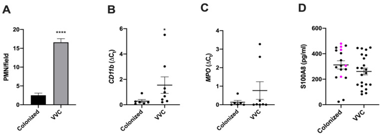
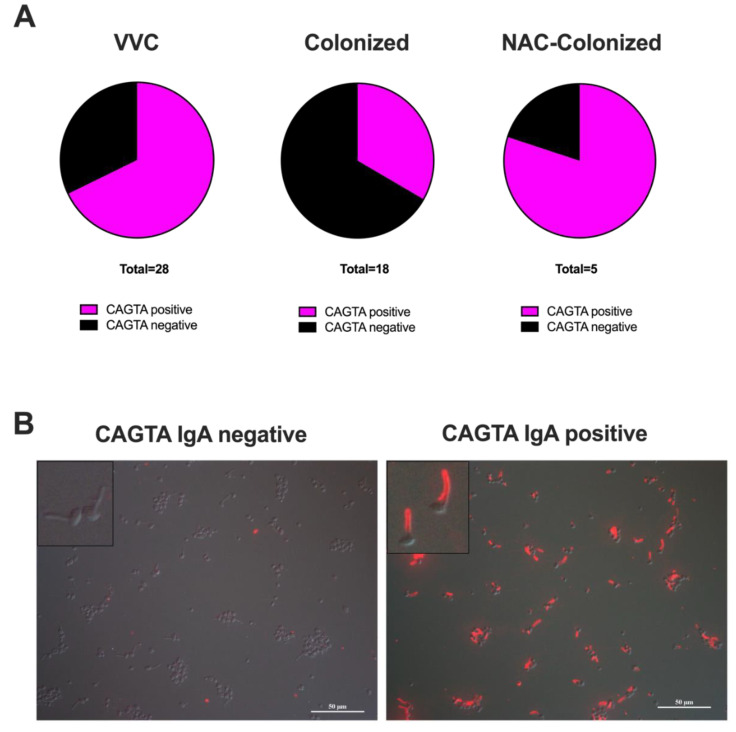
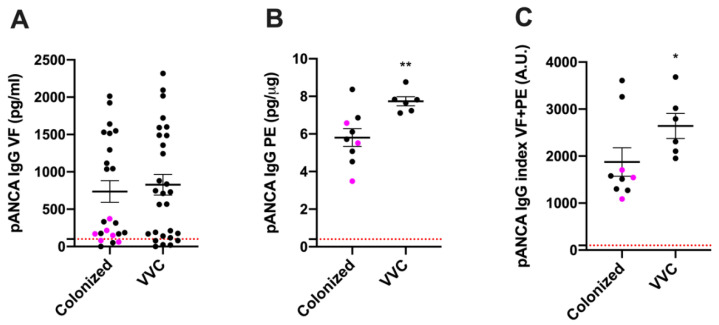
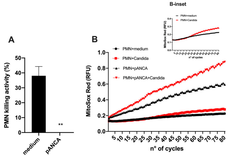
Vulvovaginal candidiasis (VVC) is primarily caused by Candida albicans and affects 75% of childbearing age women. Although C. albicans can colonize asymptomatically, disease is associated with an increased Candida burden, a loss of epithelial tolerance and a breakdown in vaginal microbiota homeostasis. VVC symptoms have been ascribed to a powerful inflammatory response associated with the infiltration of non-protective neutrophils (PMN). Here, we compared the immunological characteristics of vaginal fluids and cellular protein extracts obtained from 28 VVC women and from 23 healthy women colonized by Candida spp. We measured the levels of antibodies against fungal antigens and human autoantigens (anti-Saccharomyces cerevisiae antibodies (ASCA), C. albicans germ tube antibodies (CAGTAs) and perinuclear anti-neutrophil cytoplasmic antibodies (pANCA)), in addition to other immunological markers. Our results show that the pANCA levels detected in the cellular protein extracts from the vaginal fluids of symptomatic women were significantly higher than those obtained from healthy colonized women. Consistent with a potential physiologically relevant role for this pANCA, we found that specific anti-myeloperoxidase antibodies could completely neutralize the ex vivo killing capacity of polymorphonuclear cells. Collectively, this preliminary study suggests for the first time that pANCA are found in the pathogenic vaginal environment and can promptly impair neutrophil function against Candida, potentially preventing a protective response.
Keywords: ASCA; CAGTA; Candida; VVC; pANCA.
Conflict of interest statement
The authors declare no conflict of interest.
Figures
Similar articles
-
It Takes Two to Tango: How a Dysregulation of the Innate Immunity, Coupled With Candida Virulence, Triggers VVC Onset.Front Microbiol. 2021 Jun 7;12:692491. doi: 10.3389/fmicb.2021.692491. eCollection 2021. Front Microbiol. 2021. PMID: 34163460 Free PMC article. Review.
-
Vaginal Heparan Sulfate Linked to Neutrophil Dysfunction in the Acute Inflammatory Response Associated with Experimental Vulvovaginal Candidiasis.mBio. 2017 Mar 14;8(2):e00211-17. doi: 10.1128/mBio.00211-17. mBio. 2017. PMID: 28292981 Free PMC article.
-
An intravaginal live Candida challenge in humans leads to new hypotheses for the immunopathogenesis of vulvovaginal candidiasis.Infect Immun. 2004 May;72(5):2939-46. doi: 10.1128/IAI.72.5.2939-2946.2004. Infect Immun. 2004. PMID: 15102806 Free PMC article.
-
The acute neutrophil response mediated by S100 alarmins during vaginal Candida infections is independent of the Th17-pathway.PLoS One. 2012;7(9):e46311. doi: 10.1371/journal.pone.0046311. Epub 2012 Sep 25. PLoS One. 2012. PMID: 23050010 Free PMC article.
-
Recurrent Vulvovaginal Candidiasis: An Immunological Perspective.Microorganisms. 2020 Jan 21;8(2):144. doi: 10.3390/microorganisms8020144. Microorganisms. 2020. PMID: 31972980 Free PMC article. Review.
Cited by
-
Host-microbe interaction paradigms in acute and recurrent vulvovaginal candidiasis.Cell Host Microbe. 2024 Oct 9;32(10):1654-1667. doi: 10.1016/j.chom.2024.08.018. Cell Host Microbe. 2024. PMID: 39389030 Review.
-
Cutibacterium acnes lysate improves cellular response against Candida albicans, Escherichia coli and Gardnerella vaginalis in an in vitro model of vaginal infection.Front Cell Infect Microbiol. 2025 May 2;15:1578831. doi: 10.3389/fcimb.2025.1578831. eCollection 2025. Front Cell Infect Microbiol. 2025. PMID: 40384982 Free PMC article.
-
Nanobody-mediated neutralization of candidalysin prevents epithelial damage and inflammatory responses that drive vulvovaginal candidiasis pathogenesis.mBio. 2024 Mar 13;15(3):e0340923. doi: 10.1128/mbio.03409-23. Epub 2024 Feb 13. mBio. 2024. PMID: 38349176 Free PMC article.
-
Effects of Boric Acid Gel on Vaginal Candida albicans Infections and the Local Immune System in Mice.Front Immunol. 2022 Jul 25;13:950215. doi: 10.3389/fimmu.2022.950215. eCollection 2022. Front Immunol. 2022. PMID: 35958550 Free PMC article.
-
The impact of the Fungus-Host-Microbiota interplay upon Candida albicans infections: current knowledge and new perspectives.FEMS Microbiol Rev. 2021 May 5;45(3):fuaa060. doi: 10.1093/femsre/fuaa060. FEMS Microbiol Rev. 2021. PMID: 33232448 Free PMC article. Review.
References
Grants and funding
LinkOut - more resources
Full Text Sources
Research Materials
Miscellaneous
