. 2020 Oct 16;12(10):979.
doi: 10.3390/pharmaceutics12100979.

Conjugation of Native-Like HIV-1 Envelope Trimers onto Liposomes Using EDC/Sulfo-NHS Chemistry: Requirements and Limitations

Affiliations

Conjugation of Native-Like HIV-1 Envelope Trimers onto Liposomes Using EDC/Sulfo-NHS Chemistry: Requirements and Limitations

Ehsan Suleiman et al. Pharmaceutics. .

Abstract

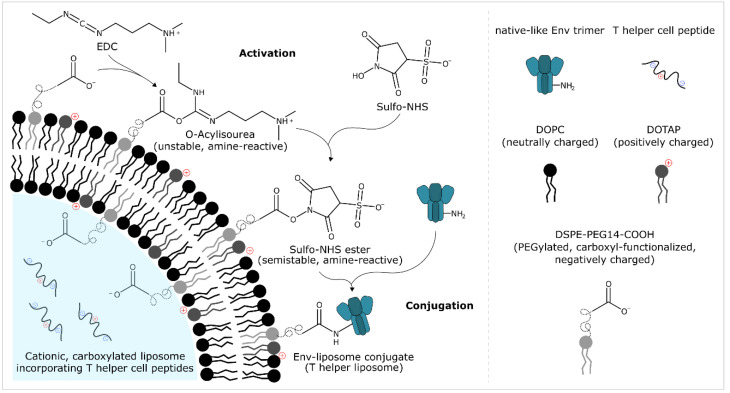
The display of native-like human immunodeficiency virus type 1 envelope (HIV-1 Env) trimers on liposomes has gained wide attention over the last few years. Currently, available methods have enabled the preparation of Env-liposome conjugates of unprecedented quality. However, these protocols require the Env trimer to be tagged and/or to carry a specific functional group. For this reason, we have investigated N-(3-Dimethylaminopropyl)-N'-ethylcarbodiimide/N-Hydroxysulfosuccinimide (EDC/Sulfo-NHS) chemistry for its potential to covalently conjugate tag-free, non-functionalized native-like Env trimers onto the surface of carboxyl-functionalized liposomes. The preservation of the liposome's physical integrity and the immunogen's conformation required a fine-tuned two-step approach based on the controlled use of β-mercaptoethanol. The display of Env trimers was strictly limited to activated liposomes of positive charge, i.e., liposomes with a positive zeta potential that carry amine-reactive Sulfo-NHS esters on their surface. In agreement with that, conjugation was found to be highly ionic strength- and pH-dependent. Overall, we have identified electrostatic pre-concentration (i.e., close proximity between negatively charged Env trimers and positively charged liposomes established through electrostatic attraction) to be crucial for conjugation reactions to proceed. The present study highlights the requirements and limitations of potentially scalable EDC/Sulfo-NHS-based approaches and represents a solid basis for further research into the controlled conjugation of tag-free, non-functionalized native-like Env trimers on the surface of liposomes, and other nanoparticles.

Keywords: EDC/Sulfo-NHS; HIV-1; covalent conjugation; intrastructural help; liposomes; native-like Env trimers; particulate display; pre-concentration; protein-liposome conjugates; tag-free conjugation; vaccines.


Conflict of interest statement

The authors declare no conflict of interest. E.S. and A.W. are employees of Polymun Scientific Immunbiologische Forschung GmbH and they declared no competing financial interests.

Figures

Figure 1
Figure 1
EDC/Sulfo-NHS-based preparation of Env-liposome conjugates (T helper liposomes): conjugation of native-like HIV-1 envelope trimers onto helper peptide-loaded liposomes. Carboxyl-functionalised liposomes are activated with EDC/Sulfo-NHS. Excess activation reagents are then removed by filtration-based separation techniques or through chemical inactivation using β-mercaptoethanol (not shown). Native-like Env trimers are then added and covalent Env-liposome conjugates are formed. Env-liposome conjugates that additionally incorporate T helper cell peptides (derived from commonly used licensed protein vaccines) are referred to as T helper liposomes. These liposomes are designed to harness intrastructural help, i.e., to recruit pre-existing, non-cognate T helper cells upon administration in order to provide help to Env-reactive/Env specific B cells and eventually shape/improve the immune response against the liposome-displayed Env trimers [43]. First steps towards the development of liposome-based vaccines capable of harnessing intrastructural help date back to the early 1990′s [61,62,63]. Recent studies that involved particle-based vaccines (virus-like particles and calcium phosphate nanoparticles) have provided additional proof of concept for this type of strategy within the context of HIV-1 [64,65,66,67,68]. Recent independent reports on liposome-based vaccines (against HIV-1, malaria, group A streptococci and ErbB-2 overexpressing breast cancer) utilizing the same principle are also very encouraging [69,70,71,72].
Figure 2
Figure 2
Conjugation efficiency and PGT145 reactivity as a function of pH and the apparent reactivity of the amine-reactive intermediate (Sulfo-NHS ester). (A) Reducing SDS-PAGE showing the extent of covalent conjugation onto cationic liposomes. (B,C). (B) Conjugation efficiency and (C) PGT145 reactivity after a hold time of 0 h as determined by ELISA. Data (ELISA) represent the mean from two analytical replicates. (AC). Liposomes were activated in 50 mM MB sucrose pH 6.1. Excess activation reagents were removed by two consecutive gel-filtration steps. Liposomes were eluted with 5 mM PB (w/15 mM NaCl and w/270 mM sucrose) with a pH of 6.5, 7.5, and 8.5, respectively. Conjugation was initiated by the addition of UFO Env after 0, 2, 4, and 6 h, respectively. The panel shows the results from a single explorative screening experiment. Abbreviations: T, total conjugation reaction mix; CF, conjugate fraction; NCF 1, non-conjugate fraction 1; NCF 2, non-conjugate fraction 2. See Section 2.8 for further information.
Figure 3
Figure 3
Concentration- and time-dependent quenching of excess EDC by β-mercaptoethanol (β-ME). (A,C) Reducing SDS-PAGE showing the extent of covalent conjugation of UFO Env trimers onto cationic liposomes as a function of (A) the used molar excess of β-ME and (C) the quenching time (i.e., the time β-ME was allowed to react with EDC before initiating the conjugation by the addition of UFO Env), respectively. (B,D) Conjugation efficiency, PGT145 reactivity and the amount of conjugated UFO Env per lipid as a function of (B) the used molar excess of β-mercaptoethanol and (D) the quenching time, respectively. (AD) Liposomes were activated in 5 mM PB pH 6.0 w/15 mM NaCl and w/270 mM sucrose. (A,B) Excess activation reagents were deactivated by the addition of a 0-, 1-, 2.5-, 5-, 10- and 20-fold molar excess β-ME over the amount of EDC used for activation. Conjugation was initiated by the addition of UFO Env after a quenching time of 10 min. (C,D) Excess activation reagents were deactivated by the addition of a 1.75-fold molar excess β-ME over the amount of EDC used for activation. Conjugation was initiated by the addition of UFO Env after a quenching time of 0.5, 1, 2.5, 5, 10, and 20 min, respectively. Data (ELISA and lipid quantification) represent the mean from two analytical replicates. The panel shows the results from a single explorative screening experiment. Abbreviations: T, total conjugation reaction mix; CF, conjugate fraction; NCF 1, non-conjugate fraction 1; NCF 2, non-conjugate fraction 2. See Section 2.8 for further information.
Figure 4
Figure 4
Liposome charge-dependent conjugation of UFO Env trimers: physico-chemical characterisation of liposomes before and after conjugation. (A,B) Zeta potential of carboxyl-functionalised liposomes with increasing fractions of (A) DOPG and (B) DOTAP. (C,D) Diameter, (i.e., Z-average diameter) and PdI of carboxyl-functionalised liposomes with increasing fractions of (C) DOPG and (D) DOTAP. Data represent the mean ± standard deviation from three analytical replicates. The panel shows the results from a single explorative screening experiment.
Figure 5
Figure 5
Liposome charge-dependent conjugation of UFO Env trimers. (A) Reducing SDS-PAGE showing the extent of covalent conjugation onto liposomes as a function of increasing molar fractions of DOPG (left panel) or DOTAP (right panel) in liposomal membranes. (B) Conjugation efficiency, PGT145 reactivity and the amount of conjugated UFO Env per lipid. Data (ELISA and lipid quantification) represent the mean from two analytical replicates. (A,B) Liposomes of the DOPG series were activated in 50 mM MB sucrose pH 6.1. Excess activation reagents were removed by gel filtration. Liposomes were eluted with 5 mM PBS pH 6.5 w/150 mM NaCl. In contrast, liposomes of the DOTAP series were activated in 5 mM PB pH 6.0 w/15 mM NaCl and w/270 mM sucrose. Excess EDC was chemically deactivated by the addition of β-ME. The panel shows the results from a single explorative screening experiment. Abbreviations: T, total conjugation reaction mix; CF, conjugate fraction; NCF 1, non-conjugate fraction 1; NCF 2, non-conjugate fraction 2. See Section 2.8 for further information.
Figure 6
Figure 6
Ionic strength-dependent conjugation of UFO Env trimers onto cationic liposomes. (A,B) (A) Diameter, (i.e., Z-average diameter), PdI and (B) zeta potential of liposomes before and after conjugation. Measurements were performed with the corresponding conjugation buffers. Data represent the mean ± standard deviation from three analytical replicates. (C) Reducing SDS-PAGE showing the extent of covalent conjugation. (D) Conjugation efficiency, PGT145 reactivity and the amount of conjugated UFO Env per lipid. Data (ELISA and lipid quantification) represent the mean from two analytical replicates. (AD) Conjugation experiments were performed in 5 mM PB pH 6.0 with a constant osmolality but with a varying ionic strength, i.e., varying concentrations of sodium chloride and sucrose. For this purpose, cationic liposomes dispersed in 5 mM PB sucrose pH 6.0 w/300 mM sucrose and liposomes of the same lipid composition but dispersed in 5 mM PBS pH 6.0 w/150 mM NaCl, were mixed to give the desired sodium chloride concentration. After activation of carboxyl groups, excess EDC was chemically deactivated by the addition of β-ME. The panel shows the results from a single explorative screening experiment. Abbreviations: T, total conjugation reaction mix; CF, conjugate fraction; NCF 1, non-conjugate fraction 1; NCF 2, non-conjugate fraction 2. See Section 2.8 for further information.
Figure 7
Figure 7
pH-dependent conjugation of UFO Env trimers onto cationic liposomes. (A) Reducing SDS-PAGE showing the extent of covalent conjugation. (B) Conjugation efficiency, PGT145 reactivity and the amount of conjugated UFO Env per lipid. Data (ELISA and lipid quantification) represent the mean from two analytical replicates. (A,B) Liposomes were activated in 5 mM PB pH 6.0 w/15 mM NaCl and w/270 mM sucrose. Excess EDC was chemically deactivated by the addition of β-ME. The desired pH was adjusted by adding 0.1 M NaOH or 0.1 M HCl right before initiation of the conjugation reaction. The indicated pH was measured at the end of the reaction, i.e., after stopping the reaction by the addition of glycine. Data on the physico-chemical properties of the liposomes before and after conjugation can be found in the supplement (Figure S6). The panel shows the results from a single explorative screening experiment. Abbreviations: T, total conjugation reaction mix; CF, conjugate fraction; NCF 1, non-conjugate fraction 1; NCF 2, non-conjugate fraction 2. See Section 2.8 for further information.
Figure 8
Figure 8
Conjugation of next-generation HIV-1 immunogens onto cationic, helper peptide-loaded liposomes. (A) Reducing SDS-PAGE showing the extent of covalent conjugation of UFO Env trimers, SOSIP Env trimers and their EDC cross-linked versions. Marker: Precision Plus Protein™ Kaleidoscope™ (Bio-Rad Laboratories, Inc., Hercules, CA, USA). (B) Conjugation efficiency, PGT145 reactivity and the amount of conjugated Env per lipid. Data (ELISA and lipid quantification) represent the mean from two analytical replicates. (A,B) Excess EDC was chemically deactivated by the addition of β-ME. The panel shows the results from a single explorative screening experiment. Data on the peptide recovery after conjugation as well as on the physico-chemical properties of the liposomes before and after conjugation can be found in the supplement (Figure S9). Abbreviations: T, total conjugation reaction mix; CF, conjugate fraction; NCF 1, non-conjugate fraction 1; NCF 2, non-conjugate fraction 2. See Section 2.8 for further information.
Figure 9
Figure 9
Antigenicity of soluble and liposome-displayed next-generation HIV-1 immunogens. Monoclonal antibody titrations of 2G12-captured Env or Env-liposome conjugates. The graphs show the binding curves of Triton X-100-treated conjugate fractions (CF), i.e., non-intact Env-liposome conjugates without non-conjugated Env. The concentration of the captured samples was determined using an 2G12-based ELISA and was the same (0.5 µg/mL) throughout all titrations. Data represent the mean from two analytical replicates.

Similar articles

Cited by

References

    1. Sanders R.W., Moore J.P. Native-like Env trimers as a platform for HIV-1 vaccine design. Immunol. Rev. 2017;275:161–182. doi: 10.1111/imr.12481. - DOI - PMC - PubMed
    1. Klasse P.J., Ozorowski G., Sanders R.W., Moore J.P. Env Exceptionalism: Why Are HIV-1 Env Glycoproteins Atypical Immunogens? Cell Host Microbe. 2020;27:507–518. doi: 10.1016/j.chom.2020.03.018. - DOI - PMC - PubMed
    1. Stephenson K.E., Wagh K., Korber B., Barouch D.H. Vaccines and Broadly Neutralizing Antibodies for HIV-1 Prevention. Annu. Rev. Immunol. 2020;38:673–703. doi: 10.1146/annurev-immunol-080219-023629. - DOI - PMC - PubMed
    1. Irvine D.J., Read B.J. Shaping humoral immunity to vaccines through antigen-displaying nanoparticles. Curr. Opin. Immunol. 2020;65:1–6. doi: 10.1016/j.coi.2020.01.007. - DOI - PMC - PubMed
    1. Thalhauser S., Peterhoff D., Wagner R., Breunig M. Critical design criteria for engineering a nanoparticulate HIV-1 vaccine. J. Control. Release. 2020;317:322–335. doi: 10.1016/j.jconrel.2019.11.035. - DOI - PubMed