. 2020 Oct 16;8(10):426.
doi: 10.3390/biomedicines8100426.

Engineering and Preclinical Evaluation of Western Reserve Oncolytic Vaccinia Virus Expressing A167Y Mutant Herpes Simplex Virus Thymidine Kinase

Affiliations

Engineering and Preclinical Evaluation of Western Reserve Oncolytic Vaccinia Virus Expressing A167Y Mutant Herpes Simplex Virus Thymidine Kinase

S M Bakhtiar Ul Islam et al. Biomedicines. .

Abstract

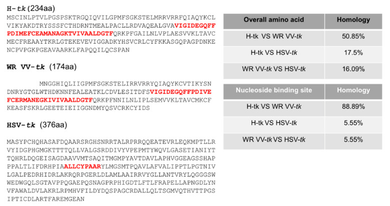
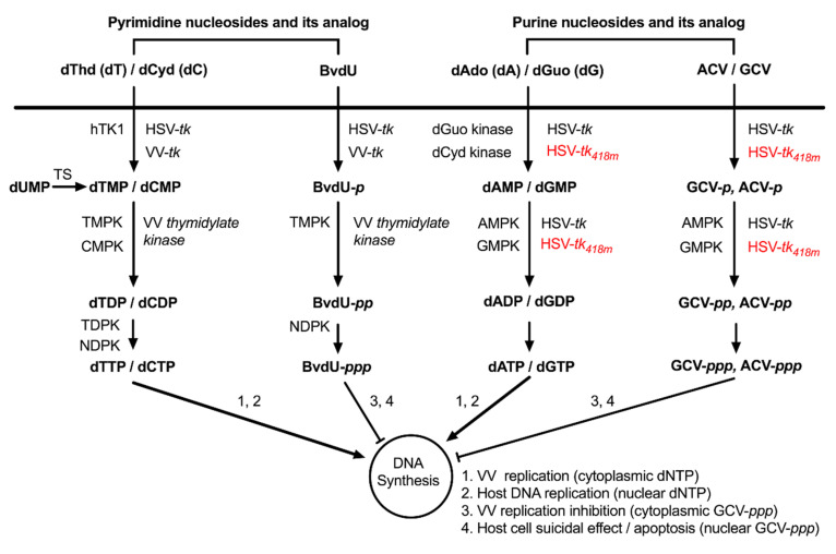
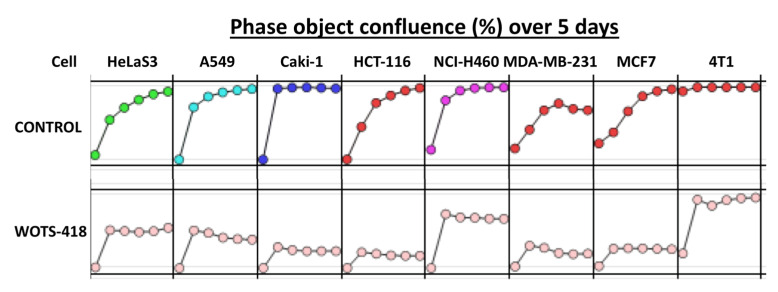
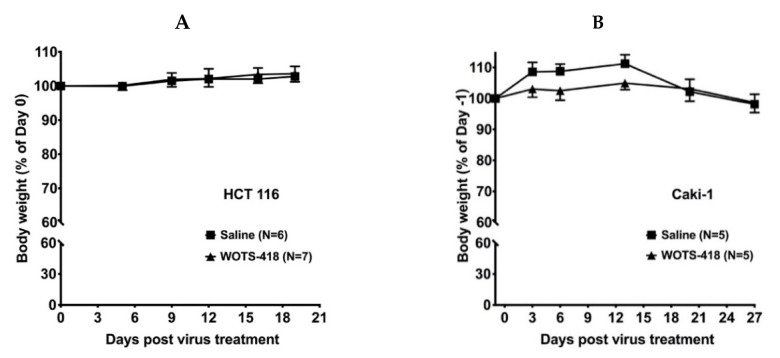
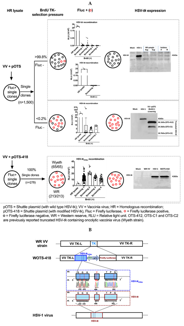
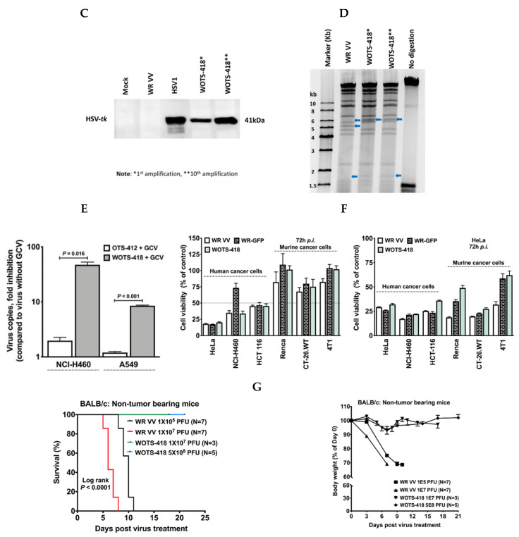
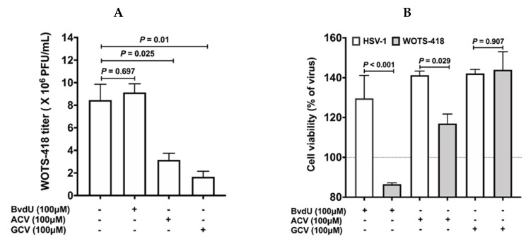
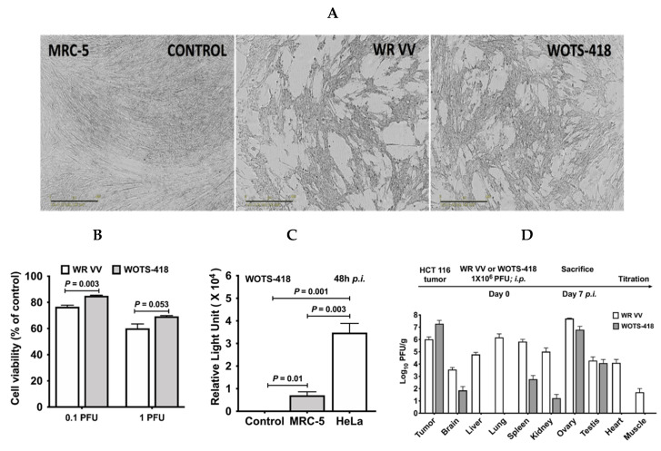
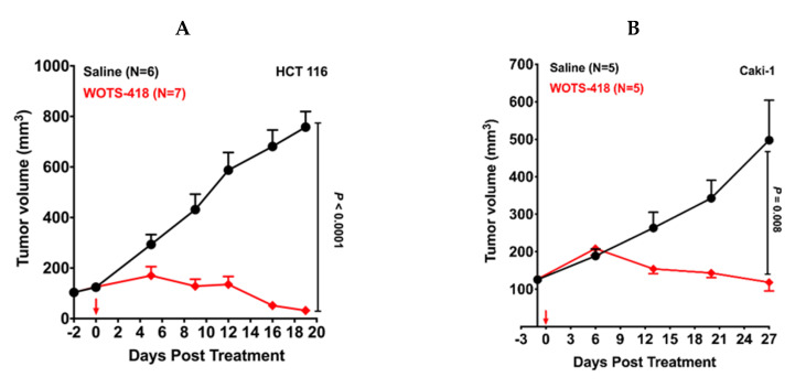
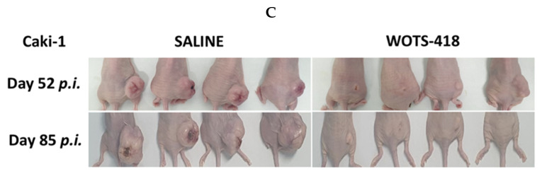
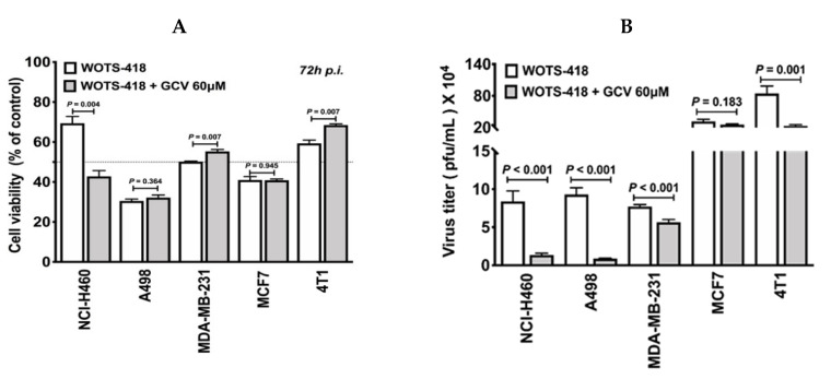
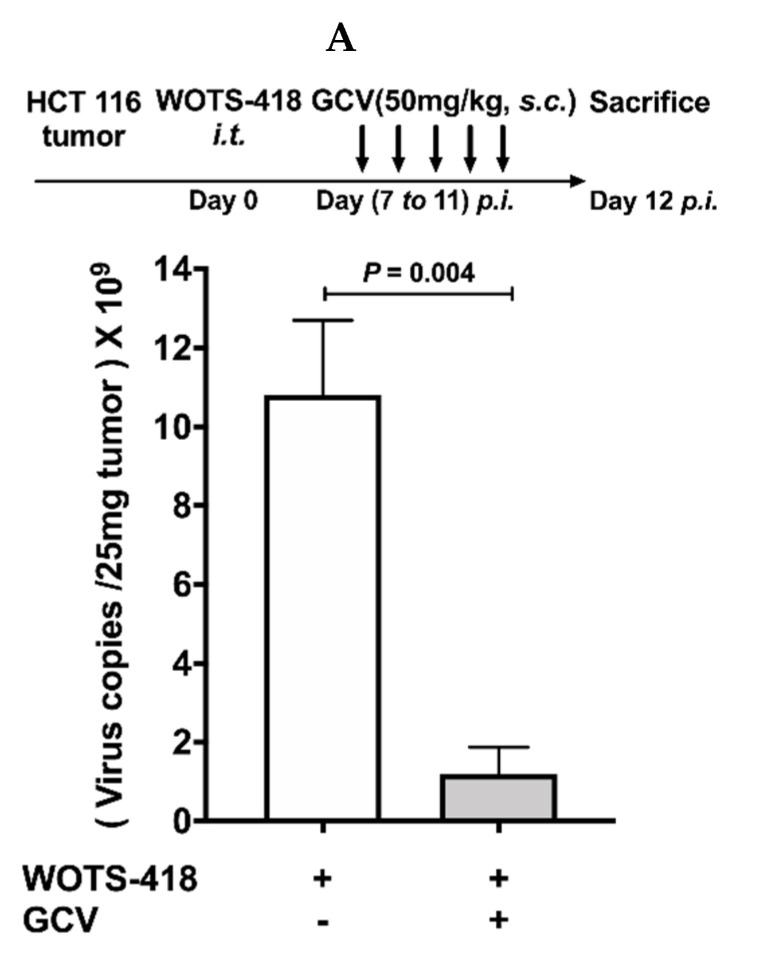
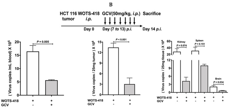
Viral replication of thymidine kinase deleted (tk-) vaccinia virus (VV) is attenuated in resting normal cells, enabling cancer selectivity, however, replication potency of VV-tk- appears to be diminished in cancer cells. Previously, we found that wild-type herpes simplex virus (HSV)-tk (HSV-tk) disappeared in most of the recombinant VV after multiple screenings, and only a few recombinant VV containing naturally mutated HSV-tk remained stable. In this study, VV-tk of western reserve (WR) VV was replaced by A167Y mutated HSV-tk (HSV-tk418m), to alter nucleoside selectivity from broad spectrum to purine exclusive selectivity. WOTS-418 remained stable after numerous passages. WOTS-418 replication was significantly attenuated in normal cells, but cytotoxicity was almost similar to that of wild type WR VV in cancer cells. WOTS-418 showed no lethality following a 5 × 10⁸ PFU intranasal injection, contrasting WR VV, which showed 100% lethality at 1 × 10⁵ PFU. Additionally, ganciclovir (GCV) but not BvdU inhibited WOTS-418 replication, confirming specificity to purine nucleoside analogs. The potency of WOTS-418 replication inhibition by GCV was > 10-fold higher than that of our previous truncated HSV-tk recombinant OTS-412. Overall, WOTS-418 demonstrated robust oncolytic efficacy and pharmacological safety which may delegate it as a candidate for future clinical use in OV therapy.

Keywords: A167Y; ganciclovir; herpes simplex virus; oncolytic viruses; thymidine kinase; western reserve vaccinia virus.


Conflict of interest statement

The authors declare no conflict of interest. T.H.H and M.C are the patent owner of the WOTS-418. The funders had no role in the design of the study; in the collection, analyses, or interpretation of data; in the writing of the manuscript, or in the decision to publish the results.

Figures

Figure A1
Figure A1
Human, WR VV and HSV1 virus tk protein sequences and comparison: Homology comparison among human thymidine kinase (NCBI GenBank: AAA61191.1), and vaccinia virus thymidine Kinase (NCBI GenBank: AAO89373.1) and herpes simplex virus thymidine kinase (GenBank: AAA45811.1) protein sequences is shown here. Overall amino acid homology and nucleoside binding side (NBS) homology is analyzed and shown in this figure (Right). NBS for each thymidine kinases are shown as red color (Left).
Figure A2
Figure A2
Nucleoside metabolism in DNA precursors synthesis (salvage pathway) by different kinases from human, vaccinia virus, and herpes simplex virus. HSV-tk418m is A167Y mutant HSV-tk armed into tk of WR VV backbone which is described earlier in the method Section 2.2. While wild type HSV-tk broadly phosphorylate purine and pyrimidine nucleosides and their analogs, A167Y mutant HSV-tk which is expressed by WOTS-418 phosphorylate purine nucleosides and it analog but not pyrimidine and its analogs. TS: Thymidylate synthase; TMPK: Thymidylate monophosphate kinase; CMPK: Cytidine monophosphate kinase; TDPK: Thiamin diphosphate kinase; NDPK: Nucleoside diphosphate kinase; TMPK: Thymidylate monophosphate kinase; NDPK: Nucleoside-diphosphate kinases; AMPK: AMP-activated protein kinase; GMPK: Guanylate monophosphate kinase; AMPK: AMP-activated protein kinase; GMPK: Guanylate monophosphate kinase.
Figure A3
Figure A3
WOTS-418 effect on multiple cancer cells were evaluated by Incucyte S3 live imaging system. Cells (1 × 10⁴) were seeded in a 96-well plate (Essen Incucyte ImageLock 96-well plate) and next day infected with WOTS-418 at 1 PFU/cell dose. Plates were incubated at 37 °C with 5% CO2 in an incubator equipped with Incucyte S3 live cell image monitoring system (Essen Bioscience, Ann Arbor, MI, USA). Phase object confluence was recorded over 5 days of 6-h intervals. The diagram is shown as a 24 h imaging interval (gap between the circles).
Figure A4
Figure A4
WOTS-418 effect on HCT 116 and Caki-1 tumor bearing murine body weight. (A) Human colorectal cancer (HCT 116) model. Detail procedure and treatment plan described in Figure 4A. (B). Human renal cancer (Caki-1) model: Detail procedure and treatment plan described in Figure 4B.
Figure 1
Figure 1
Rationale for engineering modified HSV-tk armed OVV, WOTS-418 and characterization. (A) Wild type HSV-tk transgene instability in VV backbone. Wild type HSV-tk or modified HSV-tk418m containing shuttle plasmid (pOTS or pOTS-418) was used for HR with VV. Firefly luciferase positive HR lysate were further screened with or without BrdU TK negative selection pressure. Isolated firefly luciferase positive singles plaques were further checked for HSV-tk expression by western blotting (detail screening and selection procedure described in the methods section). (B) WOTS-418 engineering map. Transgene insertion location in WR VV backbone is shown (top) and the diagram is showing the partial and representative location of the wild type HSV-tk DNA sequence (bottom). The middle panel is showing the representative modified HSV-tk418m DNA sequence, indicating the location of altered nucleotides and DNA sequence confirmation. The promoters used for HSV-tk418m and firefly luciferase were pSE/L and p 7.5, respectively. (C) Monitoring long-term HSV-tk418m gene stability. To evaluate the long-term transgene stability, WOTS-418 virus samples from two different time points (first amplification and tenth amplification) were used. A549 cells were infected with WOTS-418 (0.1 PFU/cell), WR VV and HSV-1. 24 h post infection (p.i.), cell pellets were used to measure HSV-tk expression by western blotting. WR VV: Wild type western reserve vaccinia virus (Negative control); HSV-1: Wild type HSV-1 virus (Positive control); Anti-HSV-1 thymidine Kinase; sc-28037, vN-20: SCBT, Dallas, TX, USA; Anti-GAPDH; MB001: Bioworld, Louis Park, MN, USA; Secondary antibody anti-goat; A50-101P: Bethyl, Montgomery, TX, USA; Secondary antibody anti-mouse; ADI-SAB-100-J: Enzo, Executive Blvd Farmingdale, NY, USA. Protein Marker: ExcelBand 3-Color PreStained Protein Markers, #2700 (Green BioResearch LLC, Baton Rouge, LA, USA), a mixture of blue, red, and green stained recombinant proteins (5 to 245 kDa), was used as size standards in western blotting. (D) WOTS-418 DNA stability: Samples from Figure 1C was also used for long-term DNA stability confirmation, which was evaluated by HindIII restriction digestion assay (detail procedure described in the methods section). Blue arrows indicate the alteration pattern of DNA band size after recombination. (E) GCV antiviral potency comparison between OTS-412 and WOTS-418: Quantitative analysis of viral DNA copies, measured in fold inhibition, was done following virus + GCV treatment and virus without GCV treatment. NCI-H460 and A549 cells were infected with either OTS-412 or WOTS-418 (0.1 PFU/cell) and co-treated with GCV (100 μM). 72 h p.i., samples were harvested, and viral copy numbers were measured by quantitative polymerase chain reaction (qPCR). Data presented as mean ± SEM (n = 2) and compared using two-tailed Student’s t-test (p-value). (F) WR, WR-GFP, and WOTS-418 cytotoxicity comparison (left panel). Three human cancer cells (HeLa, NCI-H460, and HCT 116) and three murine cancer cells (Renca, CT-26.WT, and 4T1) were infected with WR, WR-GFP, WOTS-418 at a dose 0.1 PFU/cell for human cancer and 1 PFU/cell for murine cancer cells. 72 h p.i. samples were evaluated for cytotoxicity by CCK-8 assay. Data are presented as mean± SEM (n = 2). WR VV, WR-GFP, and WOTS-418 virus yield in various cancer cell lines (right panel): After cytotoxicity experiment, samples were harvested and subjected to three freeze and thaw cycles. Next, samples were sonicated with a cup sonicator (100% power for 30 s, 3 times with 1-min interval) and reinfected (same volume, 30 μL) in pre-seeded HeLa cells (10,000 cells/well) in 96-well plates and incubated at 37 °C with 5% CO2. 72 h p.i., CCK-8 assay was performed to evaluate that viral cytotoxicity of the three viruses. Data are presented as mean ± SEM (n = 3). (G) WR vs. WOTS-418 safety assessment: Non-tumor bearing BALB/c syngeneic mice were treated intranasally with WR VV (1 × 10⁵ PFU and 1 × 10⁷ PFU) or WOTS-418 (1 × 10 7 PFU and 5 × 10⁸ PFU). After WR VV or WOTS-418 treatment, survival and body weight were monitored. Survival curve demonstrating overall survival and p < 0.0001 was determined by log-rank (Mantel-Cox) test (n ≥ 3).
Figure 1
Figure 1
Rationale for engineering modified HSV-tk armed OVV, WOTS-418 and characterization. (A) Wild type HSV-tk transgene instability in VV backbone. Wild type HSV-tk or modified HSV-tk418m containing shuttle plasmid (pOTS or pOTS-418) was used for HR with VV. Firefly luciferase positive HR lysate were further screened with or without BrdU TK negative selection pressure. Isolated firefly luciferase positive singles plaques were further checked for HSV-tk expression by western blotting (detail screening and selection procedure described in the methods section). (B) WOTS-418 engineering map. Transgene insertion location in WR VV backbone is shown (top) and the diagram is showing the partial and representative location of the wild type HSV-tk DNA sequence (bottom). The middle panel is showing the representative modified HSV-tk418m DNA sequence, indicating the location of altered nucleotides and DNA sequence confirmation. The promoters used for HSV-tk418m and firefly luciferase were pSE/L and p 7.5, respectively. (C) Monitoring long-term HSV-tk418m gene stability. To evaluate the long-term transgene stability, WOTS-418 virus samples from two different time points (first amplification and tenth amplification) were used. A549 cells were infected with WOTS-418 (0.1 PFU/cell), WR VV and HSV-1. 24 h post infection (p.i.), cell pellets were used to measure HSV-tk expression by western blotting. WR VV: Wild type western reserve vaccinia virus (Negative control); HSV-1: Wild type HSV-1 virus (Positive control); Anti-HSV-1 thymidine Kinase; sc-28037, vN-20: SCBT, Dallas, TX, USA; Anti-GAPDH; MB001: Bioworld, Louis Park, MN, USA; Secondary antibody anti-goat; A50-101P: Bethyl, Montgomery, TX, USA; Secondary antibody anti-mouse; ADI-SAB-100-J: Enzo, Executive Blvd Farmingdale, NY, USA. Protein Marker: ExcelBand 3-Color PreStained Protein Markers, #2700 (Green BioResearch LLC, Baton Rouge, LA, USA), a mixture of blue, red, and green stained recombinant proteins (5 to 245 kDa), was used as size standards in western blotting. (D) WOTS-418 DNA stability: Samples from Figure 1C was also used for long-term DNA stability confirmation, which was evaluated by HindIII restriction digestion assay (detail procedure described in the methods section). Blue arrows indicate the alteration pattern of DNA band size after recombination. (E) GCV antiviral potency comparison between OTS-412 and WOTS-418: Quantitative analysis of viral DNA copies, measured in fold inhibition, was done following virus + GCV treatment and virus without GCV treatment. NCI-H460 and A549 cells were infected with either OTS-412 or WOTS-418 (0.1 PFU/cell) and co-treated with GCV (100 μM). 72 h p.i., samples were harvested, and viral copy numbers were measured by quantitative polymerase chain reaction (qPCR). Data presented as mean ± SEM (n = 2) and compared using two-tailed Student’s t-test (p-value). (F) WR, WR-GFP, and WOTS-418 cytotoxicity comparison (left panel). Three human cancer cells (HeLa, NCI-H460, and HCT 116) and three murine cancer cells (Renca, CT-26.WT, and 4T1) were infected with WR, WR-GFP, WOTS-418 at a dose 0.1 PFU/cell for human cancer and 1 PFU/cell for murine cancer cells. 72 h p.i. samples were evaluated for cytotoxicity by CCK-8 assay. Data are presented as mean± SEM (n = 2). WR VV, WR-GFP, and WOTS-418 virus yield in various cancer cell lines (right panel): After cytotoxicity experiment, samples were harvested and subjected to three freeze and thaw cycles. Next, samples were sonicated with a cup sonicator (100% power for 30 s, 3 times with 1-min interval) and reinfected (same volume, 30 μL) in pre-seeded HeLa cells (10,000 cells/well) in 96-well plates and incubated at 37 °C with 5% CO2. 72 h p.i., CCK-8 assay was performed to evaluate that viral cytotoxicity of the three viruses. Data are presented as mean ± SEM (n = 3). (G) WR vs. WOTS-418 safety assessment: Non-tumor bearing BALB/c syngeneic mice were treated intranasally with WR VV (1 × 10⁵ PFU and 1 × 10⁷ PFU) or WOTS-418 (1 × 10 7 PFU and 5 × 10⁸ PFU). After WR VV or WOTS-418 treatment, survival and body weight were monitored. Survival curve demonstrating overall survival and p < 0.0001 was determined by log-rank (Mantel-Cox) test (n ≥ 3).
Figure 2
Figure 2
Evaluation of WOTS-418 (HSV-tk418) purine and pyrimidine analogs antiviral potency. (A) 143B cells (tk negative) were infected with WOTS-418 (0.1 PFU/cell) and co-treated with purine analogs (ACV and GCV), and pyrimidine analog (BvdU) at a dose 100 μM. 72 h p.i., samples were harvested, and antiviral potency to each of these analogs was evaluated by vaccinia virus plaque titration assay in U-2 OS (B) 143B cells (tk negative) were infected with WOTS-418 (0.1 PFU/cell) or HSV-1 virus (positive control) and co-treated with purine analogs (ACV and GCV), and pyrimidine analog (BvdU) at a dose 100 μM. 72 h p.i., samples were harvested and subjected to three freeze and thaw cycles. Next, samples were sonicated with a cup sonicator (100% power for 45 s, 3 times with 1-min interval) and reinfected (same volume, 100 μL) in pre-seeded HeLa cells (10,000 cells/well) in 96 well plates and incubated at 37 °C with 5% CO2. 72 h p.i., cytotoxicity assay (CCK-8) was performed. Data presented as mean ± SEM (n ≥ 2) and compared using two-tailed Student’s t-test (p-value). ACV: Acyclovir, GCV: Ganciclovir and BvdU: (E)-5-(2-Bromovinyl)-2′-deoxyuridine.
Figure 3
Figure 3
Tumor selectivity of WOTS-418. (A) Normal human cell line MRC-5 cells were seeded in 96-well plate at 3 × 10⁴ cells/well and infected with WR-VV or WOTS-418 (0.1 PFU/cell or 1 PFU/cell) to evaluate cytopathic effect and virus replication of WOTS-418. 48 h p.i., a phase contrast scan was performed using Incucyte S3 live cell imaging system (Scale bar: 400 μm). (B) Next, using the same samples from Figure 3A, cell viability was measured by cytotoxicity assay (CCK-8). Data presented as mean± SEM (n ≥ 3) and compared using two-tailed Student’s t-test (p-value). (C) To evaluate virus replication in MRC-5, a control cancer cell line, HeLa, was infected under the same conditions and after 48 h p.i., 20 μL supernatant was used to analyze the luciferase expression (E1500, Promega, WI, USA). Data presented as mean± SEM (n ≥ 3) and compared using two-tailed Student’s t-test (p-value). (D) HCT 116 tumor bearing male and female BALB/c nude mice were intraperitoneally injected with either WR VV or WOTS-418 at a dose 1 × 10⁶ PFU. 7 days p.i., mice were sacrificed, tumor and other normal organs (brain, liver, lung, spleen, kidney, ovary, testis, heart and muscle) were harvested aseptically. Weight of each organ was recorded. Samples were homogenized (Omni bead 24 rupture) and analyzed by titration (plaque assay) for vaccinia virus detection and quantification. Data are presented as Log10 PFU/gram and as a mean ± SEM (n = 3).
Figure 4
Figure 4
WOTS-418 antitumor potency in human colorectal (HCT 116) and renal (Caki-1) cancer models. (A) Human colorectal cancer (HCT 116) model: Antitumor effect following a single dose treatment of WOTS-418 was evaluated in HCT 116 human colorectal cancer model. Xenografts were established in the BALB/c athymic nude mice by injecting 5 × 10⁶ cells subcutaneously in the right flank of each mouse. After randomization and grouping, animals were treated intratumorally with either saline or WOTS-418, at a dose 1 × 10⁶ PFU on Day 0. Tumor and body weight (Appendix A in Figure A4A) were recorded at various time points and data are presented as mean ± SEM (n ≥ 6) and compared using two-tailed Student’s t-test (p-value). (B) Human renal cancer (Caki-1) model: Xenografts were established in the BALB/c athymic nude mice by injecting 1.75X10⁶ cells subcutaneously in the right flank of each mouse. After randomization and grouping, animals were treated intratumorally, with either saline or WOTS-418, at a dose1 × 106 PFU at Day 0. Tumor and body weight (Appendix A in Figure A4B) were recorded at various time points until Day 27 p.i. and data are presented as mean ± SEM (n = 5) and compared using two-tailed Student’s t-test (p-value). (C) Treated mice from Caki-1 model were kept until Day 85 p.i. to monitor further tumor progression or regression. At 52 p.i. and day 85 p.i. images were captured. The day 85 p.i. photo was taken after sacrifice. Therefore, mouse skin color was faint. Confirmation of complete response was evaluated based on the absence of pulpable tumors.
Figure 4
Figure 4
WOTS-418 antitumor potency in human colorectal (HCT 116) and renal (Caki-1) cancer models. (A) Human colorectal cancer (HCT 116) model: Antitumor effect following a single dose treatment of WOTS-418 was evaluated in HCT 116 human colorectal cancer model. Xenografts were established in the BALB/c athymic nude mice by injecting 5 × 10⁶ cells subcutaneously in the right flank of each mouse. After randomization and grouping, animals were treated intratumorally with either saline or WOTS-418, at a dose 1 × 10⁶ PFU on Day 0. Tumor and body weight (Appendix A in Figure A4A) were recorded at various time points and data are presented as mean ± SEM (n ≥ 6) and compared using two-tailed Student’s t-test (p-value). (B) Human renal cancer (Caki-1) model: Xenografts were established in the BALB/c athymic nude mice by injecting 1.75X10⁶ cells subcutaneously in the right flank of each mouse. After randomization and grouping, animals were treated intratumorally, with either saline or WOTS-418, at a dose1 × 106 PFU at Day 0. Tumor and body weight (Appendix A in Figure A4B) were recorded at various time points until Day 27 p.i. and data are presented as mean ± SEM (n = 5) and compared using two-tailed Student’s t-test (p-value). (C) Treated mice from Caki-1 model were kept until Day 85 p.i. to monitor further tumor progression or regression. At 52 p.i. and day 85 p.i. images were captured. The day 85 p.i. photo was taken after sacrifice. Therefore, mouse skin color was faint. Confirmation of complete response was evaluated based on the absence of pulpable tumors.
Figure 5
Figure 5
GCV suicidal effect on WOTS-418 infected cancer cells evaluation of viability and virus replication inhibition in vitro. (A) Impact of GCV on WOTS-418 infected cancer cells (NCI-H460, A498, MDA-MB-231, MCF7, and 4T1) was evaluated by cell viability assay. Cells (3 × 10³/well) were seeded in 96-cell plate and were infected with WOTS-418 at 1 PFU/cell the following day. After two hours, infection media was replaced with low dose GCV (60 μM) and incubated for 72 h at 37 °C with 5% CO2. Cell viability was measured by cell counting kit (CCK-8, Dojindo, Kumamoto, Japan). Data presented as a mean ± SEM (n = 3) and compared using two-tailed Student’s t-test (p-value). (B) Impact of low dose GCV (60 μM) on WOTS-418 plaque formation was monitored in multiple neoplastic cancer cell lines. Samples for Figure 5A were harvested, cycled through freezing and thawing three times, and sonicated with a cup sonicator (100% power for 30 s, three times with 1-min interval). Titration was performed in U2-OS cell line according to vaccinia virus titration protocol. Data presented as mean ± SEM (n ≥ 4) and compared using two-tailed Student’s t-test (p-value).
Figure 6
Figure 6
Dynamics of GCV antiviral potency for controlling WOTS-418 replication in animal model. (A) In vivo assessment of GCV antiviral potency to WOTS-418 replication was evaluated in an HCT 116 tumor bearing mouse model by qPCR analysis. Mice were injected subcutaneously with 5 × 10⁶ HCT 116 cells/mouse in the right flank of the BALB/c nu/nu mice. When palpable tumors were appeared, WOTS-418 was injected intratumorally at 1 × 10⁶ PFU dose and seven days post virus injection, subcutaneous GCV treatment (50 mg/Kg) performed daily for five times (D 7 p.i. to D 11 p.i.). Tumors harvested and homogenized using a bead rupture 24 (Omni international, Kennesaw, GA, USA) set to 3 cycles for 30 s at 4 m/s with 30 s intervals. WOTS-418 virus was quantified by qPCR. Data presented as mean ± SEM (n ≥ 2) and compared using two-tailed Student’s t-test (p-value). (B) In a separate in vivo study, GCV antiviral potency to WOTS-418 replication was evaluated in an HCT 116 tumor bearing mouse model and quantified by qPCR analysis. 5 × 10⁶ HCT 116 cells/mouse were injected subcutaneously in the right flank of the BALB/c nu/nu mice. When palpable tumors were appeared, WOTS-418 was injected intraperitoneally at 1 × 10⁶ PFU dose. 7 days following WOTS-418 injection, mice were given an intraperitoneal GCV treatment (50 mg/Kg) performed daily for seven times (D 7 p.i. to D 13 p.i.). Mice were sacrificed and blood samples were harvested in BD vacutainer blood collection tubes (BD, Franklin Lakes, NJ, USA). Other tissues (kidney, spleen, brain, and tumor) were harvested, weighed, and homogenized using bead rupture 24 (Omni international, Kennesaw, GA, USA) set to three cycles of 30 s at 4 m/s with 30 s intervals. WOTS-418 virus was quantified by qPCR. Data presented as mean ± SEM (n ≥ 2) and compared using two-tailed Student’s t-test (p-value).
Figure 6
Figure 6
Dynamics of GCV antiviral potency for controlling WOTS-418 replication in animal model. (A) In vivo assessment of GCV antiviral potency to WOTS-418 replication was evaluated in an HCT 116 tumor bearing mouse model by qPCR analysis. Mice were injected subcutaneously with 5 × 10⁶ HCT 116 cells/mouse in the right flank of the BALB/c nu/nu mice. When palpable tumors were appeared, WOTS-418 was injected intratumorally at 1 × 10⁶ PFU dose and seven days post virus injection, subcutaneous GCV treatment (50 mg/Kg) performed daily for five times (D 7 p.i. to D 11 p.i.). Tumors harvested and homogenized using a bead rupture 24 (Omni international, Kennesaw, GA, USA) set to 3 cycles for 30 s at 4 m/s with 30 s intervals. WOTS-418 virus was quantified by qPCR. Data presented as mean ± SEM (n ≥ 2) and compared using two-tailed Student’s t-test (p-value). (B) In a separate in vivo study, GCV antiviral potency to WOTS-418 replication was evaluated in an HCT 116 tumor bearing mouse model and quantified by qPCR analysis. 5 × 10⁶ HCT 116 cells/mouse were injected subcutaneously in the right flank of the BALB/c nu/nu mice. When palpable tumors were appeared, WOTS-418 was injected intraperitoneally at 1 × 10⁶ PFU dose. 7 days following WOTS-418 injection, mice were given an intraperitoneal GCV treatment (50 mg/Kg) performed daily for seven times (D 7 p.i. to D 13 p.i.). Mice were sacrificed and blood samples were harvested in BD vacutainer blood collection tubes (BD, Franklin Lakes, NJ, USA). Other tissues (kidney, spleen, brain, and tumor) were harvested, weighed, and homogenized using bead rupture 24 (Omni international, Kennesaw, GA, USA) set to three cycles of 30 s at 4 m/s with 30 s intervals. WOTS-418 virus was quantified by qPCR. Data presented as mean ± SEM (n ≥ 2) and compared using two-tailed Student’s t-test (p-value).

Similar articles

Cited by

References

    1. Park B.-H., Hwang T., Liu T.-C., Sze D.Y., Kim J.-S., Kwon H.-C., Oh S.Y., Han S.-Y., Yoon J.-H., Hong S.-H., et al. Use of a targeted oncolytic poxvirus, JX-594, in patients with refractory primary or metastatic liver cancer: A phase I trial. Lancet Oncol. 2008;9:533–542. doi: 10.1016/S1470-2045(08)70107-4. - DOI - PubMed
    1. Hwang T.-H., Moon A., Burke J., Ribas A., Stephenson J., Breitbach C.J., Daneshmand M., De Silva N., Parato K., Diallo J.-S., et al. A mechanistic proof-of-concept clinical trial with JX-594, a targeted multi-mechanistic oncolytic poxvirus, in patients with metastatic melanoma. Mol. Ther. J. Am. Soc. Gene Ther. 2011;19:1913–1922. doi: 10.1038/mt.2011.132. - DOI - PMC - PubMed
    1. Downs-Canner S., Guo Z.S., Ravindranathan R., Breitbach C.J., O’Malley M.E., Jones H.L., Moon A., McCart J.A., Shuai Y., Zeh H.J., et al. Phase 1 Study of Intravenous Oncolytic Poxvirus (vvDD) in Patients with Advanced Solid Cancers. Mol. Ther. 2016;24:1492–1501. doi: 10.1038/mt.2016.101. - DOI - PMC - PubMed
    1. Kim S.G., Hwang T.H. 2597 Phase 2 trial of Pexa-Vec (pexastimogene devacirepvec; JX-594), an oncolytic and immunotherapeutic vaccinia virus, in patients with metastatic, refractory renal cell carcinoma (RCC) Eur. J. Cancer. 2015;51:S510. doi: 10.1016/S0959-8049(16)31415-0. - DOI
    1. Park S.H., Breitbach C.J., Lee J., Park J.O., Lim H.Y., Kang W.K., Moon A., Mun J.-H., Sommermann E.M., Maruri Avidal L., et al. Phase 1b Trial of Biweekly Intravenous Pexa-Vec (JX-594), an Oncolytic and Immunotherapeutic Vaccinia Virus in Colorectal Cancer. Mol. Ther. 2015;23:1532–1540. doi: 10.1038/mt.2015.109. - DOI - PMC - PubMed

LinkOut - more resources