. 2020 Oct 16;9(10):1008.
doi: 10.3390/antiox9101008.

The Protective Role of Bioactive Quinones in Stress-induced Senescence Phenotype of Endothelial Cells Exposed to Cigarette Smoke Extract

Affiliations

The Protective Role of Bioactive Quinones in Stress-induced Senescence Phenotype of Endothelial Cells Exposed to Cigarette Smoke Extract

Ilenia Cirilli et al. Antioxidants (Basel). .

Abstract

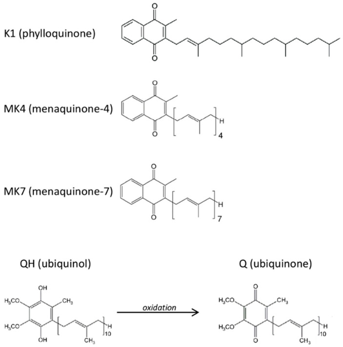
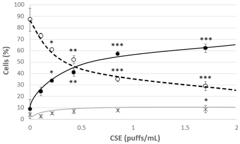
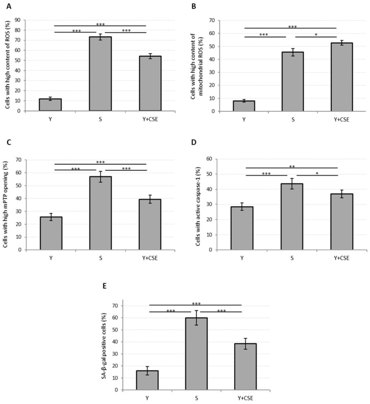
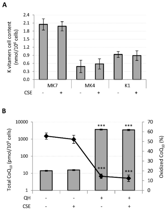
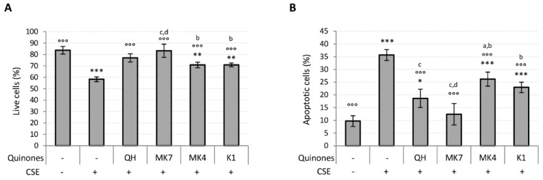
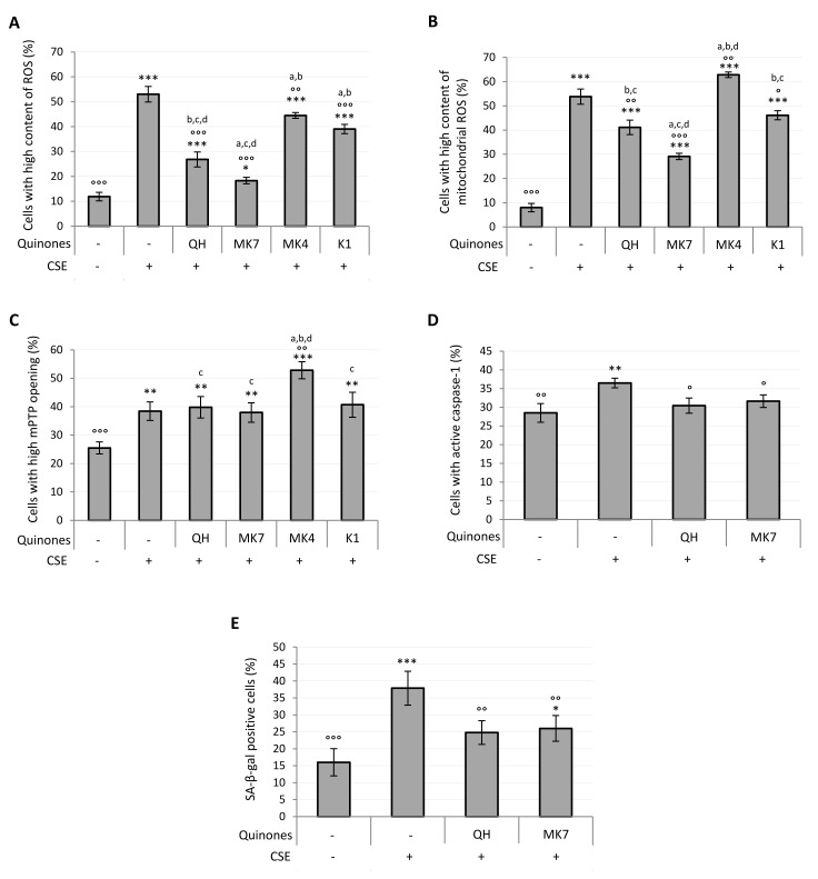
Endothelial dysfunction represents the initial stage in atherosclerotic lesion development which occurs physiologically during aging, but external factors like diet, sedentary lifestyle, smoking accelerate it. Since cigarette smoking promotes oxidative stress and cell damage, we developed an in vitro model of endothelial dysfunction using vascular cells exposed to chemicals present in cigarette smoke, to help elucidate the protective effects of anti-inflammatory and antioxidant agents, such as ubiquinol and vitamin K, that play a fundamental role in vascular health. Treatment of both young and senescent Human Umbilical Vein Endothelial Cells (HUVECs) for 24 h with cigarette smoke extract (CSE) decreased cellular viability, induced apoptosis via reactive oxygen species (ROS) imbalance and mitochondrial dysfunction and promoted an inflammatory response. Moreover, the senescence marker SA-β-galactosidase was observed in both young CSE-exposed and in senescent HUVECs suggesting that CSE exposure accelerates aging in endothelial cells. Supplementation with 10 µM ubiquinol and menaquinone-7 (MK7) counteracted oxidative stress and inflammation, resulting in improved viability, decreased apoptosis and reduced SA-β-galactosidase, but were ineffective against CSE-induced mitochondrial permeability transition pore opening. Other K vitamins tested like menaquinone-4 (MK4) and menaquinone-1 (K1) were less protective. In conclusion, CSE exposure was able to promote a stress-induced senescent phenotype in young endothelial cells likely contributing to endothelial dysfunction in vivo. Furthermore, the molecular changes encountered could be offset by ubiquinol and menaquinone-7 supplementation, the latter resulting the most bioactive K vitamin in counteracting CSE-induced damage.

Keywords: aging; cigarette smoke; endothelial dysfunction; menaquinone; mitochondrial dysfunction; oxidative stress; ubiquinol; vitamin K.


Conflict of interest statement

The authors declare no conflict of interest.

Figures

Figure 1
Figure 1
Molecular structures of the tested quinones.
Figure 2
Figure 2
Cell viability after 24 h treatment of young HUVECs with increasing doses of CSE. O Live cells, ● Apoptotic cells, X Dead cells. Significance was calculated with respect to young untreated cells (0 puffs/mL CSE). * p ≤ 0.05, ** p ≤ 0.01, *** p ≤ 0.001.
Figure 3
Figure 3
Oxidative stress, mitochondrial functionality, caspase-1 activation and senescence in young HUVECs exposed to CSE (0.425 puffs/mL for 24 h). The histograms depict the results obtained from untreated young (Y) and untreated senescent (S) HUVECs and young HUVECs exposed to CSE (Y + CSE), that show the percentage of cells with: (A) high ROS content, (B) high mitochondrial ROS content, (C) high mPTP opening, (D) active caspase-1, (E) SA-β-galactosidase positive cells. * p ≤ 0.05, ** p ≤ 0.01, *** p ≤ 0.001.
Figure 4
Figure 4
Effect of CSE exposure (0.425 puffs/mL for 24 h) on quinones bioavailability and CoQ10 oxidative status in young HUVECs. K vitamin cellular content (A) and CoQ10 cellular content and oxidative status (B), were evaluated in young HUVECs treated with 10 µM of quinones alone or in association with CSE exposure. + and − represent exposure or not exposure, respectively, to the indicated substances. Untreated and CSE exposed cells were used as controls. *** p ≤ 0.001 vs. untreated cells.
Figure 5
Figure 5
Effect of quinones supplementation on viability loss caused by CSE exposure (0.425 puffs/mL for 24 h) in young HUVECs. Cells were supplemented with 10 µM quinones for 24 h followed by replacement with CSE medium containing the same quinones and incubated for a further 24 h. (A) Percentage of live cells after treatment, (B) percentage of apoptotic cells after treatment. + and – represent exposure or not exposure, respectively, to the indicated substances. °°° p ≤ 0.001 vs. CSE-treated cells; * p ≤ 0.05, ** p ≤ 0.01, *** p ≤ 0.001 vs. untreated young HUVECs; a vs QH; b vs MK7; c vs MK4; d vs K1.
Figure 6
Figure 6
Effect of quinones supplementation on oxidative stress, mitochondrial functionality, caspase-1 activation and senescence in young HUVECs exposed to CSE (0.425 puffs/mL for 24 h). Cells were supplemented with 10 μM quinones followed by replacement with CSE medium containing the same quinones and incubated for a further 24 h (except in the case of SA-β-galactosidase activity where cells were incubated for 48 h). Percentage of cells with: (A) high ROS content, (B) high mitochondrial ROS content, (C) high mPTP opening, (D) active caspase-1, (E) SA-β-galactosidase positive cells. + and – represent exposure or not exposure, respectively, to the indicated substances. ° p ≤ 0.05, °° p ≤ 0.01, °°° p ≤ 0.001 vs. CSE-treated cells; * p ≤ 0.05, ** p ≤ 0.01, *** p ≤ 0.001 vs. untreated HUVECs; a vs QH; b vs MK7; c vs MK4; d vs K1.

Similar articles

Cited by

References

    1. Roth G.A., Johnson C., Abajobir A., Abd-Allah F., Abera S.F., Abyu G., Ahmed M., Aksut B., Alam T., Alam K., et al. Global, regional, and national burden of cardiovascular diseases for 10 causes, 1990 to 2015. J. Am. Coll. Cardiol. 2017;70:1–25. doi: 10.1016/j.jacc.2017.04.052. - DOI - PMC - PubMed
    1. Ezzati M., Obermeyer Z., Tzoulaki I., Mayosi B.M., Elliott P., Leon D.A. Contributions of risk factor trends and medical care to cardiovascular mortality trends. Nat. Rev. Cardiol. 2015;12:508–530. doi: 10.1038/nrcardio.2015.82. - DOI - PMC - PubMed
    1. Rodgman A., Perfetti T.A. The Chemical Components of Tobacco and Tobacco Smoke. 2nd ed. CRC Press; Boca Raton, FL, USA: 2013. pp. 1221–1297.
    1. Farhat N., Thorin-Trescases N., Voghel G., Villeneuve L., Mamarbachi M., Perrault L.P., Carrier M., Thorin E. Stress-induced senescence predominates in endothelial cells isolated from atherosclerotic chronic smokers. Can. J. Physiol. Pharmacol. 2008;86:761–769. doi: 10.1139/Y08-082. - DOI - PMC - PubMed
    1. Li D., Mehta J.L. Intracellular signaling of LOX-1 in endothelial cell apoptosis. Circ. Res. 2009;104:566–568. doi: 10.1161/CIRCRESAHA.109.194209. - DOI - PubMed

LinkOut - more resources