

Complete inhibition of ABCB1 and ABCG2 at the blood-brain barrier by co-infusion of erlotinib and tariquidar to improve brain delivery of the model ABCB1/ABCG2 substrate [11C]erlotinib
- PMID: 33081568
- PMCID: PMC8221757
- DOI: 10.1177/0271678X20965500
Complete inhibition of ABCB1 and ABCG2 at the blood-brain barrier by co-infusion of erlotinib and tariquidar to improve brain delivery of the model ABCB1/ABCG2 substrate [11C]erlotinib
Abstract
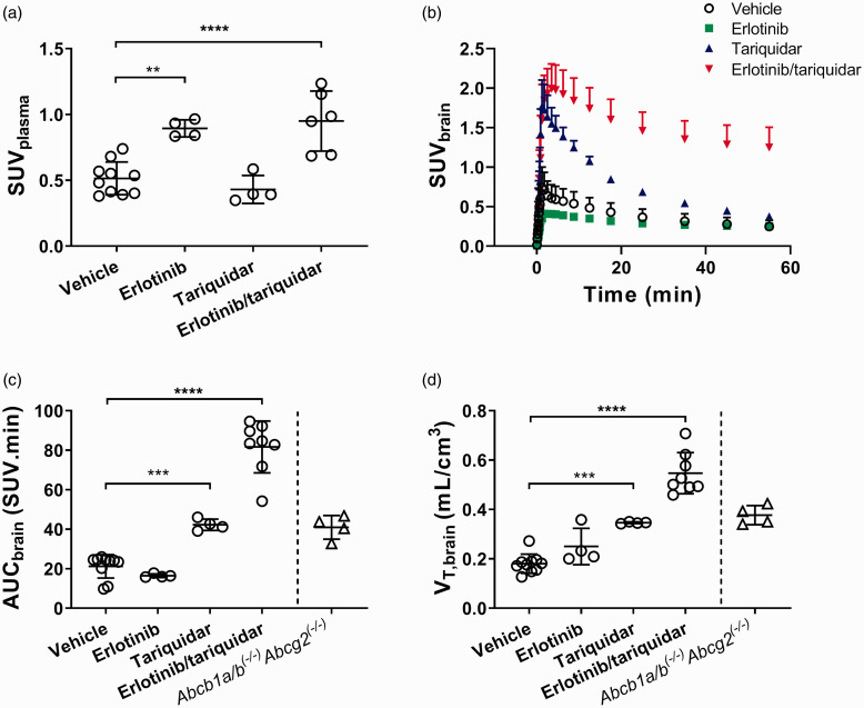
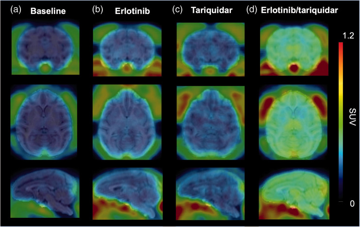
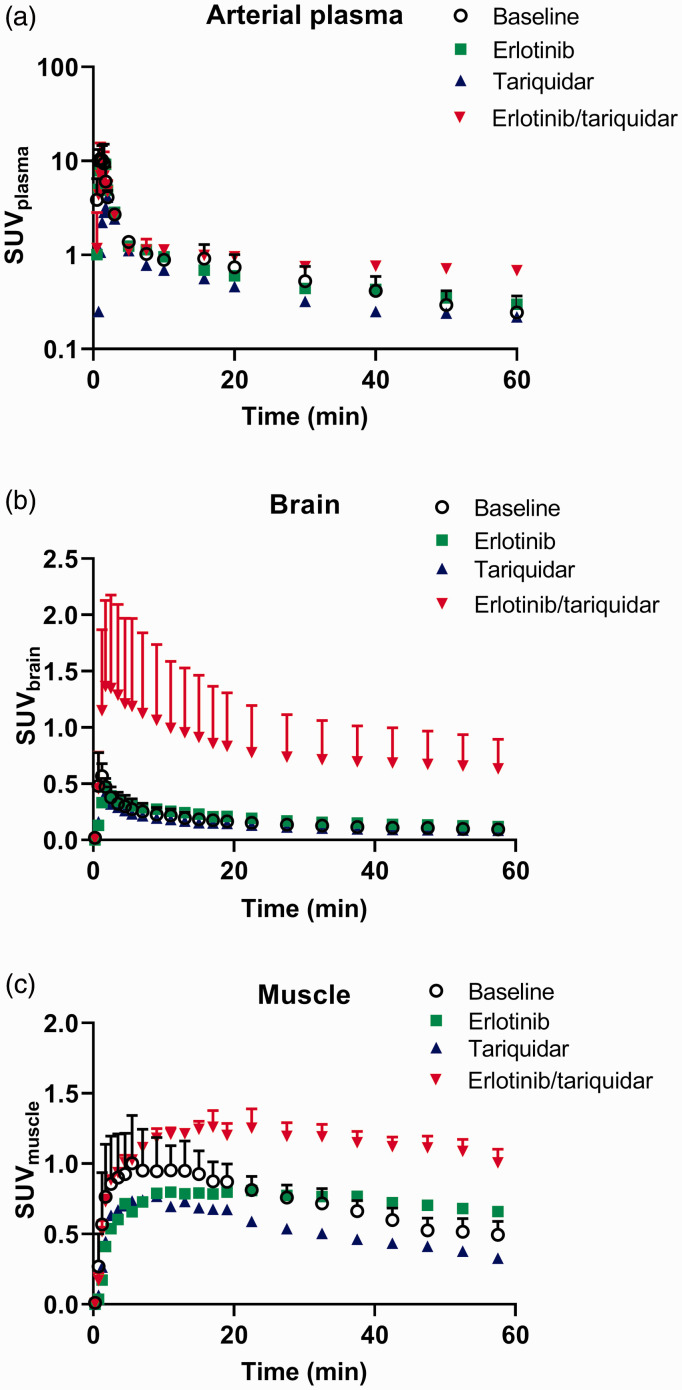
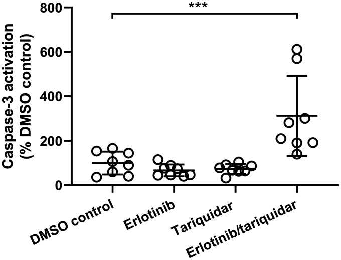
P-glycoprotein (ABCB1) and breast cancer resistance protein (ABCG2) restrict at the blood-brain barrier (BBB) the brain distribution of the majority of currently known molecularly targeted anticancer drugs. To improve brain delivery of dual ABCB1/ABCG2 substrates, both ABCB1 and ABCG2 need to be inhibited simultaneously at the BBB. We examined the feasibility of simultaneous ABCB1/ABCG2 inhibition with i.v. co-infusion of erlotinib and tariquidar by studying brain distribution of the model ABCB1/ABCG2 substrate [11C]erlotinib in mice and rhesus macaques with PET. Tolerability of the erlotinib/tariquidar combination was assessed in human embryonic stem cell-derived cerebral organoids. In mice and macaques, baseline brain distribution of [11C]erlotinib was low (brain distribution volume, VT,brain < 0.3 mL/cm3). Co-infusion of erlotinib and tariquidar increased VT,brain in mice by 3.0-fold and in macaques by 3.4- to 5.0-fold, while infusion of erlotinib alone or tariquidar alone led to less pronounced VT,brain increases in both species. Treatment of cerebral organoids with erlotinib/tariquidar led to an induction of Caspase-3-dependent apoptosis. Co-infusion of erlotinib/tariquidar may potentially allow for complete ABCB1/ABCG2 inhibition at the BBB, while simultaneously achieving brain-targeted EGFR inhibition. Our protocol may be applicable to enhance brain delivery of molecularly targeted anticancer drugs for a more effective treatment of brain tumors.
Keywords: Blood–brain barrier; P-glycoprotein; brain delivery; breast cancer resistance protein; transporter inhibition.
Figures
References
-
- Abbott NJ, Patabendige AA, Dolman DE, et al. Structure and function of the blood-brain barrier. Neurobiol Dis 2010; 37: 13–25. - PubMed
-
- Durmus S, Hendrikx JJ, Schinkel AH.Apical ABC transporters and cancer chemotherapeutic drug disposition. Adv Cancer Res 2015; 125: 1–41. - PubMed
-
- Kodaira H, Kusuhara H, Ushiki J, et al. Kinetic analysis of the cooperation of P-glycoprotein (P-gp/Abcb1) and breast cancer resistance protein (bcrp/Abcg2) in limiting the brain and testis penetration of erlotinib, flavopiridol, and mitoxantrone. J Pharmacol Exp Ther 2010; 333: 788–796. - PubMed
Publication types
MeSH terms
Substances
LinkOut - more resources
Full Text Sources
Research Materials
Miscellaneous
