. 2020 Oct;10(10):200197.
doi: 10.1098/rsob.200197. Epub 2020 Oct 21.

PGE2 upregulates gene expression of dual oxidase in a lepidopteran insect midgut via cAMP signalling pathway

Affiliations

PGE2 upregulates gene expression of dual oxidase in a lepidopteran insect midgut via cAMP signalling pathway

Seyedeh Minoo Sajjadian et al. Open Biol. 2020 Oct.

Abstract

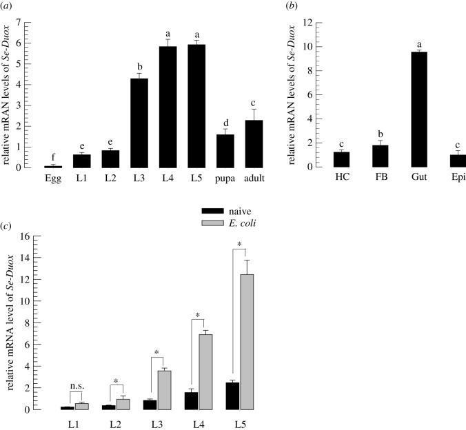
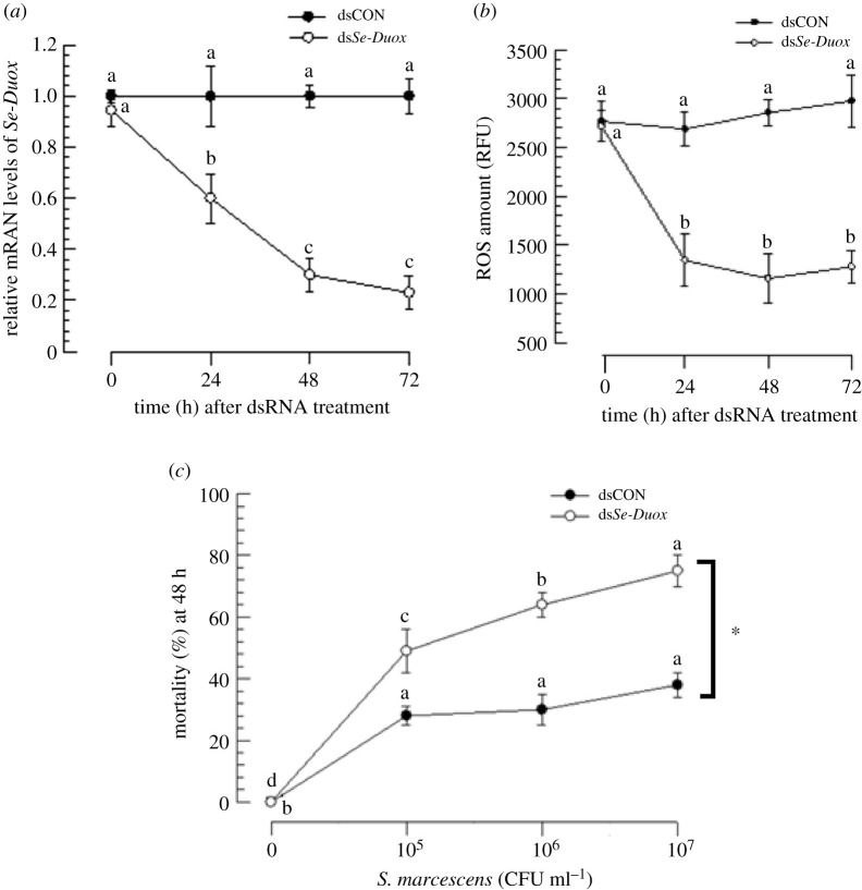
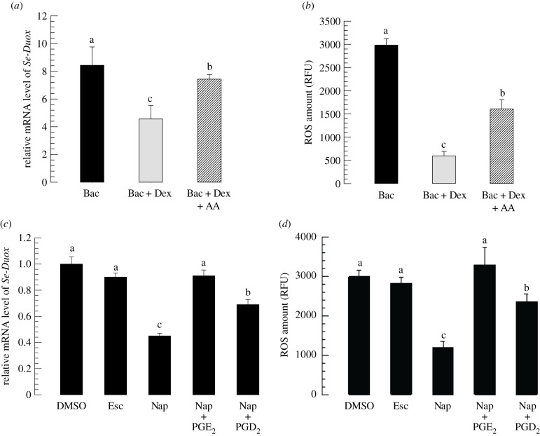
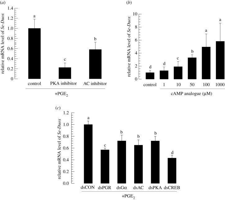
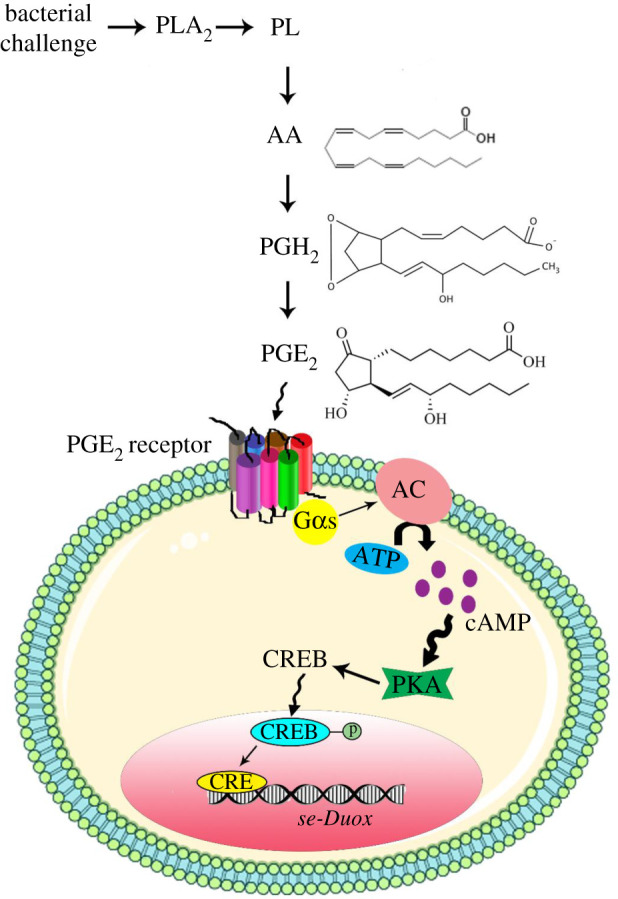
In insect midgut, prostaglandins (PGs) play a crucial role in defending bacterial and malarial pathogens. However, little is known about the PG signalling pathway in the midgut. A dual oxidase (Se-Duox) with presumed function of catalysing reactive oxygen species (ROS) production in the midgut was identified in beet armyworm, Spodoptera exigua. Se-Duox was expressed in all developmental stages, exhibiting relatively high expression levels in the midgut of late larval instars. Se-Duox expression was upregulated upon bacterial challenge. RNA interference (RNAi) of Se-Duox expression significantly suppressed ROS levels in the midgut lumen. The suppression of ROS levels increased insecticidal activity of Serratia marcescens after oral infection. Interestingly, treatment with a PLA2 inhibitor prevented the induction of Se-Duox expression in response to bacterial challenge. On the other hand, addition of its catalytic product rescued the induction of Se-Duox expression. Especially, PG synthesis inhibitor significantly suppressed Se-Duox expression, while the addition of PGE2 or PGD2 rescued the inhibition. Subsequent PG signals involved cAMP and downstream components because specific inhibitors of cAMP signal components such as adenylate cyclase (AC) and protein kinase A (PKA) significantly inhibited Se-Duox expression. Indeed, addition of a cAMP analogue stimulated Se-Duox expression in the midgut. Furthermore, individual RNAi specific to PGE2 receptor (a trimeric G-protein subunit), AC, PKA or cAMP-responsive element-binding protein resulted in suppression of Se-Duox expression. These results suggest that PGs can activate midgut immunity via cAMP signalling pathway by inducing Se-Duox expression along with increased ROS levels.

Keywords: Spodoptera exigua; cAMP; dual oxidase; gut immunity; prostaglandin; reactive oxygen species.


Conflict of interest statement

We declare we have no competing interests.

Figures

Figure 1.
Figure 1.
Molecular characterization of Se-Duox. (a) Functional domain analysis of Se-Duox. Domains were predicted using HMMER (https://www.ebi.ac.uk) and Pfam (http://pfam.xfam.org). Predicted domains include ‘Pox’ for peroxidase, ‘EF’ for calcium-binding EF hand, ‘Ferric-reduct’ for ferric chelate reductase, ‘FAD’ for FAD-binding domain and ‘NAD’ for NAD-binding domain. (b) Phylogenetic analysis of Se-Duox with other insect dual oxidases based on their amino acid sequences. The tree was generated by the neighbour-joining method using MEGA 6.0. Bootstrapping values were obtained with 1000 repetitions to support branch and clustering. Amino acid sequences were retrieved from GenBank. Accession numbers of genes are shown in electronic supplementary material, table S2.
Figure 2.
Figure 2.
Expression profile of Se-Duox. (a) Expression patterns of Se-Duox in different developmental stages: egg, first to fifth instar larvae (‘L1–L5’), pupa and adult. (b) Expression patterns of Se-Duox in indicated tissues of L5D2 larvae, including haemocyte (HC), fat body (FB), midgut (Gut) and epidermis (Epi). (c) Induction of Se-Duox expression in response to bacterial challenge. L1–L5 larvae were fed with E. coli (2 × 10⁴)-treated artificial diet for 8 h. Midguts were then dissected to study expression patterns of Se-Duox. A ribosomal gene, RL32, was used as a reference gene. Each treatment was replicated three times with independent tissue preparations. Different letters above standard deviation bars indicate significant differences among means at Type I error = 0.05 (LSD test).
Figure 3.
Figure 3.
RNAi of Se-Duox expression and subsequent influence on ROS level and larval susceptibility to S. marcescens. (a) Effects of RNAi on Se-Duox expression at different time points in midguts of S. exigua larvae (L5). One microgram of gene-specific dsRNA (dsSe-Duox) was injected into each larva. CpBV302, a viral gene, was used to generate control dsRNA (dsCON). (b) Inhibitory effects of RNAi specific to Se-Duox expression on total ROS levels in the midgut. To induce Duox expression and ROS, after dsRNA treatments, larvae were fed with E. coli (2 × 10⁴ cells) treated artificial diet for 12 h. (c) Effects of Se-Duox RNAi on pathogenicity of S. marcescens against L4 larvae of S. exigua. RNAi-treated larvae were exposed to different concentrations of S. marcescens. Mortality was recorded at 48 h post feeding. Each treatment was replicated three times. Each replication used 10 larvae. Different letters above standard deviation bars indicate significant difference among means in each treatment and control at Type I error = 0.05 (LSD test). Asterisk indicates the statistical difference between control and treatment at 107 CFU ml−1 dose.
Figure 4.
Figure 4.
Effects of PLA2 inhibitor and COX and LOX inhibitors on expression levels of Se-Duox. (a) Inhibitory effect of a PLA2 inhibitor, dexamethasone (DEX), on Se-Duox expression in L5 larvae. For bacterial challenge, larvae were fed with E. coli (2 × 10⁴ cells/larva) treated artificial diet for 12 h. (b) Effects of DEX on ROS levels in gut lumen. To induce ROS, E. coli (2 × 10⁴ cells)-treated artificial diet was fed to larvae at 8 h post-injection of DEX. At 12 h after bacterial treatment, intestinal ROS levels were measured. (c) Influence of naproxene (‘Nap’, a COX inhibitor) and esculetin (‘Esc’, a LOX inhibitor) on expression of Se-Duox. (d) Effects of Nap and Esc on ROS levels in gut lumen. Each inhibitor was injected into larvae at a final concentration of 10 µg per larva. At 8 h post-injection of inhibitor, E. coli (2 × 10⁴ cells)-treated artificial diet were fed to larvae to induce Se-Duox expression and ROS. To rescue Se-Duox expression and ROS production, PGD2 or PGE2 was injected at 1 µg/larva. Each treatment was replicated three times. Each replication used 15 larvae. Different letters indicate significant differences among means at Type I error = 0.05 (LSD test).
Figure 5.
Figure 5.
Identification of CRE and CREB associated with Se-Duox. (a) Promoter analysis of dual oxidase gene of S. exigua using PROMO and GPMiner programs. The upstream region from ATG start codon of Se-Duox was analysed for promoter components. (b) Functional domain analysis and phylogenetic analysis of Se-CREB with other lepidopteran CREBs based on their amino acid sequences. Domains were predicted using HMMER (https://www.ebi.ac.uk) and Pfam (http://pfam.xfam.org). Predicted domains include ‘pKID’ for phosphorylated kinase-inducible-domain and ‘bZIP’ for basic leucine zipper. The tree was generated by the neighbour-joining method using MEGA 6.0. Bootstrapping values were obtained with 1000 repetitions to support branch and clustering. Amino acid sequences were retrieved from GenBank. Accession numbers of genes are shown in electronic supplementary material, table S3. (c) Expression patterns in indicated tissues of L5D2 larvae, including haemocyte (HC), fat body (FB), midgut (Gut) and epidermis (Epi). A ribosomal gene, RL32, was used as a reference gene. Each treatment was replicated three times with independent tissue preparations. Different letters above standard deviation bars indicate significant differences among means at Type I error = 0.05 (LSD test).
Figure 6.
Figure 6.
Se-Duox expression is regulated by PKA signalling pathway. (a) Inhibitory effects of PKA and AC inhibitors on Se-Duox expression in fifth instar larvae (L5). (b) Dose-dependent effect of cAMP analogue on Se-Duox expression in L5. For bacterial challenge and induction of Duox expression, at 8 h post-injection of inhibitors or cAMP analogue, larvae were fed with E. coli (2 × 10⁴ cells)-treated artificial diet for 12 h. (c) Downregulatory effects of RNAi specific to PKA signalling pathway components on expression levels of Se-Duox. At 48 h after dsRNA injection, larvae were fed with E. coli (2 × 10⁴ cells)-treated artificial diet for 12 h to induce Duox expression. Each treatment was replicated three times. Each replication used five larvae. Different letters indicate significant differences among means at Type I error = 0.05 (LSD test).
Figure 7.
Figure 7.
A schematic of Se-Duox expression under the control of PKA signalling pathway. In response to bacterial challenge, PGE2 receptor on the cell surface is activated. AC can increase the amount of cAMP in the cytosol which induces PKA. Induced PKA causes CREB phosphorylation and subsequent translocation to the nucleus. CREB in the nucleus acts as a transcription factor and upregulates Se-Duox expression.

Similar articles

Cited by

References

    1. Wu K, Yang B, Huang W, Dobens L, Song H, Ling E. 2016. Gut immunity in lepidopteran insects. Dev. Comp. Immunol. 64, 65–74. (10.1016/j.dci.2016.02.010) - DOI - PubMed
    1. Terra WR, Dias RO, Oliveira PL, Ferreira C, Venancio TM. 2018. Transcriptomic analyses uncover emerging roles of mucins, lysosome/secretory addressing and detoxification pathways in insect midguts . Curr. Opin. Insect Sci. 29, 34–40. (10.1016/j.cois.2018.05.015) - DOI - PubMed
    1. Engel P, Moran NA. 2013. The gut microbiota of insects-diversity in structure and function. FEMS Microbiol. Rev. 37, 699–735. (10.1111/1574-6976.12025) - DOI - PubMed
    1. Lee KA, et al. 2018. Inflammation-modulated metabolic reprogramming is required for DUOX-dependent gut immunity in Drosophila. Cell Host Microbe 23, 338–352. (10.1016/j.chom.2018.01.011) - DOI - PubMed
    1. Lemaitre B, Miguel-Aliaga I. 2013. The digestive tract of Drosophila melanogaster. Annu. Rev. Genet. 47, 377–404. (10.1146/annurev-genet-111212-133343) - DOI - PubMed

Publication types

LinkOut - more resources