Review
. 2020 Oct;10(10):200279.
doi: 10.1098/rsob.200279. Epub 2020 Oct 21.

The structure and function of deubiquitinases: lessons from budding yeast

Affiliations
Review

The structure and function of deubiquitinases: lessons from budding yeast

Harsha Garadi Suresh et al. Open Biol. 2020 Oct.

Abstract

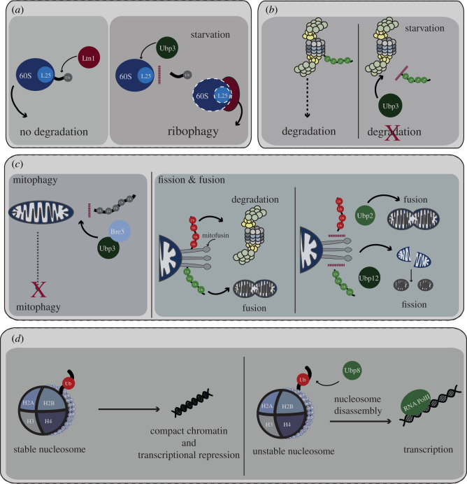
Protein ubiquitination is a key post-translational modification that regulates diverse cellular processes in eukaryotic cells. The specificity of ubiquitin (Ub) signalling for different bioprocesses and pathways is dictated by the large variety of mono-ubiquitination and polyubiquitination events, including many possible chain architectures. Deubiquitinases (DUBs) reverse or edit Ub signals with high sophistication and specificity, forming an integral arm of the Ub signalling machinery, thus impinging on fundamental cellular processes including DNA damage repair, gene expression, protein quality control and organellar integrity. In this review, we discuss the many layers of DUB function and regulation, with a focus on insights gained from budding yeast. Our review provides a framework to understand key aspects of DUB biology.

Keywords: deubiquitinases; protein degradation; ubiquitin signalling.


Conflict of interest statement

We declare we have no competing interests.

Figures

Figure 1.
Figure 1.
The yeast DUBs. Shown are schematics of the DUB proteins in S. cerevisiae categorized by family, illustrating key functional domains (legend at base of the figure). Any human homologues are noted to the right of the protein diagrams. The size of the DUB proteins is indicated on the left of the schematics and domain sizes are indicated according to scale. ‘aa’ refers to amino acids.
Figure 2.
Figure 2.
Mechanisms of substrate recognition by DUBs. (a) Overview of DUB recognition of Ub chains. A generic substrate ‘X’ with an attached Ub chain is illustrated on the left, with the proximal Ub being substrate bound, while the distal Ub is unmodified. The cartoons in the middle of the panel show that DUBs can recognize their substrates either through interaction with a specific target protein that they deubiquitinate or through interactions with the Ub chain itself. Schematics on the right illustrate endo or exo-cleavage activity associated with DUBs (the exo activity can originate at the distal or proximal end of a Ub chain). (b) Types of Ub chain linkages. The various types of Ub chain linkages are illustrated. The M1 and K63 chains, which adopt open conformations, and K6, K11, K27, K29, K33 and K48-linked chains, which adopt closed conformations, are grouped together. Mono-ubiquitination and branched Ub chains may also occur. (ch) DUB mechanisms for substrate recognition. (c) Ub–S1 site interaction. DUBs that show little or no specificity for Ub chain linkages, such as Rpn11 and several USP/Ubp DUBs, contact Ub through an S1 site only. (d) Ub–S1–S1′ interactions. DUBs may contact Ub through both the S1 site and an S1′ site which can impart additional linkage specificity. For example, the RPN11-related DUB AMSH-LP achieves linkage specificity for K11-linked Ub chains through contacts made by its S1′ site. (e) S1′ site interaction with target. DUBs such as Ubp8 contain an S1′ site that recognizes a target protein, an interaction that does not involve Ub. (f) Ub–S1–S1′–S2 interactions. OTUD2 is an example of a DUB with an S2 domain in its catalytic site, which can accommodate longer K11-linked Ub chains. (g) DUB–Ub interactions outside of catalytic domain. Some DUBs such as MINDY-1 contain Ub-binding sites outside of their catalytic domain. The diagram illustrates the case of MINDY-2, in which tandem MIU (tMIU) domains work together to mediate the recognition of longer K48-linked Ub chains. (h) Multiple Ub-binding domains. IsoT achieves substrate specificity for K48-linked chains through several interactions with Ub chains using its four Ub-binding domains: ZnF-UBP, USP/UBP domain and two UBA domains, which are inserted into its catalytic domain.
Figure 3.
Figure 3.
Functions for DUBs in protein turnover and Ub homeostasis. (a,d) DUB roles in Ub homeostasis. The diagrams show the roles for the DUBs Doa4 (d), Ubp6 and Rpn11 (a) in ubiquitin homeostasis. Doa4 functions to recycle Ub from ubiquitinated substrates that are targeted to the vacuole. Ubp6 and Rpn11 are both associated with the proteasome and remove Ub from proteins targeted for proteasomal degradation. The inset panel illustrates upregulation of Ubp6 and Doa4 to increase Ub recycling during periods of stress. An inhibitor of Doa4, Rfu1, is downregulated in response to stress, enhancing the activity of Doa4. (b) DUBs in proteasome assembly. The key role for Ubp6 in proteasome assembly is illustrated. During proteasome assembly, components of the proteasome (particularly the RP with several Ub-binding domains/motifs) can interact with ubiquitinated substrates, hindering the formation of the complete proteasome. Ubp6 functions to remove Ub from substrates during the assembly process, facilitating maturation of the proteasome. (c) DUB roles in endocytosis and cargo turnover. The diagram illustrates the function of the ART complex and Rsp5 Ub ligase in ubiquitination of defective membrane proteins, (cargo) targeting them for degradation in the vacuole. The Ubp2 and Ubp15 DUBs positively regulate ART/Rsp5 by deubiquitinating the complex, thus contributing to cargo turnover and the maintenance of plasma membrane integrity.
Figure 4.
Figure 4.
DUB functions in protein quality control. (a) DUB roles in regulation of misfolded proteins. The diagram illustrates targeting of misfolded proteins to the proteasome by the DUBs Ubp2 and Ubp3. Misfolded proteins are ubiquitinated by the Ydj1–Rsp5 complex, resulting in the addition of K63-linked Ub chains. As proteasomal targeting requires K48-linked Ub chains, Ubp2/Ubp3 and an unknown E3 ligase function to remove these K63-linked chains and remodel to K48-linked chains. The misfolded proteins are then degraded by the proteasome. (b) Role for DUBs in ER-associated protein homeostasis. The left panel illustrates the interplay of the ER-associated E3 ligase Doa10 and DUB Ubp1 in the pre-insertional ER associated degradation (prERAD) response. By recognizing C-terminal hydrophobic motifs (blue circle on peptide), prERAD tags pre-inserted proteins for degradation that have remained on the cytosolic leaflet of the ER for too long. If proteins that have a GPI-anchor sequence fail to translocate to the ER in a timely manner, they are ubiquitinated by the E3 ligase Doa10. Molecular chaperones (brown crescents) aid in both translocation of proteins into the ER and targeting of non-inserted GPI-anchored proteins to Doa10-mediated ubiquitination and subsequent degradation. Ubp1-mediated deubiquitination allows these ubiquitinated substrates to avoid immediate degradation and as such promotes translocation into the ER. However, if their translocation remains unsuccessful then Doa10-mediated degradation dominates to degrade the prERAD substrates. Thus, Ubp1 acts as a ‘molecular timer’ deciding the fate of prERAD substrates. The right panel diagrams the function of the Otu1 DUB in ER-associated degradation (ERAD) of substrates that are polyubiquitinated by Hrd1. The Cdc48 ATPase (black shape) is recruited to the ER membrane and uses ATP hydrolysis to pull the polypeptide substrate out of the membrane. The complex of Cdc48 ATPase and substrate leaves the membrane, and the DUB Otu1 trims the Ub chain, allowing release of the substrate from Cdc48, and its subsequent degradation by the proteasome.
Figure 5.
Figure 5.
DUB roles in the degradation of protein complexes. (a) Ribophagy. Regulation of ribophagy by the Ubp3 DUB under conditions of starvation is illustrated. Ribophagy involves ubiquitination of the 60S ribosomal protein subunit Rpl25 (L25) by the E3 ligase Ltn1. Ubiquitination acts as a signal to protect the ribosome from degradation (left panel). Under conditions of starvation, Ubp3 deubiquitinates the Rpl25, promoting degradation of the ribosome (illustrated by dashed lines). (b) Proteaphagy. Ubp3 also plays a role in selective autophagy of the RP (blue) and CP (grey) subunits of the proteasome, a process called proteaphagy. Under conditions of starvation, Ubp3 deubiquitinates the 20S core of the proteasome, preventing its degradation. (c) Mitochondrial homeostasis. Regulation of mitochondrial homeostasis by the Ubp3, Ubp2 and Ubp12 DUBs is illustrated. The left panel illustrates deubiquitination of mitochondrial proteins by Ubp3 in complex with cofactor Bre5, preventing autophagic degradation of the mitochondria or mitophagy. The right panel diagrams the role of DUBs Ubp2 and Ubp12 in mitochondrial fission and fusion. Mitochondrial fusion is mediated by mitofusin, Fzo1 (grey stalks), which is ubiquitinated at two locations by distinct E3 ligases. Mdm30 ubiquitinates and stabilizes mitofusion (green Ub chain) thus promoting mitochondrial fusion. Ubp12 can remove these activating Ub chains and promote mitochondrial fragmentation. An unknown E3 ligase can ubiquitinate mitofusion at a distinct site (red Ub chain), and this modification can promote mitofusin degradation. Ubp2 can remove this destabilizing Ub chain and thus promote mitochondrial fusion. (d) Transcriptional regulation. Shown is a model for nucleosome stability in yeast regulated by the DUB Ubp8. The nucleosome on the left carries a Ub modification on its histone H2B subunit, which prevents eviction of H2A–H2B, stabilizing the nucleosome and repressing transcription. The nucleosome on the right is rendered unstable owing to the removal of Ub by Ubp8, leading to its disassembly by histone chaperones and/or other regulatory complexes, and activation of transcription by RNAPII (RNA PolII).

Similar articles

Cited by

References

    1. Komander D. 2009. The emerging complexity of protein ubiquitination. Biochem. Soc. Trans. 37(Pt 5), 937–953. (10.1042/BST0370937) - DOI - PubMed
    1. Capili AD, Lima CD. 2007. Taking it step by step: mechanistic insights from structural studies of ubiquitin/ubiquitin-like protein modification pathways. Curr. Opin. Struct. Biol. 17, 726–735. (10.1016/j.sbi.2007.08.018) - DOI - PMC - PubMed
    1. Komander D, Clague MJ, Urbe S. 2009. Breaking the chains: structure and function of the deubiquitinases. Nat. Rev. Mol. Cell Biol. 10, 550–563. (10.1038/nrm2731) - DOI - PubMed
    1. Li W, Ye Y. 2008. Polyubiquitin chains: functions, structures, and mechanisms. Cell Mol. Life Sci. 65, 2397–2406. (10.1007/s00018-008-8090-6) - DOI - PMC - PubMed
    1. Nijman SM, Luna-Vargas MPA, Velds A, Brummelkamp TR, Dirac AMG, Sixma TK, Bernards R. et al. 2005. A genomic and functional inventory of deubiquitinating enzymes. Cell 123, 773–786. (10.1016/j.cell.2005.11.007) - DOI - PubMed

Publication types

MeSH terms

Grants and funding

LinkOut - more resources