. 2020 Oct 28;287(1937):20202055.
doi: 10.1098/rspb.2020.2055. Epub 2020 Oct 21.

Krüppel-like factor/specificity protein evolution in the Spiralia and the implications for cephalopod visual system novelties

Affiliations

Krüppel-like factor/specificity protein evolution in the Spiralia and the implications for cephalopod visual system novelties

Kyle J McCulloch et al. Proc Biol Sci. .

Abstract

The cephalopod visual system is an exquisite example of convergence in biological complexity. However, we have little understanding of the genetic and molecular mechanisms underpinning its elaboration. The generation of new genetic material is considered a significant contributor to the evolution of biological novelty. We sought to understand if this mechanism may be contributing to cephalopod-specific visual system novelties. Specifically, we identified duplications in the Krüppel-like factor/specificity protein (KLF/SP) sub-family of C2H2 zinc-finger transcription factors in the squid Doryteuthis pealeii. We cloned and analysed gene expression of the KLF/SP family, including two paralogs of the DpSP6-9 gene. These duplicates showed overlapping expression domains but one paralog showed unique expression in the developing squid lens, suggesting a neofunctionalization of DpSP6-9a. To better understand this neofunctionalization, we performed a thorough phylogenetic analysis of SP6-9 orthologues in the Spiralia. We find multiple duplications and losses of the SP6-9 gene throughout spiralian lineages and at least one cephalopod-specific duplication. This work supports the hypothesis that gene duplication and neofunctionalization contribute to novel traits like the cephalopod image-forming eye and to the diversity found within Spiralia.

Keywords: Spiralia; cephalopod; eye evolution; lens; neofunctionalization; transcription factor.


Conflict of interest statement

We declare we have no competing interests.

Figures

Figure 1.
Figure 1.
Domain architecture, expression, and phylogeny of KLF/SP family members (a) KLF/SP proteins are defined by a conserved triple 2-cysteine 2-histidine (C2H2) DNA-binding domain. The Buttonhead box (Btd) distinguishes SP transcription factors from KLFs. (b) Normalized expression of KLF/SP family in the developing eye and optic lobe in D. pealeii, stages 19, 21, 23, 25, and 27 in biological triplicate (original dataset from [6]). DpSP6-9a and DpSP6-9b are highly expressed throughout development. (c) Bayesian inference of KLF/SP phylogeny (MrBayes). Many deeper nodes are poorly supported, making it difficult to draw firm conclusions about the relationships of many KLF/SP subgroups. Circles represent posterior probabilities above 0.5 (white), 0.7 (grey), and 0.95 (black) on branches leading to the nodes of major labelled and coloured subclades. Subgroup naming conventions from previous phylogenetic studies have been used. Red asterisks mark D. pealeii sequences in the tree.
Figure 2.
Figure 2.
DpSP6-9 paralog expression during development. (a) D. pealeii embryo schematics, anterior and posterior views. After stage 23, most organs remain recognizable in similar locations on the embryo. In situ hybridization of DpSP6-9a mRNA (b–f’) and DpSP6-9b mRNA (g–k’), stages 19 to 27. Expression is nearly identical for both genes in the nervous system, gills, arms, and anterior chamber organ. DpSP6-9a is highly expressed around the lens (black arrowheads), the cerebral ganglion (white arrowheads), and the palliovisceral ganglion (cyan arrowheads). DpSP6-9b expression domain is similar but lower in these corresponding areas (arrowheads), and differentially highly expressed in the mantle (yellow asterisks). Scale bars, 200 µm; ac organ, anterior chamber organ.
Figure 3.
Figure 3.
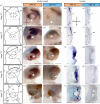
DpSP6-9a but not DpSP6-9b is highly expressed in lentigenic cells of the developing eye. (a) Schematics showing lateral views of the developing eye, anterior is left. At stage 19, the retina placode is exposed and being internalized by a surrounding lip of cells (arrows). The vesicle is closed at stage 21 and the lip cells generate the lens and anterior segment. ol, optic lobe; lip, lip of cells that internalizes the retinal placode; l, lens, r, retina; ac, anterior chamber organ; a, arms. (b–u) Lateral view and cryosections of DpSP6-9a and DpSP6-9b. White arrowheads highlight the region of lip and developing lentigenic cells. In sections, anterior of the animal is up, medial is left. Expression in the retinal placode can be seen for both genes, r, retina. At stage 19, DpSP6-9a is highly expressed on both sides of the lip as it closes (arrows), where DpSP6-9b is restricted medially (b–e). At stage 21, the anterior of the eye vesicle expresses DpSP6-9a with minimal expression of DpSp6-9b r, retina; as, anterior segment (f–i). Later DpSP6-9a is strongly expressed in the lentigenic cells. Again DpSP6-9b is reduced (j–u). Asterisk, panel (t) is a stage 28 embryo; scale bars, 200 µm.
Figure 4.
Figure 4.
Synteny and cephalopod-specific phylogeny reveal SP6-9 evolutionary history. (a) Bayesian tree shows support for SP6-9c as the outgroup to SP6-9a and b in Cephalopoda. Branch lengths show divergence in SP6-9b and c while little sequence change has occurred within the SP6-9a clade across cephalopods. Circles represent posterior probabilities above 0.5 (white), 0.7 (grey), and 0.95 (black). (b) Mapping SP6-9 paralog genomic locations shows cephalopod genomic architecture is distinct from other spiralians. Cephalopod SP6-9a genes have introns while SP6-9b and SP6-9c do not. The four paralogs in Crassostrea virginica may represent a recent genomic duplication or genome assembly error. Genomic backbones in black are all to the same scale, and zoomed-in gene lengths are all to scale with one another, except where breaks are indicated with double vertical bars. Taxon colours correspond to colours used in the trees in figure 5 and electronic supplementary material figure S3.
Figure 5.
Figure 5.
SP6-9 has a complex pattern of duplication across Spiralia and Cephalopoda. (a) The most parsimonious pattern of duplication events mapped onto a current spiralian phylogeny. At minimum, separate duplications occurred in Rotifera, Platyhelminthes, Annelida, the branch leading to Phoronida & Bryozoa, and Conchifera. Annelida and Mollusca subtrees are collapsed and detailed in (b) and (c). (b) Annelida: amino acid tree evidence indicates two duplications within Sedentaria after the split of Orbiniidae and Sabellidae, followed by a loss and subsequent duplication event in Clitellata. Basally branching annelids have a single paralog only, suggesting a single paralog at the base of Annelida. (c) Mollusca: this hypothesis suggests a duplication event following the split of Aculifera. At minimum, there was a duplication in Cephalopoda, a loss in Protobranchia and a loss and duplication in Hypsogastropoda. Phylogenies based on [–61]. In all panels: star, gene duplication; X, gene loss; images from Phylopic.org, or drawn by K. McCulloch, with credit for Capitella and Platynereis B. Duygu Özpolat, and credit for abalone and owl limpet to Taro Maeda.

Similar articles

Cited by

References

    1. Ohno S. 1970. Evolution by gene duplication. Berlin, Germany: Springer.
    1. Taylor JS, Raes J. 2004. Duplication and divergence: the evolution of new genes and old ideas. Annu. Rev. Genet. 38, 615–643. (10.1146/annurev.genet.38.072902.092831) - DOI - PubMed
    1. Lynch M, Conery JS. 2000. The evolutionary fate and consequences of duplicate genes. Science 290, 1151–1155. (10.1126/science.290.5494.1151) - DOI - PubMed
    1. Ritschard EA, Whitelaw B, Albertin CB, Cooke IR, Strugnell JM, Simakov O. 2019. Coupled genomic evolutionary histories as signatures of organismal innovations in cephalopods. Bioessays 41, 1900073 (10.1002/bies.201900073) - DOI - PubMed
    1. da Fonseca RR, et al. . 2020. A draft genome sequence of the elusive giant squid, Architeuthis dux. Gigascience 9, giz152 (10.1093/gigascience/giz152) - DOI - PMC - PubMed

Publication types

Substances

LinkOut - more resources