. 2020 Oct 20;18(1):145.
doi: 10.1186/s12915-020-00884-3.

Bioengineered human skeletal muscle capable of functional regeneration

Affiliations

Bioengineered human skeletal muscle capable of functional regeneration

J W Fleming et al. BMC Biol. .

Abstract

Background: Skeletal muscle (SkM) regenerates following injury, replacing damaged tissue with high fidelity. However, in serious injuries, non-regenerative defects leave patients with loss of function, increased re-injury risk and often chronic pain. Progress in treating these non-regenerative defects has been slow, with advances only occurring where a comprehensive understanding of regeneration has been gained. Tissue engineering has allowed the development of bioengineered models of SkM which regenerate following injury to support research in regenerative physiology. To date, however, no studies have utilised human myogenic precursor cells (hMPCs) to closely mimic functional human regenerative physiology.

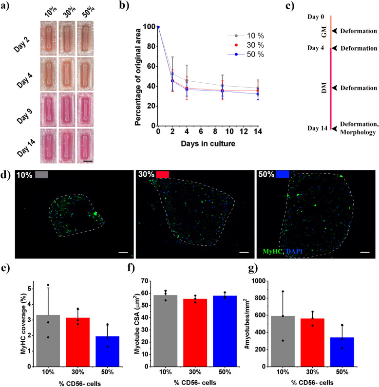
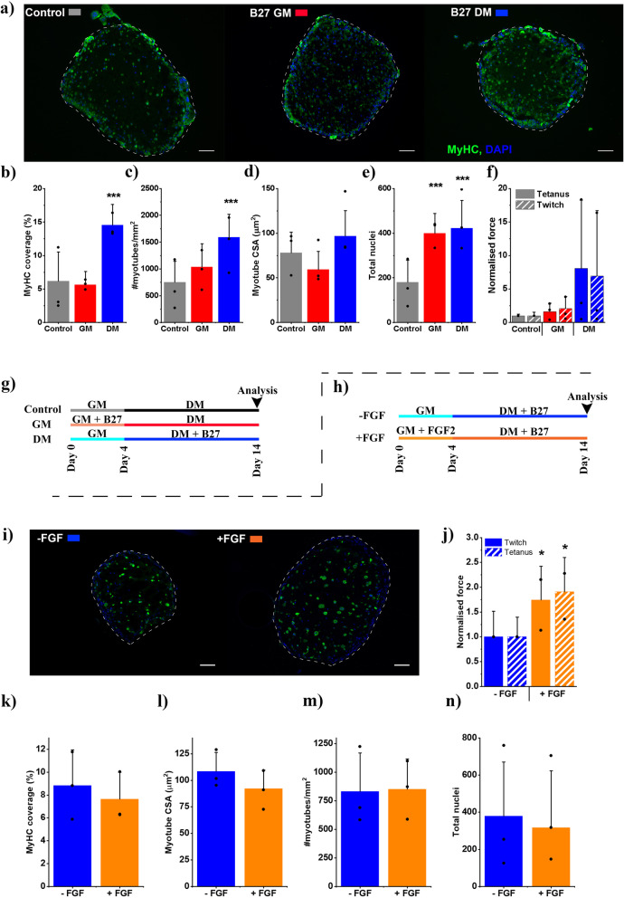
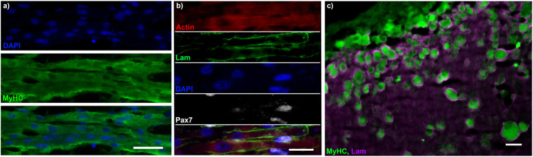
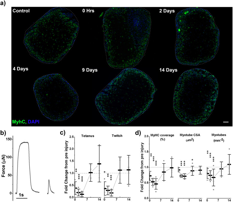
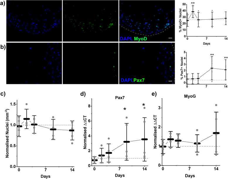
Results: Here we address some of the difficulties associated with cell number and hMPC mitogenicity using magnetic association cell sorting (MACS), for the marker CD56, and media supplementation with fibroblast growth factor 2 (FGF-2) and B-27 supplement. Cell sorting allowed extended expansion of myogenic cells and supplementation was shown to improve myogenesis within engineered tissues and force generation at maturity. In addition, these engineered human SkM regenerated following barium chloride (BaCl2) injury. Following injury, reductions in function (87.5%) and myotube number (33.3%) were observed, followed by a proliferative phase with increased MyoD+ cells and a subsequent recovery of function and myotube number. An expansion of the Pax7+ cell population was observed across recovery suggesting an ability to generate Pax7+ cells within the tissue, similar to the self-renewal of satellite cells seen in vivo.

Conclusions: This work outlines an engineered human SkM capable of functional regeneration following injury, built upon an open source system adding to the pre-clinical testing toolbox to improve the understanding of basic regenerative physiology.

Keywords: Regeneration; Satellite cell; Skeletal muscle; Tissue engineering.


Conflict of interest statement

The authors declare that they have no competing interests.

Figures

Fig. 1
Fig. 1
Remixing of CD56-sorted populations leads to robust engineered muscles. Throughout percentages refer to the percentage of CD56- cells remixed to create the human cell population. Black in all graphs denotes 10%, red 30% and blue 50% CD56− .a Photographs of engineered muscles across time showing deformation. Scale bar – 5 mm. b Deformation over time. c Experimental scheme showing time points of analysis. d Representative micrographs stained for MyHC – green and Nuclei (DAPI) – blue. Scale bar 100 μm. e–g Graphs displaying; MyHC percentage coverage, myotube cross-sectional area (CSA) and myotubes per mm2. All graphs display mean ± S. D, individual repeat means are displayed as points. No statistically significant comparisons were identified, n = 9 samples across 3 repeats
Fig. 2
Fig. 2
Media supplementation of remixed engineered muscles increases morphological maturity and increases functional capacity. a Representative micrographs stained for myosin heavy chain (MyHC) – green and Nuclei (DAPI) – blue. Scale bar 100 μm. b–f Graphs displaying; percentage MyHC coverage, myotubes per mm2, myotube cross-sectional area (CSA), nuclei per section and normalised force (normalised to control), respectively. Mean tetanus force at control 45.9μN, 0.96 kPa; twitch force 22.7μN, 0.048 kPa. All graphs display mean ± S. D. g, h Experimental schemes showing conditions coloured similarly to graphs. Scheme (g) applies to af whilst scheme (h) applies to in. i Representative micrographs stained for myosin heavy chain (MyHC) – green and Nuclei (DAPI) – blue. Scale bar 100 μm. j Normalised force (normalised to -FGF condition) mean tetanic force for -FGF condition 14.3μN, 0.10 kPa; twitch 6.2μN, 0.35 kPa. kn Graphs displaying; Percentage MyHC coverage, myotubes per mm2, myotube cross-sectional area (CSA), nuclei per section respectively. All graphs display mean ± S. D, individual repeat means are displayed as dots. Statistical significance from control (af) and -FGF (in) is denoted as *p ≤ 0.05, ***p ≤ 0.001, n = 9 samples across 3 repeats
Fig. 3
Fig. 3
Human engineered muscles display laminin organisation and Pax7+ nuclei. a Micrograph of longitudinal cryosection of engineered muscle. Stained for nuclei (DAPI, blue) and myotubes (MyHC, green). b Micrograph of longitudinal cryosection of engineered muscle. Stained for Actin (red), Laminin (green), DNA (DAPI, blue) and Pax7 (white). c Cross-section of engineered muscle imaged on the extreme periphery of the section. MyhC (green) and Laminin (magenta). All scale bars represent 25 μm
Fig. 4
Fig. 4
Human engineered muscles regenerate functionally and morphologically following injury. a Representative micrographs of engineered muscle cross-sections. Stained for MyHC (green) and Nuclei (DAPI, blue). Scale bar represents 100 μm. b Representative force traces used for tetanus and twitch force measurements. c Normalised force measurements across recovery. Means at control; tetanus – 101.6 μN, 0.19 kPa; twitch – 36.6μN, 0.072 kPa. d Normalised morphological measures across recovery. Means at control; percentage MyHC coverage – 13.0%, myotube cross-sectional area (CSA) – 132.6 μm2, myotubes per mm2 –971.6. bd All graphs display control normalised means ± S. D, individual repeat means are displayed as points. Dashed line represents level at control, normalised to 1, on all graphs. Statistical significance from control is denoted as *p ≤ 0.05, **p ≤ 0.01 and ***p ≤ 0.001, n = 15 samples across 5 repeats
Fig. 5
Fig. 5
Dynamics of cell populations following injury. a Representative images showing staining left to right, Nuclei (DAPI, blue), MyoD (green) and overlay image. Scale bar represents 25 μm. Graph displays percentage of MyoD+ nuclei across recovery. b Representative images showing staining left to right, Nuclei (DAPI, blue), Pax7 (green) and overlay image. Scale bar represents 25 μm. Graph displays percentage of Pax7+ nuclei across recovery. c Normalised nuclei per mm2 across recovery, mean value at control – 1471 mm− 2. d RT-PCR data displayed as ΔΔCT values for myogenic genes Pax7 and MyoG across recovery. ae All graphs display means ± S. D, individual repeat means are displayed as points. Dashed line represents level at control on all graphs. Statistical significance from control is denoted as *p ≤ 0.05, **p ≤ 0.01 and ***p ≤ 0.001, n = 15 samples across 5 repeats

Similar articles

Cited by

References

    1. Järvinen TA, Järvinen M, Kalimo H. Regeneration of injured skeletal muscle after the injury. Muscles Ligaments Tendons J. 2013;3:337–345. doi: 10.32098/mltj.04.2013.16. - DOI - PMC - PubMed
    1. Mauro A. Satellite cell of skeletal muscle fibers. J Biophys Biochem Cytol. 1961;9:493–495. doi: 10.1083/jcb.9.2.493. - DOI - PMC - PubMed
    1. Lepper C, Partridge TA, Fan CM. An absolute requirement for pax7-positive satellite cells in acute injury-induced skeletal muscle regeneration. Development. 2011;138:3639–3646. doi: 10.1242/dev.067595. - DOI - PMC - PubMed
    1. Hindi SM, Kumar A. TRAF6 regulates satellite stem cell self-renewal and function during regenerative myogenesis. J Clin Invest. 2016;126:151–168. doi: 10.1172/JCI81655. - DOI - PMC - PubMed
    1. Seale P, Sabourin LA, Girgis-Gabardo A, Mansouri A, Gruss P, Rudnicki MA. Pax7 is required for the specification of myogenic satellite cells. Cell. 2000;102:777–786. doi: 10.1016/S0092-8674(00)00066-0. - DOI - PubMed

Publication types