. 2020 Oct 20;53(1):48.
doi: 10.1186/s40659-020-00316-0.

Daphnetin inhibits proliferation and inflammatory response in human HaCaT keratinocytes and ameliorates imiquimod-induced psoriasis-like skin lesion in mice

Affiliations

Daphnetin inhibits proliferation and inflammatory response in human HaCaT keratinocytes and ameliorates imiquimod-induced psoriasis-like skin lesion in mice

Jintao Gao et al. Biol Res. .

Abstract

Background: Psoriasis is a common chronic inflammatory skin disease. Keratinocytes hyperproliferation and excessive inflammatory response contribute to psoriasis pathogenesis. The agents able to attenuate keratinocytes hyperproliferation and excessive inflammatory response are considered to be potentially useful for psoriasis treatment. Daphnetin exhibits broad bioactivities including anti-proliferation and anti-inflammatory. This study aims to evaluate the anti-psoriatic potential of daphnetin in vitro and in vivo, and explore underlying mechanisms.

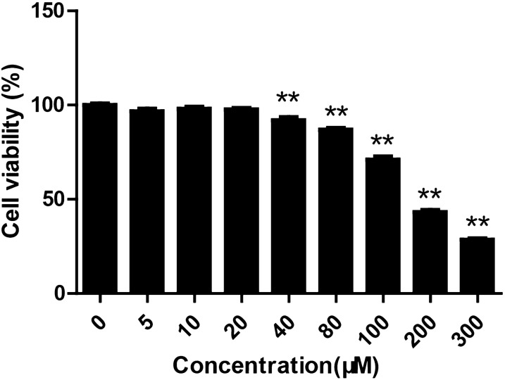
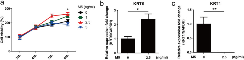
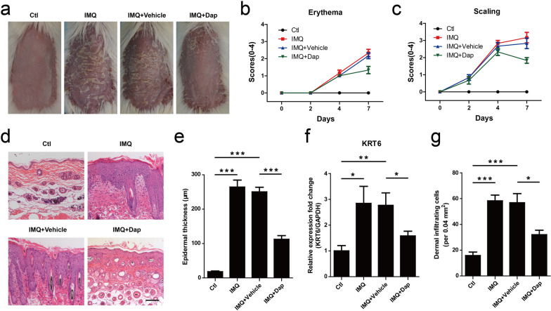
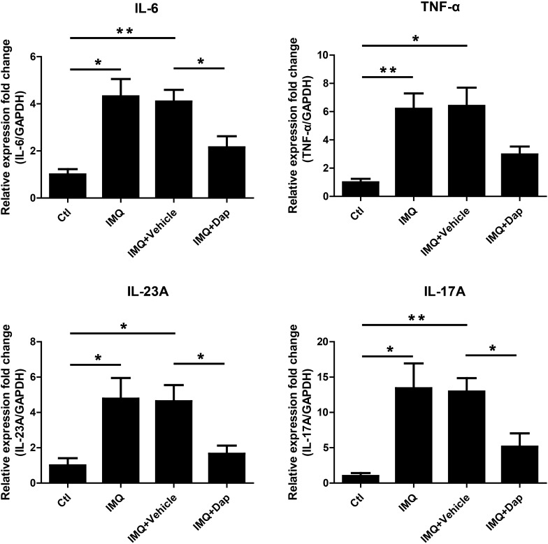
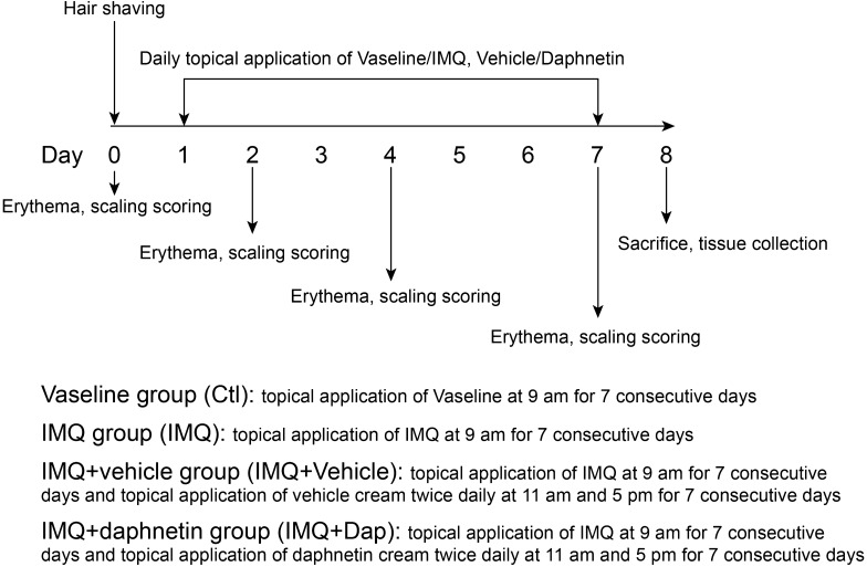
Methods: HaCaT keratinocytes was stimulated with the mixture of IL-17A, IL-22, oncostatin M, IL-1α, and TNF-α (M5) to establish psoriatic keratinocyte model in vitro. Cell viability was measured using Cell Counting Kit-8 (CCK-8). Quantitative Real-Time PCR (qRT-PCR) was performed to measure the mRNA levels of hyperproliferative marker gene keratin 6 (KRT6), differentiation marker gene keratin 1 (KRT1) and inflammatory factors IL-1β, IL-6, IL-8, TNF-α, IL-23A and MCP-1. Western blotting was used to detect the protein levels of p65 and p-p65. Indirect immunofluorescence assay (IFA) was carried out to detect p65 nuclear translocation. Imiquimod (IMQ) was used to construct psoriasis-like mouse model. Psoriasis severity (erythema, scaling) was scored based on Psoriasis Area Severity Index (PASI). Hematoxylin and eosin (H&E) staining was performed to examine histological change in skin lesion. The expression of inflammatory factors including IL-6, TNF-α, IL-23A and IL-17A in skin lesion was measured by qRT-PCR.

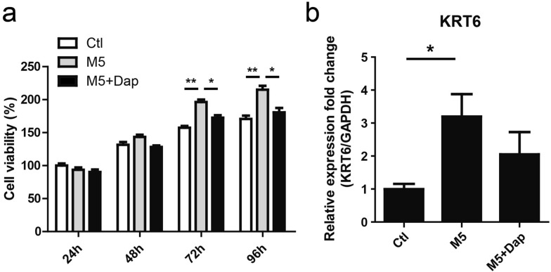
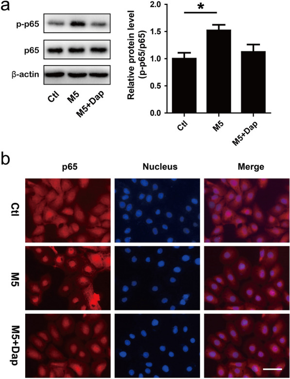
Results: Daphnetin attenuated M5-induced hyperproliferation in HaCaT keratinocytes. M5 stimulation significantly upregulated mRNA levels of IL-1β, IL-6, IL-8, TNF-α, IL-23A and MCP-1. However, daphnetin treatment partially attenuated the upregulation of those inflammatory cytokines. Daphnetin was found to be able to inhibit p65 phosphorylation and nuclear translocation in HaCaT keratinocytes. In addition, daphnetin significantly ameliorate the severity of skin lesion (erythema, scaling and epidermal thickness, inflammatory cell infiltration) in IMQ-induced psoriasis-like mouse model. Daphnetin treatment attenuated IMQ-induced upregulation of inflammatory cytokines including IL-6, IL-23A and IL-17A in skin lesion of mice.

Conclusions: Daphnetin was able to attenuate proliferation and inflammatory response induced by M5 in HaCaT keratinocytes through suppression of NF-κB signaling pathway. Daphnetin could ameliorate the severity of skin lesion and improve inflammation status in IMQ-induced psoriasis-like mouse model. Daphnetin could be an attractive candidate for future development as an anti-psoriatic agent.

Keywords: Daphnetin; Inflammatory response; NF-κb signaling pathway; Proliferation; Psoriasis.


Conflict of interest statement

The authors declare no competing interests.

Figures

Fig. 1
Fig. 1
The effect of daphnetin on cell viability of HaCaT keratinocytes. HaCaT keratinocytes were seeded in 96-well plates and treated with different concentrations of daphnetin (0, 5, 10, 20, 40, 35, 80, 100, 200 and 300 μM) for 96 h. CCK-8 was used to measure cell viability. **P < 0.01
Fig. 2
Fig. 2
The effect of M5 on the proliferation of HaCaT keratinocytes. a HaCaT keratinocytes were seeded in 96-well plates and stimulated with M5 (0, 1, 2.5 and 5 ng/ml) for 24–96 h. CCK-8 was used to measure cell viability. *P < 0.05, M5 (0 ng/ml) group vs M5 (2.5 ng/ml) group. b, c HaCaT keratinocytes were seeded in 12-well plates and stimulated with M5 (2.5 ng/ml) for 72 h. Cells were harvested and RNA was extracted for qRT-PCR analysis of KRT6 and KRT1 mRNA levels. GAPDH served as an internal reference. *P < 0.05. **P < 0.01
Fig. 3
Fig. 3
The effect of daphnetin on the expression of inflammatory cytokines in M5-stimulated HaCaT keratinocytes. HaCaT keratinocytes were seeded in 12-well plates and treated with daphnetin (Dap, 20 μM) or not for 2 h, and subsequently stimulated with M5 cytokines (2.5 ng/ml) or not for 24 h. Cells were harvested and RNA was extracted for qRT-PCR analysis of IL-1β, IL-6, IL-8, TNF-α, IL-23A and MCP-1 mRNA levels. GAPDH served as an internal reference. *P < 0.05. **P < 0.01. DMSO (Ctl), DMSO + M5 (M5), daphnetin + M5 (M5 + Dap)
Fig. 4
Fig. 4
The effect of daphnetin on the proliferation of HaCaT keratinocytes stimulated with M5. a HaCaT keratinocytes were seeded in 96-well plates and treated with daphnetin (Dap, 20 μM) or DMSO (Ctl) for 2 h, and subsequently stimulated with M5 cytokines (2.5 ng/ml) or not for 24–96 h. CCK-8 was used to measure cell viability. *P < 0.05. **P < 0.01. b HaCaT keratinocytes were seeded in 12-well plates and treated with daphnetin (Dap, 20 μM) or DMSO for 2 h, and subsequently stimulated with M5 cytokines (2.5 ng/ml) or not for 72 h. Cells were harvested and RNA was extracted for qRT-PCR analysis of KRT6 mRNA level. GAPDH served as an internal reference. *P < 0.05. DMSO (Ctl), DMSO + M5 (M5), daphnetin + M5 (M5 + Dap)
Fig. 5
Fig. 5
The effects of daphnetin on p65 phosphorylation and nuclear translocation in M5-stimulated HaCaT keratinocytes. HaCaT keratinocytes were treated with daphnetin (Dap, 20 μM) for 24 h, and then stimulated with M5 (5 ng/ml) for 30 min. a Cells were harvested and total protein was extracted for Western blotting assay to detect p65 and p-p65 protein levels. β-actin served as an internal reference. *P < 0.05. b Cells were fixed and IFA was carried out to detect p65 (red) distribution. Nuclei were stained with Hoechst dye 33,258 (blue). Bar, 50 μm. DMSO (Ctl), DMSO + M5 (M5), daphnetin + M5 (M5 + Dap)
Fig. 6
Fig. 6
The effect of daphnetin on skin lesion in IMQ-induced psoriasis-like mouse model. a The macroscopic appearance of mouse back skin on day 8. b Erythema and (c) scaling was scored on days 0, 2, 4, and 7 based on the PASI. d H&E staining of the mouse skin. Bar, 100 μm. e Epidermal thickness was calculated by Image-pro Plus 6.0 software. ***P < 0.001. f qRT-PCR was performed to measure the expression of KRT6 in skin lesion. GAPDH served as an internal reference. *P < 0.05. **P < 0.01. g Dermal cellular infiltrates were quantitated with H&E staining images. *P < 0.05. ***P < 0.001
Fig. 7
Fig. 7
The effect of daphnetin on the expression of inflammatory cytokines in IMQ-induced psoriasis-like skin lesion. qRT-PCR was performed to measure the expression of IL-6, TNF-α, IL-23A and IL-17A in skin lesion. GAPDH served as an internal reference. *P < 0.05. **P < 0.01
Fig. 8
Fig. 8
The diagram depicting the experimental design for animal treatment

Similar articles

Cited by

References

    1. Nestle FO, Kaplan DH, Barker J. Psoriasis. New Engl J Med. 2009;361:496–509. doi: 10.1056/NEJMra0804595. - DOI - PubMed
    1. Boehncke WH, Schön MP. Psoriasis. Lancet. 2015;386:983–994. doi: 10.1016/S0140-6736(14)61909-7. - DOI - PubMed
    1. Hawkes JE, Chan TC, Krueger JG. Psoriasis pathogenesis and the development of novel targeted immune therapies. J Allergy Clin Immunol. 2017;140:645–653. doi: 10.1016/j.jaci.2017.07.004. - DOI - PMC - PubMed
    1. Albanesi C, De Pità O, Girolomoni G. Resident skin cells in psoriasis: a special look at the pathogenetic functions of keratinocytes. Clin Dermatol. 2007;25:581–588. doi: 10.1016/j.clindermatol.2007.08.013. - DOI - PubMed
    1. Albanesi C, Scarponi C, Giustizieri ML, Girolomoni G. Keratinocytes in inflammatory skin diseases. Curr Drug Targets Inflamm Allergy. 2005;4:329–334. doi: 10.2174/1568010054022033. - DOI - PubMed