

Reversible splenial lesion syndrome associated with SARS-CoV-2 infection in two children
- PMID: 33082059
- PMCID: PMC7553133
- DOI: 10.1016/j.braindev.2020.10.002
Reversible splenial lesion syndrome associated with SARS-CoV-2 infection in two children
Abstract
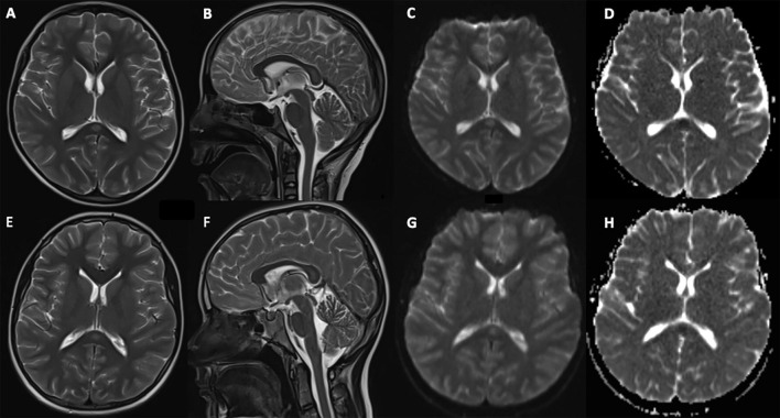
Background: Reversible splenial lesion syndrome (RESLES) is characterized by a temporary lesion in the splenium of the corpus callosum, emerging related to encephalitis, seizures, antiepileptic drug withdrawal, or metabolic disturbances. Among RESLES, mild encephalitis/encephalopathy with reversible splenial lesion (MERS) has been defined as a distinct clinicoradiologic syndrome associated with viral infections.
Case presentation: We report two children with multisystem inflammatory syndrome-children related to severe acute respiratory syndrome coronavirus 2 (SARS-CoV-2) who developed RESLES during the disease course. Encephalopathy was the main central nervous system symptom. Both of the children showed a rapid recovery, and brain magnetic resonance imaging revealed complete resolution of the splenial lesion within 1 week.
Conclusion: The complete resolution of the splenial lesion and rapid recovery from encephalopathy in RESLES associated with SARS CoV-2 were similar to observed in MERS.
Keywords: Child; Coronavirus; Corpus callosum; Encephalitis.
Copyright © 2020 The Japanese Society of Child Neurology. Published by Elsevier B.V. All rights reserved.
Figures
References
-
- World Health Organization (WHO) Coronavirus disease (COVID-19) Situation Report 2020-104 [Internet]. [cited 2020 May 7]. Available at https://www.who.int/docs/default-source/coronaviruse/situation-reports/2....
-
- World Health Organization (WHO) Coronavirus Disease (COVID-19) Dashboard-Turkey [Internet]. [cited 2020 Sep 7]. Avaliable at https://covid19.who.int/region/euro/country/tr.
Publication types
MeSH terms
Substances
Supplementary concepts
LinkOut - more resources
Full Text Sources
Other Literature Sources
Medical
Miscellaneous
