. 2021 Jan;19(1):99-111.
doi: 10.1158/1541-7786.MCR-20-0169. Epub 2020 Oct 20.

The Relative Expression of ERα Isoforms ERα66 and ERα36 Controls the Cellular Response to 24R,25-Dihydroxyvitamin D3 in Breast Cancer

Affiliations

The Relative Expression of ERα Isoforms ERα66 and ERα36 Controls the Cellular Response to 24R,25-Dihydroxyvitamin D3 in Breast Cancer

Anjali Verma et al. Mol Cancer Res. 2021 Jan.

Abstract

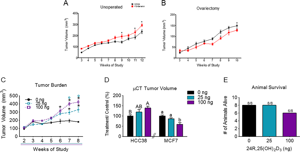
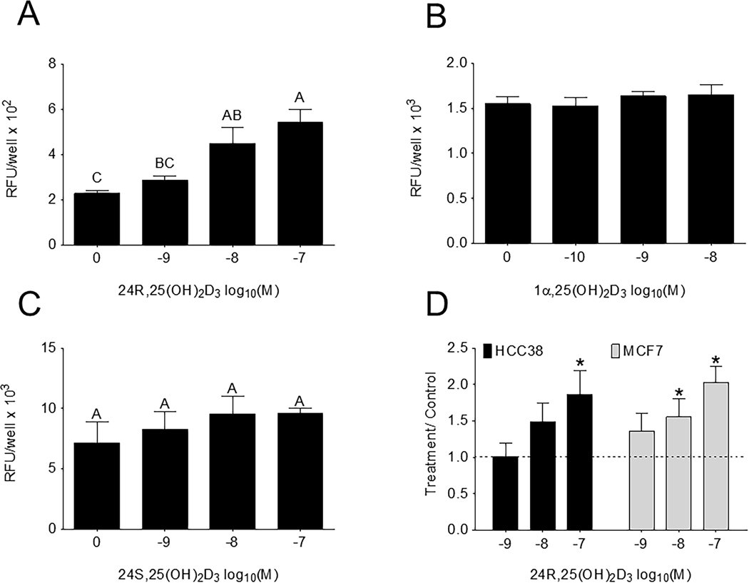
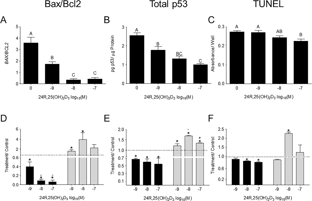
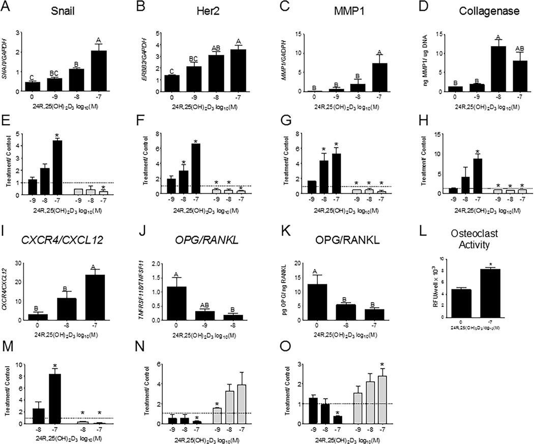
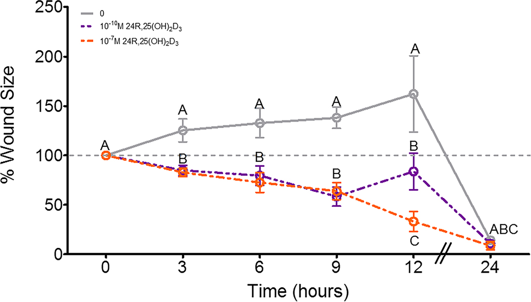
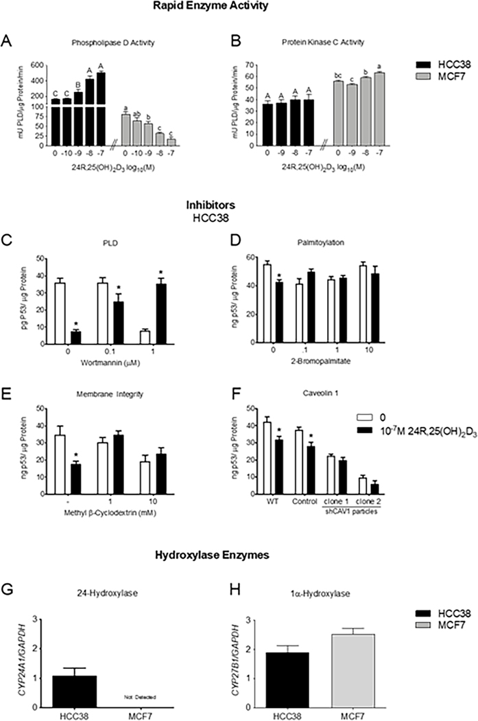
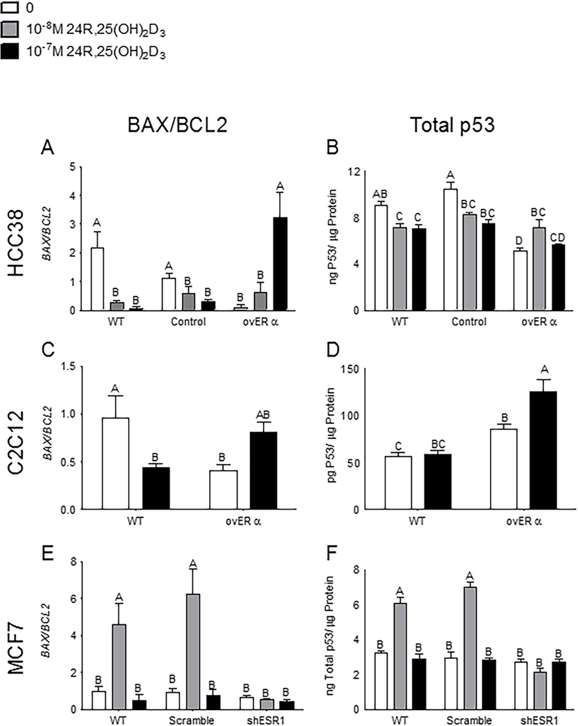
Vitamin D3 and its metabolites have antitumorigenic properties in vitro and in vivo; however, clinical trials and retrospective studies on the effectiveness of vitamin D3 oral supplementation against cancer have been inconclusive. One reason for this may be that clinical trials ignore the complex vitamin D metabolome and the many active vitamin D3 metabolites present in the body. Recent work by our lab showed that 24R,25(OH)2D3, a vitamin D3 metabolite that is active in chondrocyte proliferation and differentiation, has antitumorigenic properties in estrogen receptor alpha-66 (ERα66)-positive (ER+) breast cancer, but not in ERα66-negative (ER-) breast cancer. Here we show that 24R,25(OH)2D3 is protumorigenic in an in vivo mouse model (NOD.Cg-PrkdcscidIl2rgtm1Wjl /SzJ (NSG) mice) of ER- breast cancer, causing greater tumor growth than in mice treated with vehicle alone. In vitro results indicate that the effect of 24R,25(OH)2D3 is via a membrane-associated mechanism involving ERs and phospholipase D. 24R,25(OH)2D3 increased proliferation and reduced apoptosis in ERα66-negative HCC38 breast cancer cells, and stimulated expression of metastatic markers. Overexpressing ESRI, which encodes ERα66, ERα46, and ERα36, reduced the proapoptotic response of ERα66- cells to 24R,25(OH)2D3, possibly by upregulating ERα66. Silencing ESR1 in ERα66+ cells increased apoptosis. This suggests 24R,25(OH)2D3 is differentially tumorigenic in cancers with different ERα isoform profiles. Antiapoptotic actions of 24R,25(OH)2D3 require ERα36 and proapoptotic actions require ERα66. IMPLICATIONS: These results suggest that 24R,25(OH)2D3, which is a major circulating metabolite of vitamin D, is functionally active in breast cancer and that the regulatory properties of 24R,25(OH)2D3 are dependent upon the relative expression of ERα66 and ERα36.


Conflict of interest statement

Conflicts of Interest: Nothing to disclose.

Figures

Figure 1:
Figure 1:. Breast cancer cell lines differentially respond to 24R,25(OH)2D3.
[A] Unoperated female NSG mice implanted with HCC38 xenograft mammary fat pad tumors were intraperitoneally injected with 2.5mg/kg 17β-estradiol or a vehicle twice a week for 12 weeks. Tumor burden was measured twice a week throughout the course of the study and plotted against time. [B] A similar study was done with ovariectomized NSG mice. HCC38 xenografts were treated with the same dose (2.5mg/kg) or 17β-estradiol) or a vehicle and tumors were measured twice a week until harvest at 10 weeks. The n=6 animals per group for these studies. *indicates statistical significance against week-matched vehicle tumors at α=0.05 with two-way ANOVA. [C] NSG mice with HCC38 mammary fat pad xenograft tumors were intraperitoneally injected with 25 or 100 ng of 24R,25(OH)2D3 3 times a week for 8 weeks. Tumor burden was measured with digital calipers and plotted against time. Animals given 24R,25(OH)2D3 had increased tumor burden as measured by digital calipers. *indicates significance as compared to vehicle-treated animals within the same time point. O indicates significance as compared to animals treated with 25 ng of 24R,25(OH)2D3 within the same time point. [D] Animals given 24R,25(OH)2D3 had increased tumor burden as measured by μCT as compared to tumor burden in vehicle-treated mice. Capital letters were used to indicate significance within HCC38 xenograft tumor groups, while lower-case letters were used to indicate significance within MCF7 tumor groups. Groups that do not share a letter are statistically significant. [E] After 8 weeks, 8 animals from the vehicle group, 8 animals from the low-dose (25 ng per injection) 24R,25(OH)2D3 group, and 6 animals from the high-dose (100 ng per injection) 24R,25(OH)2D3 group survived out of an original n of 8. Animals in the high-dose group did not have a statistically significant reduced survival rate as compared to animals in low dose or vehicle groups.
Figure 2:
Figure 2:. 24R,25(OH)2D3 specifically induces proliferation in HCC38 cells.
HCC38 monolayer cultures were serum-starved for 48 hours, treated with 24R,25(OH)2D3, 24S,25(OH)2D3, or 1α,25(OH)2D3 for 15 minutes and assayed 24 hours later for EdU incorporation. [A] 24R,25(OH)2D3, but not [B] 24S,25(OH)2D3 nor [C] 1α,25(OH)2D3, induced proliferation in HCC38 cell monolayers 24 hours after a 15-minute treatment. [D]. Data shown are from single representative experiments of two or more repeats and are presented as the mean ± standard error of six independent cultures per treatment group. Treatment over control was calculated as the average fold-change of treatment as compared to vehicle-treated cultures in three or more experiments and analyzed with a paired t-test against normalized vehicle controls. *indicates significance at P<0.05 as compared to vehicle-treated cultures. High-dose 24R,25(OH)2D3 induced proliferation in HCC38 and MCF7 cell monolayers 2-fold over control.
Figure 3:
Figure 3:. 24R,25(OH)2D3 prevents apoptosis in HCC38 cells, but induces apoptosis in MCF7 cells.
HCC38 monolayer cultures were treated with 24R,25(OH)2D3 for 15 minutes, and harvested 12 hours later for gene expression and 24 hours later for protein and TUNEL staining. Cultures were assessed for apoptosis as measured by [A, D] BAX/BCL2 gene expression, [B, E] total p53 protein, and [C, F] TUNEL staining. [A-C] 24R,25(OH)2D3 reduced apoptosis in HCC38 cells. Groups that share a letter are not significant at p<0.05. [D-F] HCC38 or MCF7 monolayer cultures were treated with 24R,25(OH)2D3 for 15 minutes and assessed for apoptosis 12–24 hours later. Data shown are from single representative experiments of two or more repeats and are presented as the mean ± standard error of six independent cultures per treatment group. Treatment over control was calculated as the average fold-change of treatment as compared to vehicle-treated cultures in three or more experiments and analyzed with a paired t-test against normalized vehicle controls. *indicates significance at P<0.05 as compared to vehicle-treated cultures.
Figure 4:
Figure 4:. 24R,25(OH)2D3 increases epithelial-to-mesenchymal transition and metastatic markers in HCC38 cultures.
HCC38 and MCF7 monolayer cultures were treated with 24R,25(OH)2D3 for 15 minutes and harvested 12 hours later for gene expression. Effect of dose-dependent 24R,25(OH)2D3 on gene expression of [A, E] snail, [B, F] her2, [C, G] MMP1, [D, H] collagenase protein, [I, M] CXCR4/ CXCL12 ratios, [J, N] OPG/ RANKL ratios, and [K, O] OPG/RANKL protein. In [A-D, I-L] HCC38 cells 24R,25(OH)2D3 increases these gene markers, but reduces them in MCF7 cells [E-H, M-O]. [L] 24R,25(OH)2D3 increases osteoclast activity in osteoclasts treated with HCC38 conditioned media from cultures treated with 24R,25(OH)2D3. Groups that share a letter are not significant at p<0.05. Data shown are from single representative experiments of two or more repeats and are presented as the mean ± standard error of six independent cultures per treatment group. Treatment over control was calculated as the average fold-change of treatment as compared to vehicle-treated cultures in three or more experiments and analyzed with a paired t-test against normalized vehicle controls. *indicates significance at P<0.05 as compared to vehicle-treated cultures.
Figure 5:
Figure 5:. Effect of 24R,25(OH)2D3 on cell migration in HCC38 cell monolayers.
HCC38 monolayer cultures were treated with 24R,25(OH)2D3 for 15 minutes and incubated with fresh media. A scratch was made across the culture with a 1mL pipette tip, and monolayer cultures were observed over 24 hours for wound closure. . Data shown are from single representative experiments of two or more repeats and are presented as the mean ± standard error of six independent cultures per treatment group.
Figure 6:
Figure 6:. 24R,25(OH)2D3 reduces total p53 levels through a caveolae-associated, PLD-dependent mechanism.
HCC38 or MCF7 monolayer cultures were treated with 24R,25(OH)2D3 for 15 minutes, and assessed for [A] PLD activity, which is decreased, or [B] PKC activity. In a separate set of experiments, HCC38 monolayer cultures were treated with inhibitors for 30 minutes before treatment with 24R,25(OH)2D3 for 15 minutes and assessed for total p53 content. 24R,25(OH)2D3 reduced p53 levels in HCC38 cell monolayers, but this effect was prevented by treatment with [C] wortmannin to inhibit PLD activity, [D] 2-bromopalmitate to inhibit palmitoylation of nuclear receptors trafficked to the membrane, [E] methyl beta-cyclodextrin to deplete the membrane of cholesterol, and [F] short-hairpin RNA to silence caveolin-1. In a separate set of experiments, HCC38 and MCF7 breast cancer cells were cultured to confluence and harvested after 12 hours for gene expression. Cells were assessed for [G] CYP24A1 or [H] CYP27B1 expression and normalized to GAPDH as a housekeeping gene. . Data shown are from single representative experiments of two or more repeats and are presented as the mean ± standard error of six independent cultures per treatment group.
Figure 7:
Figure 7:. Expression and silencing of ERα isoforms change the 24R,25(OH)2D3 response.
Overexpressing all ERα isoforms reversed the 24R,25(OH)2D3 anti-apoptotic effect in [A, B] HCC38 cells and [C, D] C2C12 cells. 24R,25(OH)2D3 prevented apoptosis as measured by [A] BAX/BCL2 and [B] total p53 levels in wild-type HCC38 and HCC38 cells transfected with an empty control vector, but induced apoptosis in ERα-transfected HCC38 cells. In C2C12 muscle cells, 24R,25(OH)2D3 prevented or did not induce apoptosis in wild-type cells but induced apoptosis in ERα-transfected cells as measured by [C] BAX/BCL2 and had no effect on apoptosis as measured by [D] total p53 levels in wild-type cells. Silencing the ERα66 isoform reversed the pro-apoptotic effect of 24R,25(OH)2D3 on [E, F] MCF7 cells. 24R,25(OH)2D3 induced apoptosis as measured by [E] BAX/BCL2 and [F] total p53 levels in wild-type and scramble-control MCF7 cells, but had no effect on apoptosis in ERα66-knocked down MCF7 cells. . Data shown are from single representative experiments of two or more repeats and are presented as the mean ± standard error of six independent cultures per treatment group.

Similar articles

Cited by

References

    1. Freedman DM, Dosemeci M.& McGlynn K.Sunlight and mortality from breast, ovarian, colon, prostate, and non-melanoma skin cancer: A composite death certificate based case-control study. Occup. Environ. Med 59, 257–62 (2002). - PMC - PubMed
    1. Abbas S.et al. Serum 25-hydroxyvitamin D and risk of post-menopausal breast cancer--results of a large case-control study. Carcinogenesis 29, 93–99 (2008). - PubMed
    1. Garland CF et al. The role of vitamin D in cancer prevention. Am. J. Public Health 96, 252–61 (2006). - PMC - PubMed
    1. Poole EM et al. Postdiagnosis supplement use and breast cancer prognosis in the After Breast Cancer Pooling Project. Breast Cancer Res. treatment 139, 529–37 (2013). - PMC - PubMed
    1. Scragg R.et al. Monthly high-dose vitamin D supplementation and cancer risk. JAMA Oncol. 4, e182178 (2018). - PMC - PubMed

Publication types