. 2020 Oct 20;11(5):e01612-20.
doi: 10.1128/mBio.01612-20.

A Giant Extracellular Matrix Binding Protein of Staphylococcus epidermidis Binds Surface-Immobilized Fibronectin via a Novel Mechanism

Affiliations

A Giant Extracellular Matrix Binding Protein of Staphylococcus epidermidis Binds Surface-Immobilized Fibronectin via a Novel Mechanism

Henning Büttner et al. mBio. .

Abstract

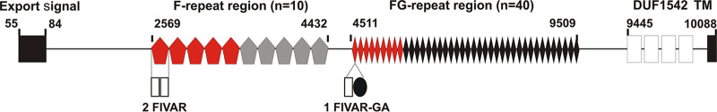
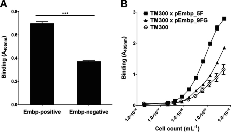
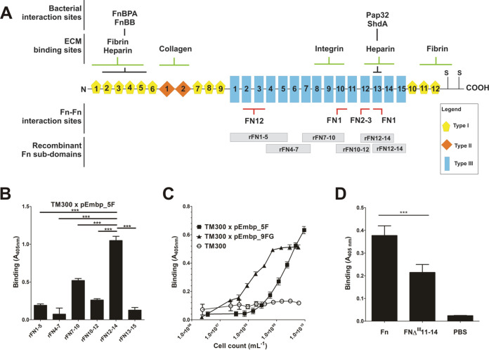
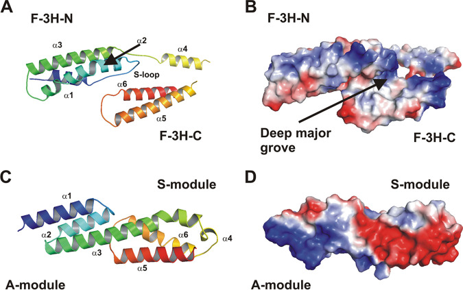
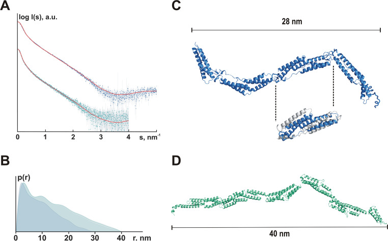
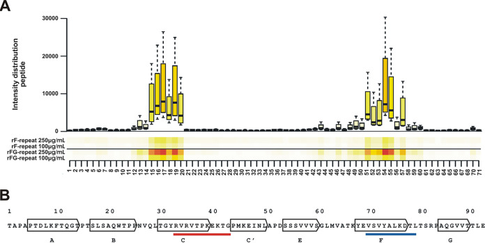
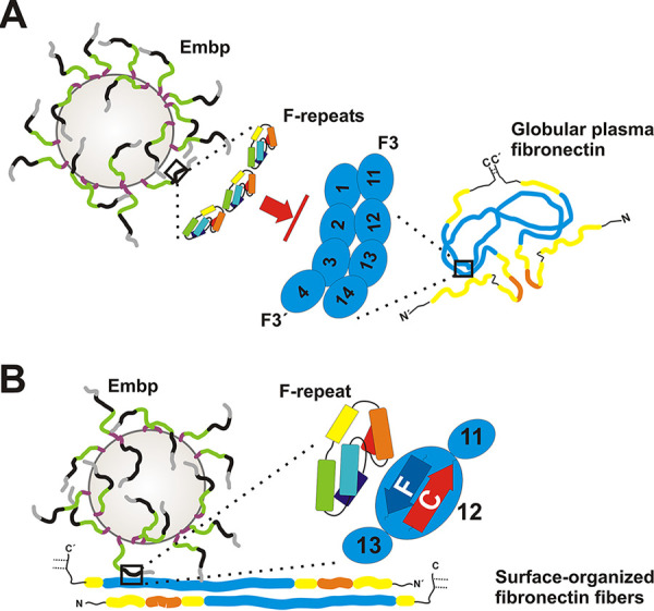
Although it is normally an innocuous part of the human skin microbiota, Staphylococcus epidermidis has emerged as a major nosocomial pathogen, and implanted foreign materials are an essential risk factor for the development of an infection. The extraordinary efficiency of S. epidermidis to colonize artificial surfaces is particularly related to the ability to form biofilms. Biofilm formation itself critically depends on stable pathogen binding to extracellular host matrix components, e.g. fibronectin (Fn), covering inserted devices in vast amounts. Extracellular matrix binding protein (Embp) and its subdomains referred to as the F-repeat and the FG-repeat are critical for adherence of S. epidermidis to surface-immobilized Fn. Embp-Fn interactions preferentially occur with surface-bound, but not folded, globular Fn via binding to the F3 domain. High-resolution structure analysis of F- and FG-repeats revealed that both repeats are composed of two tightly connected triple α-helix bundles, exhibiting an elongated but rather rigid structural organization in solution. Both F- and FG-repeat possess Fn-binding capacity via interactions with type III subdomain FN12, involving residues within the C and F β-sheet. FN12 essentially supports stability of the globular Fn state, and thus these findings reasonably explain why Embp-mediated interaction of S. epidermidis necessitates Fn surface immobilization. Thus, Embp employs an uncharacterized bacterial Fn-binding mechanism to promote staphylococcal adherence.IMPORTANCEStaphylococcus epidermidis is a leading pathogen in implant-associated hospital infections. The pathogenesis critically depends on bacterial binding to ECM components, specifically fibronectin (Fn). The cell surface-localized, 1-MDa extracellular matrix binding protein (Embp) is essentially characterized by 10 F- and 40 FG-repeats. These repetitive units, each characterized by two α-helical bundles, organize themselves in a rigid, elongated form. Embp binds preferentially to surface-localized but not soluble Fn, with both F- and FG-repeats being sufficient for Fn binding and resulting bacterial adherence. Binding preferentially involves Fn type III domain, specifically residues of FN12 β-sheets C and F. Both play key role in stabilizing the globular Fn conformation, explaining the necessity of Fn surface immobilization for a subsequent interaction with Embp. In comparison to many other bacterial Fn-binding proteins using the Fn N terminus, Embp employs a previously undescribed mechanism supporting the adhesion of S. epidermidis to surface-immobilized Fn.

Keywords: Staphylococcus; biofilms; fibronectin binding; surface proteins; surface structures.


Figures

FIG 1
FIG 1
Schematic representation of the Embp architecture. The 1-MDa Embp carries two major repetitive regions consisting of repeats each encompassing 170 and 125 amino acids (aa), respectively. The 170-aa repeat is referred to as F-repeat and is present in 10 copies (indicated by pentagons). The 125-aa repeat, referred to as FG-repeat, can be found in 40 copies (each indicated by a diamond). Previous bioinformatics analysis (22) identified 22 Found in Various Architectures (FIVAR) modules within the F-repeat region (indicated by open boxes). One F-repeat is represented by two FIVAR modules. The FG-repeat region is predicted to contain 38 G-related Albumine binding (GA) modules (indicated by a filled circle), each associated with one FIVAR module. Each FG-repeat represents a pair of GA and FIVAR modules. Experimental evidence demonstrates that the predicted modular architecture does not match the actual structural organization derived from X-ray crystallography (Fig. 5). An N-terminal export signal containing an YSIRK motif, C-terminal domains of unknown function (DUF1542) and a putative transmembrane region (TM) are bioinformatics predictions. Pentagons and diamonds filled red indicate five F-repeats and nine FG-repeats that were fused to the export signal and the putative cell wall binding region for in trans expression in staphylococci. Upper numbers indicate amino acid positions referring to the Embp amino acid sequence.
FIG 2
FIG 2
Recruitment of soluble fibronectin to Embp- or FnBPA-expressing S. epidermidis. S. epidermidis 1585Δembp, 1585Δembp × pFNBA4, and 1585Pxyl/tetembp were incubated with fibronectin. After washing, cell surface-localized Fn was detected using rabbit anti-Fn IgG and Alexa 488-conjugated anti-rabbit IgG. Bacteria were stained with DAPI (white) and Fn (green). While S. epidermidis 1585Δembp × pFNBA4 is able to recruit soluble Fn to the cell surface indicated by green fluorescence signal (C), no Fn is detected on the surface of Embp expressing 1585Pxyl/tetembp (A); S. epidermidis 1585Δembp served as a negative control (B). Scale bar, 5 μm.
FIG 3
FIG 3
Adherence of Embp-producing staphylococci to full-length Fn. A 96-well microtiter plate coated with 10-μg/ml full-length Fn was incubated for 1 h with staphylococci. After washing, bacteria were indirectly quantified by a colorimetric reaction. (A) Comparison of the Fn adherence of S. epidermidis 1585 and its isogenic embp deletion mutant grown under embp inducing conditions (50) shows a clear reduction in the bacterial load, although background binding is still observed that might be attributed to Embp-independent cell surface structures. (B) Binding of S. carnosus TM300 × pEmbp_5F and S. carnosus TM300 × pEmbp_9FG (expressing five F- or nine FG-repeats, respectively) to surface-immobilized Fn. Both, F- and FG-repeats support binding to Fn. Wild-type S. carnosus TM300 served as a control. ***, P < 0.0001 (Student t test).
FIG 4
FIG 4
(A) Schematic representation of the cellular fibronectin monomer. Fibronectin is a 250-kDa multidomain glycoprotein found in various body fluids and various tissues. The type I domain (yellow pentagons) consist of 12 repeats of about 40 aa. Repeats F16 and F17 are intersected by two type II repeats (F21 and F22, each consisting of 60 aa; orange rhomboid), forming the type II domain. In total, at least 15 Fn type III repeats (each consisting of 90 aa) form the type III domain. The secondary structure of type I and type II repeats are stabilized by disulfide bonds. Their absence in type III repeats is related to the elasticity and plasticity of the Fn type III domain. A globular Fn conformation is stabilized by several Fn domain interactions between the two strands of the Fn dimer (e.g., FN12–FN2-3, FN1–FN10, and FN1–FN13; indicated by red bars). Interaction sites with bacterial adhesins (FnBPA [S. aureus], FnBB [S. dysgalactiae], Pap32 [B. henselae], ShdA [S. enterica]), or host extracellular matrix components are indicated by black and green bars, respectively. The positions of recombinant Fn subdomains are indicated by gray boxes. S, position of cysteine residues involved in covalent Fn dimer formation. Extra domains A and B and a variable domain present in plasma Fn are not shown. The figure was adapted from Kubow et al. (46). (B) Adherence of S. carnosus expressing five F-repeats to overlapping Fn type III subdomains. Recombinant Fn type III subdomains rFN7-10, rFN4-7, rFN7-10, rFN10-12, rFN12-14, and rFN13-15 (see panel A) were immobilized on an Immobilizer microtiter plate and incubated with TM300 × pEmbp_5F (108/ml) for 1 h. The unmodified surface served as a control. Adherent bacteria were detected using a polyclonal rabbit anti-S. epidermidis serum and alkaline phosphatase-conjugated anti-rabbit IgG. Bars represent bacterial binding (expressed as the absorption at 405 nm) after background subtraction. Differences between binding to rFN12-14 were significantly different compared to all other recombinant Fn fragments tested (P < 0.0001, Student t test). (C) Adherence of S. carnosus × pEmbp_5F and S. carnosus × pEmbp_9FG to surface-immobilized rFN12-14. A 96-well Immobilizer microtiter plate surface (Nunc, Roskilde, Denmark) was coated with recombinant fibronectin subdomains. Increasing numbers of S. carnosus TM300 × pEmbp_5F and S. carnosus TM300 × pEmbp_9FG were incubated for 1 h on the surface. Adherent bacteria were detected using a polyclonal rabbit anti-S. epidermidis serum and alkaline phosphatase-conjugated anti-rabbit IgG. S. carnosus TM300 wild type served as a control. (D) Adherence of S. epidermidis 1585Pxyl/tetembp to Fn and FnΔIII11-14. Full-length Fn and an isoform lacking type III subdomains FN11 to FN14 (FnΔIII11-14) were purified from supernatants of HEK293 cells transiently transfected with FN-YPet/pHLSec2 or FNΔIII11–14/pHLSec2. Fn isoforms were immobilized on a microtiter plate and incubated with S. epidermidis 1585Pxyl/tetembp grown under embp-inducing conditions. After washing, adherent bacteria were detected using a polyclonal rabbit anti-S. epidermidis serum and alkaline phosphatase conjugated anti-rabbit IgG antibody. ***, significant (P < 0.0001) difference (Student t test).
FIG 5
FIG 5
Structural analysis of Embp. (A) Cartoon plot of the F-repeat showing the three helix-bundle arrangement. N-terminal helix bundle F-3H-N (aa 2569 to 2652) and C-terminal helix bundle F-3H-C (aa 2660–2738) are connected by a short linear linker (L2, aa 2653 to 2659). Within F-3H-N, helix α1 and α2 are connected by a Sandwich-loop (S-loop) which potentially is of fundamental importance for the structural integrity of the F-repeat as a compact unit. Helix α2 shows a remarkable almost 45° kink at residue Ala-2607. The third helix α3 (aa 2629 to 2652) follows in the opposite direction after a short loop (L1, aa 2624 to 2628). Helix α3 is slightly bended to allow a tight interaction between all three helices. Within F-3H-C, the first helix α4 (aa 2660 to 2673) is followed by the second helix α5 (aa 2685 to 2709). The connecting loop L3 (aa 2674 to 2684) is distorted and not defined by the electron density. Helix α5 (aa 2685 to 2709) and helix α6 (aa 2720 to 2738) are connected by loop L4 (aa 2710 to 2719). The model is colored according to the sequence, from blue at the N terminus to red at the C terminus. The other structural elements are assigned accordingly. (B) An electrostatic surface potential representation in the same orientation as in panel A reveals a prominent deep major groove flanked by helix α2 (from F-3H-N) and helix α4, α5, and α6 (from F-3H-C). (C) Cartoon plot of the FG-repeat showing the three helix-bundle arrangement composed of an A- and S-module. At the junction between the A- and S-module, the completely conserved residues, Leu-6798, Gln-6802, and Leu-6829 (from A-module), create a hydrophobic core with highly conserved hydrophobic residues, Met-6833, Ile-6882, and Ile-6887 (from S-module), which are further stabilized by a direct hydrogen bond between Gln-6802 and Ile-6882. Therefore, both three-helix bundles of the FG-repeat are connected rather tightly. Helix α4 is running perpendicular to all other helices and connects helix α3 and helix α5. These structural features are consistent with those described for EbhA-R7-R8 (29) from S. aureus Ebh. The model is colored according to the sequence, from blue at the N terminus to red at the C terminus. The central long helix α3 connects the A- and S-module and is colored in green. (D) An electrostatic surface potential representation in the same orientation as in panel C reveals a remarkable strong dipole character of the FG-repeat. The N-terminal region of the A-module is strongly positively charged, whereas the C-terminal region (S-module) is strongly negatively charged.
FIG 6
FIG 6
Organization of F- and FG-repeats in solution. (A) Experimental SAXS data from the Embp F-repeats construct (blue dots with error bars) and the Embp FG-repeat construct (cyan dots with error bars) fitted (red lines) with scattering computed from the rigid body models shown in Fig. S4A and B, respectively. For the F-repeats, the radius of gyration Rg estimated from Guinier approximation was 7.0 ± 0.2 nm, and the maximum dimension Dmax was 28 nm; for the FG-repeats, the Rg was 10.7 ± 0.5 nm, and the Dmax was 40 nm. (B) Pair distance distribution functions computed from the F-repeats (blue, Dmax = 28 nm) and the FG-repeats (cyan, Dmax = 40 nm) SAXS data. (C) Rigid body model of four F-repeats. Despite using several modeling approaches, it was not possible to obtain a satisfactory fit at higher angles (2 to 5 nm−1), although the overall shape resembled the respective ab initio model. Assuming that the structure of the individual F-repeats in solution was different from that in the crystal, the F-repeat structure was refined to fit the higher angle scattering data using the program SREFLEX (69). The RMSD between the original structure and the refined model (cutout: gray, F-repeat according to crystal structure analysis; blue, SREFLEX-refined F-repeat model) was 0.57 nm. RANCH (31) was then used to generate a model consisting of four repeats of the refined structure (best fit, χ2 = 2.24). Fits were computed by CRYSOL (68). (D) Rigid body model of six FG-repeats. RANCH (31) was used to generate a model consisting of six FG-repeats (best fit, χ2 = 1.07). Fits were computed by CRYSOL (68). For the rigid body model, no refinement of the 6GV5 model was required, suggesting that the crystal structure of the domain is preserved in solution.
FIG 7
FIG 7
Identification of rF- and rFG-repeat binding sites within Fn type III repeat 12. (A) One-amino-acid overlapping 10-mers were immobilized on a microchip. The surface was then probed with fluorescence labeled rF- and rFG-repeats. Both recombinant Embp fragments demonstrated binding to almost the same peptides as shown in the heatmap. (B) Mapping of rF- and rFG-binding peptides onto the amino acid sequence of FN12. Arrows indicate seven β-sheets of FN12 (A to G) (32). The red underlined sequence indicates a projection of peptides with rF- and rFG-repeat binding activity located within the C β-sheet (aa 33 to 43). The blue underlined sequence indicates a projection of peptides with rF-repeat and rFG-repeat binding activity located within the F β-sheet (aa 70 to 79). The amino acid numbering refers to the FN12 sequence, as outlined in Sharma et al. (32).
FIG 8
FIG 8
Graphical summary of Embp-mediated S. epidermidis interactions with fibronectin. S. epidermidis displays FG-repeat (green)- and F-repeat (black)-containing Embp on the cell surface of S. epidermidis (4), according to SAXS data most likely organized in elongated fibers. (A) Embp-mediated S. epidermidis interactions with globular Fn. The compact globular architecture of the soluble Fn-dimer is stabilized via intermolecular interactions, essentially involving FN2-3 of one Fn molecule (zoom-in right; F3′) and FN12 of the second molecule (zoom-in right, F3) (33). F-repeats (zoom-in left) or FG-repeats (not shown) possess Fn-binding activity through interactions with FN12. F-repeat and FG-repeat binding sites in FN12 are blocked by intramolecular Fn-Fn interactions, preventing Embp-mediated binding to globular Fn and its recruitment to the bacterial cell surface. (B) Embp-mediated S. epidermidis interactions with immobilized Fn. During surface deposition, Fn dimers become elongated by resolving intramolecular interactions and additional structural rearrangements of the F3 domain (73). As a consequence, F-repeat and FG-repeat binding sites within FN12 become accessible (i.e., within β-strands C and F; zoom-in), thus allowing S. epidermidis to adhere to Fn-conditioned surfaces. This process is fostered by additional, as-yet-uncharacterized S. epidermidis interactions with Fn. The figure is not drawn to scale.

Similar articles

Cited by

References

    1. Otto M. 2009. Staphylococcus epidermidis the “accidental” pathogen. Nat Rev Microbiol 7:555–567. doi:10.1038/nrmicro2182. - DOI - PMC - PubMed
    1. Spanu T, Sanguinetti M, Ciccaglione D, D’Inzeo T, Romano L, Leone F, Fadda G. 2003. Use of the VITEK 2 system for rapid identification of clinical isolates of staphylococci from bloodstream infections. J Clin Microbiol 41:4259–4263. doi:10.1128/jcm.41.9.4259-4263.2003. - DOI - PMC - PubMed
    1. Becker K, Both A, Weisselberg S, Heilmann C, Rohde H. 2020. Emergence of coagulase-negative staphylococci. Expert Rev Anti Infect Ther 18:349–366. doi:10.1080/14787210.2020.1730813. - DOI - PubMed
    1. Schommer NN, Christner M, Hentschke M, Ruckdeschel K, Aepfelbacher M, Rohde H. 2011. Staphylococcus epidermidis uses distinct mechanisms of biofilm formation to interfere with phagocytosis and activation of mouse macrophage-like cells 774A.1. Infect Immun 79:2267–2276. doi:10.1128/IAI.01142-10. - DOI - PMC - PubMed
    1. O’Toole G, Kaplan HB, Kolter R. 2000. Biofilm formation as microbial development. Annu Rev Microbiol 54:49–79. doi:10.1146/annurev.micro.54.1.49. - DOI - PubMed

Publication types

MeSH terms

Substances

LinkOut - more resources