. 2020 Oct 20;11(5):e02092-20.
doi: 10.1128/mBio.02092-20.

The ZIP Code of Vesicle Trafficking in Apicomplexa: SEC1/Munc18 and SNARE Proteins

Affiliations

The ZIP Code of Vesicle Trafficking in Apicomplexa: SEC1/Munc18 and SNARE Proteins

Hugo Bisio et al. mBio. .

Abstract

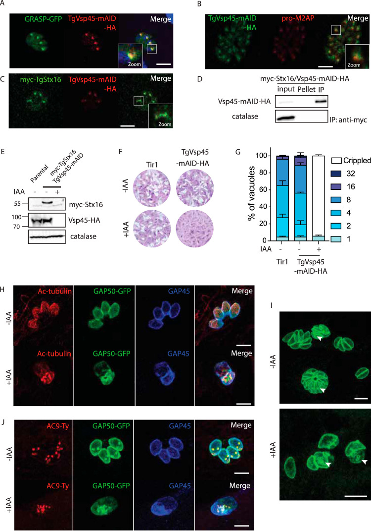
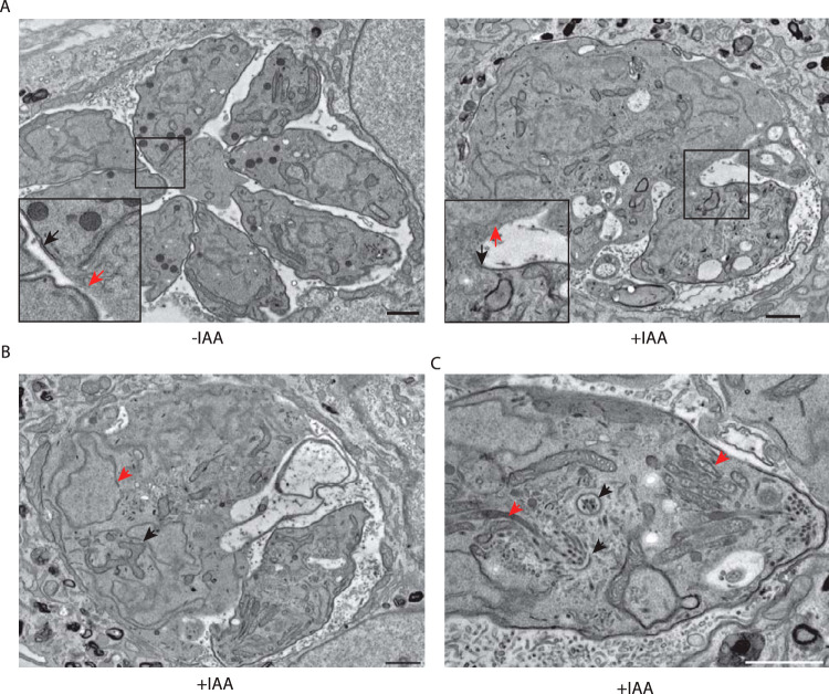
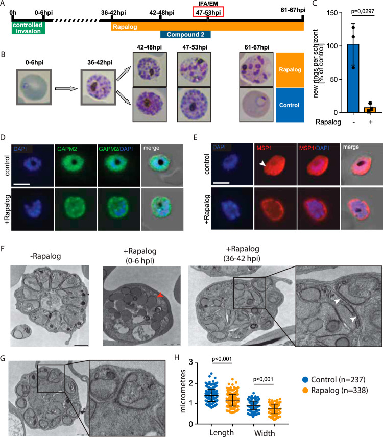
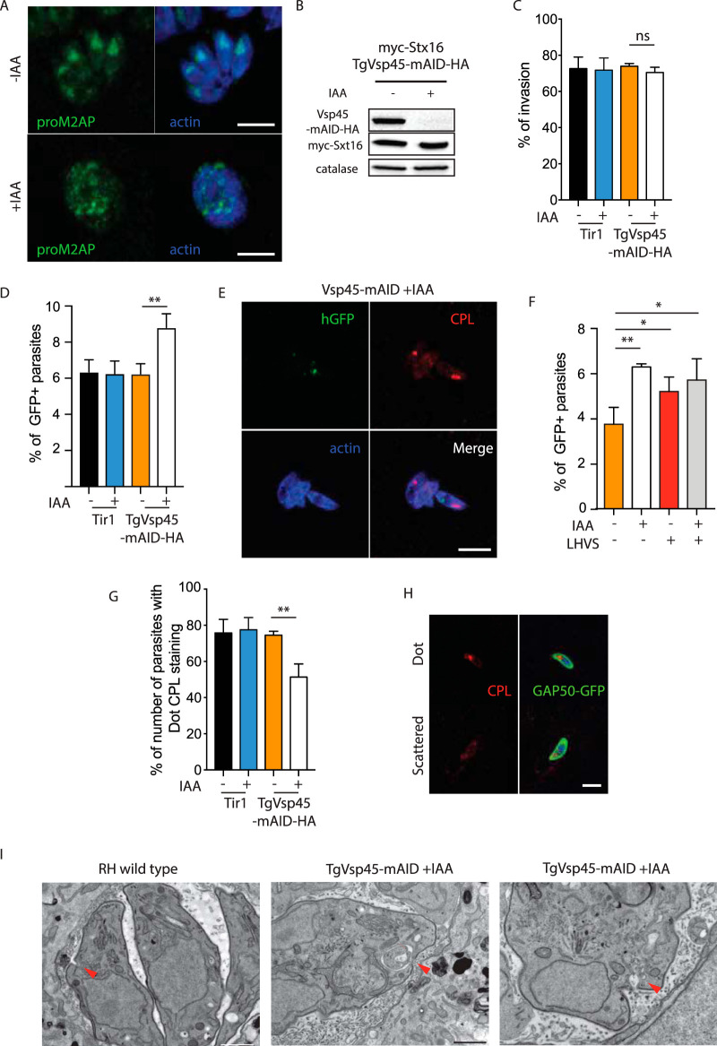
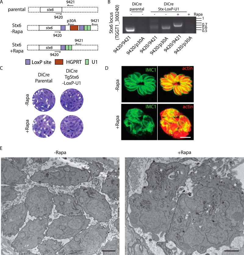
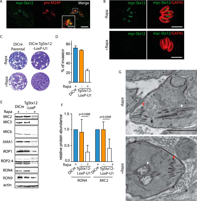
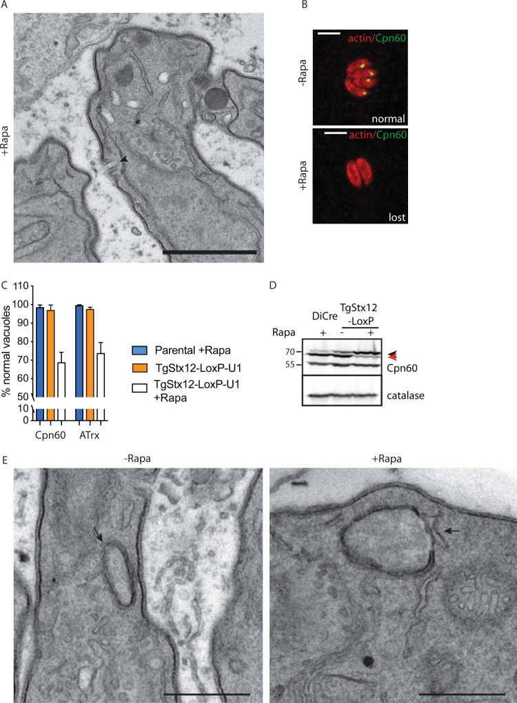
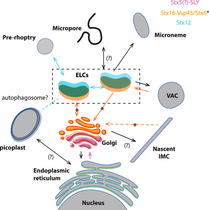
Apicomplexans are obligate intracellular parasites harboring three sets of unique secretory organelles termed micronemes, rhoptries, and dense granules that are dedicated to the establishment of infection in the host cell. Apicomplexans rely on the endolysosomal system to generate the secretory organelles and to ingest and digest host cell proteins. These parasites also possess a metabolically relevant secondary endosymbiotic organelle, the apicoplast, which relies on vesicular trafficking for correct incorporation of nuclear-encoded proteins into the organelle. Here, we demonstrate that the trafficking and destination of vesicles to the unique and specialized parasite compartments depend on SNARE proteins that interact with tethering factors. Specifically, all secreted proteins depend on the function of SLY1 at the Golgi. In addition to a critical role in trafficking of endocytosed host proteins, TgVps45 is implicated in the biogenesis of the inner membrane complex (alveoli) in both Toxoplasma gondii and Plasmodium falciparum, likely acting in a coordinated manner with Stx16 and Stx6. Finally, Stx12 localizes to the endosomal-like compartment and is involved in the trafficking of proteins to the apical secretory organelles rhoptries and micronemes as well as to the apicoplast.IMPORTANCE The phylum of Apicomplexa groups medically relevant parasites such as those responsible for malaria and toxoplasmosis. As members of the Alveolata superphylum, these protozoans possess specialized organelles in addition to those found in all members of the eukaryotic kingdom. Vesicular trafficking is the major route of communication between membranous organelles. Neither the molecular mechanism that allows communication between organelles nor the vesicular fusion events that underlie it are completely understood in Apicomplexa. Here, we assessed the function of SEC1/Munc18 and SNARE proteins to identify factors involved in the trafficking of vesicles between these various organelles. We show that SEC1/Munc18 in interaction with SNARE proteins allows targeting of vesicles to the inner membrane complex, prerhoptries, micronemes, apicoplast, and vacuolar compartment from the endoplasmic reticulum, Golgi apparatus, or endosomal-like compartment. These data provide an exciting look at the "ZIP code" of vesicular trafficking in apicomplexans, essential for precise organelle biogenesis, homeostasis, and inheritance.

Keywords: Apicomplexa; Plasmodium falciparum; SEC1/Munc18; SNARE; Toxoplasma gondii; Vps45; apicomplexan parasites; apicoplast; exocytosis; inner membrane complex; membrane fusion; microneme; pellicle; rhoptry; small GTPases; syntaxin.


Figures

FIG 1
FIG 1
Toxoplasma gondii SLY mediates ER to Golgi vesicle transport. (A) Indirect immunofluorescence assay (IFA) showing the endogenously C-terminally tagged SLY localized to the Golgi. GRASP-GFP, parasite Golgi marker. (B) Western blot showing auxin (IAA)-induced SLY-mAID-HA degradation in intracellular parasites within 2 hours. Catalase, loading control. (C) Plaque assay of the TgSLY-mAID-HA and Tir1 parental strain in the presence or absence of IAA. TgSLY-mAID-HA displays a severe defect in the lytic cycle in the presence of IAA. (D) Parasites lacking TgSLY are impaired in intracellular replication. Error bars represent the ± standard deviation (SD) from three independent experiments. (E) IFA of TgSLY-mAID-HA parasites ± IAA (24 hours). GRASP-GFP failed to localize to the Golgi upon TgSLY degradation. GAP45, marker of the parasite pellicle. Parasites were allowed to grow for 24 hours in the absence of IAA and incubated with IAA for another 24 hours. (F) Electron micrographs reveal severe morphological defects of the Golgi apparatus and endoplasmic reticulum in SLY1-iKD parasites (marked with asterisk) upon auxin treatment (+IAA) compared to the typical Golgi and ER marked with arrowheads in the control (–IAA). Some organelles are highlighted as a, apicoplast; c, conoid; n, nucleus; m, mitochondrion; and r, rhoptry. Scale bars = 1 μm. (G) IFA of TgSLY-mAID-HA parasites ± IAA (24 hours). Trafficking of the GPI-anchored SAG1 protein to the parasite plasma membrane is affected in the absence of TgSLY. GAP45, marker of the parasite pellicle. Parasites were allowed to grow for 24 hours in the absence of IAA and incubated with IAA for another 24 hours. (H) IFA of TgSLY-mAID-HA parasites ± IAA (24 hours). Trafficking of the microneme localized MIC2 protein is affected in the absence of SLY. GAP45, marker of the parasite pellicle. Parasites were allowed to grow for 24 hours in the absence of IAA and incubated with IAA for another 24 hours. (I) IFA of TgSLY-mAID-HA parasites ± IAA (24 hours). Trafficking of GRA1 protein to the dense granules is affected in the absence of TgSLY. GAP45, marker of the parasite pellicle. Parasites were allowed to grow for 24 hours in the absence of IAA and incubated with IAA for another 24 hours. (J) IFA of TgSLY-mAID-HA parasites ± IAA (24 hours). Trafficking of Cpn60 protein to the apicoplast is not affected in the absence of TgSLY. Actin, parasite cytosol. Parasites were allowed to grow for 24 hours in the absence of IAA and incubated with IAA for another 24 hours. Scale bars for all IFA = 7 μm.
FIG 2
FIG 2
Vsp45 is implicated in the IMC formation of Toxoplasma gondii (A) C-terminal epitope tagging of TgVsp45 at the endogenous locus partially colocalizes with the Golgi marker GRASP-GFP. (B) C-terminal epitope tagging of Vsp45 at the endogenous locus partially colocalizes with the ELC marker proM2AP. (C) IFA showing the colocalization of the C-terminal tagged Vsp45-HA and N-terminal tagged myc-Stx16. (D) Vsp45-mAID-Ha coimmunoprecipitates with myc-Stx16. (E) Vps45 knockdown leads to a decrease in N-terminal tagged myc-Stx16 protein steady-state levels as shown by the Western blot. Catalase is used as loading control. (F) Plaque assay of TgVps45-mAID-HA and the Tir1 parental strain in the presence or absence of IAA. TgVps45-mAID-HA knockdown but not the parental Tir1 strain displays a severe growth defect in the presence of IAA. (G) TgVps45-mAID-HA knockdown ± IAA. Parasite morphology is grossly affected following depletion of Vps45. Error bars represent the ± SD for three independent experiments. (H) IFA of VspVps45-mAID-HA parasites ± IAA (24 hours). GAP50-GFP and subpellicular microtubules are not formed in the absence of Vps45. GAP50, IMC; GAP45, parasite periphery. (I) IFA of Vsp45-mAID-HA parasites ± IAA (24 hours). Parasites depleted in Vsp45 exhibit an asynchronous block in the IMC formation. GAP50-GFP, IMC marker. (J) IFA of TgVps45-mAID-HA parasites ± IAA (24 hours). Conoid biogenesis is not impaired in Vps45-depleted parasites. Apical cap, AC9-Ty. GAP50, IMC; GAP45, parasite periphery. Scale bars for all IFA = 7 μm.
FIG 3
FIG 3
Ultrastructure of TgVps45-depleted parasites showed perturbations in IMC biogenesis. (A) Electron micrographs reveal severe morphological defects in Vps45-iKD parasites associated with a specific lack of newly formed IMC. Regions of the plasma membrane that lack association with the IMC are indicated with red arrows, while mature pellicle is indicated with black arrows. (B) Electron microscopy confirms correct karyokinesis (red arrowhead) and normal mitochondrion (black arrowhead). (C) Electron microscopy reveals correct conoid biogenesis (black arrowheads) and rhoptry docking (red arrowheads) in the absence of Vps45. Scale bars = 1 μm.
FIG 4
FIG 4
PfVps45 plays a crucial role in IMC biogenesis. (A) Schematic illustration of the experiment to conditionally inactivate PfVPS45 in schizonts. (B) Giemsa smears demonstrate that parasites lacking functional PfVps45 are arrested at the schizont stage upon induction of knock sideways (rapalog) at 36 to 42 hpi. (C) Quantification of newly formed rings per schizont after the removal of compound 2. Parasites ± rapalog (25 hours). Bars show the SD for three independent experiments. (D) IFA of PfVps45 KS parasites ± rapalog (11 hours). IMC biogenesis is inhibited in parasites when Vps45 is mis-localized. GAPM2, parasite IMC; DAPI, nuclei. Scale bar = 5 μm. (E) IFA of PfVps45 KS parasites ± rapalog (11 hours). Schizont plasma membrane fails to engulf merozoites when Vps45 is mis-localized. MSP1, parasite plasma membrane; DAPI, nuclei. An arrowhead points to fully formed merozoites with engulfed plasma membrane in wild-type conditions. Scale bar = 5 μm. (F) Electron micrographs (TEM) of ± rapalog-treated Vps45 KS parasites (47 to 53 hpi). Early treatment at the ring stage (0 to 6 hpi) with rapalog leads to arrest of the parasites in the trophozoite stage. Later treatment at the early schizont stage (36 to 42 hpi) leads to a partial IMC biogenesis defect. IMC biogenesis initiates but failed to elongate (right inset). White arrowheads show IMC, while the red arrowhead shows endocytic vesicles. Scale bars = 1 μm. (G) Schizonts formed in the presence of rapalog possess an enlarged residual body and smaller merozoites (right inset). Scale bar = 1 μm. (H) Quantification of the size of merozoites formed in the presence or absence of rapalog. Bars show the SD.
FIG 5
FIG 5
TgVps45 is essential for ingestion and digestion of cytosolic host proteins. (A) IFA of TgVps45-mAID-HA parasites ± IAA (24 hours). ELC homeostasis is grossly affected in the absence of TgVps45. proM2AP, ELC; actin, parasite cytosol. (B) IAA-induced TgVsp45-mAID-HA degradation in extracellular parasites within 2 hours. Catalase, loading control. (C) Parasites lacking TgVps45 are not impaired in invasion. Error bars represent the ± SD for three independent experiments. (D) Quantification of ingestion of host cytosolic GFP 7 minutes postinfection in WT or Vps45-mAID-HA knockdown ± IAA parasites. The percentages of GFP-positive tachyzoites from three independent experiments are shown. (E) IFA of Vps45-mAID-HA parasites + IAA (30 minutes postinfection of GFP-positive host cells). Colocalization of ingested GFP with the VAC was rarely observed. CPL, VAC; actin, parasite cytosol. (F) Quantification of ingestion of host cytosolic GFP 30 minutes postinfection in Vps45-mAID-HA knockdown ± IAA parasites ± LHVS. The percentages of GFP-positive tachyzoites from three independent experiments are shown. (G to H) The VAC morphology is dynamic and can appear as a single dot or a scattered pattern. The morphology of VAC is affected following depletion of TgVps45 (G). Error bars represent the ± SD for three independent experiments. Representative images are shown in panel H. CPL, VAC; GAP50, parasite IMC. (I) Electron microscopy reveals an enlarged micropore in Vps45 conditional knockdown (cKD) parasites. Arrowhead, micropore. Scale bars = 1 μm. Scale bars for all IFA = 7 μm.
FIG 6
FIG 6
TgStx6 is essential for ELC trafficking. (A) Schematic representation of the strategy used to generate the recombinant TgStx6 strain. (B) PCR demonstrates correct integration and excision upon rapamycin treatment. 9323/9324, 459 bp (wild-type locus); 9323/p30A, 531 bp; 9323/9324, 674 bp (Cre excised locus). (C) Stx6-LoxP-U1 cKD but not the parental DiCre strain displays a severe growth defect in the presence of rapamycin. (D) IFA of TgStx6-LoxP-U1 cKD ± rapamycin. Parasite morphology is grossly affected following depletion of TgStx6. IMC1, inner membrane complex; actin, parasite cytosol. Scale bars = 7 μm. (E) Electron microscopy reveals gross morphological defects in Stx6 cKD parasites associated with a specific lack of newly formed IMC. Scale bars = 1 μm.
FIG 7
FIG 7
TgStx12 is essential for microneme and rhoptry function. (A) Indirect immunofluorescence assay (IFA) showing the endogenously N-terminally tagged Stx12 localized to the ELC. ProM2AP, parasite ELC marker. (B) Rapamycin-induced Stx12 downregulation in intracellular parasites within 24 hours. GAP45, parasite pellicle. (C) The Stx12-LoxP-U1 knockdown but not the parental DiCre strain displays a severe growth defect in the presence of rapamycin. (D) Parasites lacking Stx12 show a significantly impaired invasion capacity. Error bars represent the ± SD for three independent experiments. (E) The content of microneme and rhoptry localized proteins is reduced upon depletion of Stx12. Catalase, loading control. (F) The quantification of RON4 and MIC2 are shown. Error bars represent the ± SD for three independent experiments. (G) Electron microscopy reveals no gross morphological defects of micronemes and mild abnormalities of rhoptries in parasites depleted in Stx12. Micronemes and rhoptries are indicated with red and white arrowheads, respectively. Scale bars = 1 μm. Scale bars for all IFA = 7 μm.
FIG 8
FIG 8
TgStx12 is essential for protein transport into the apicoplast. (A) Electron microscopy reveals no gross morphological defects of the micropore (indicated with an arrowhead) in parasites depleted in Stx12. (B) Apicoplast is shown to be lost in a subpopulation of Stx12 cKD parasites upon 72 hours of treatment with rapamycin. Representative images are shown. Cpn60 or ATrx were used as apicoplast markers. Scale bars = 7 μm. (C) Quantification of apicoplast upon depletion of Stx12 for 72 hours with rapamycin. Error bars represent the ± SD for three independent experiments. (D) Pro-Cpn60 (marked with a black arrowhead) accumulates upon depletion of Stx12 for 72 hours with rapamycin. Processed Cpn60 is marked with a read arrowhead. (E) Electron microscopy reveals no gross morphological defects of the apicoplast (indicated with an arrow) in parasites depleted in Stx12. Scale bars in A and E = 1 μm.
FIG 9
FIG 9
Model of vesicle trafficking in apicomplexan parasites. The model presented in this figure represents a combined adaptation of the trafficking models presented in references and . Organelles are colored depending on the localization of proteins studied here. Putative fusion events involving these proteins are indicated with colors in the arrows. The directionality of the transport has been deducted considering that q-SNARES usually localize on the target membranes (2, 3). Proteins transported through the secretory pathway are synthesized in the rough ER. Vesicles shed from the ER are transported and fuse with the cis-Golgi, likely through the action of SLY1. Stx5 is likely an interactor of SLY1, as demonstrated in model organisms (64), mediating the fusion of these vesicles. The IMC is produced from the trans-Golgi compartment, while recycling of trafficking molecules needed for IMC biogenesis likely involves Vsp45 (likely mediated by its interacting partner Stx16) and Stx6. Transport between the ELC to the trans-Golgi involves Vsp45, likely its interacting partner Stx16, and Stx6. Endocytosis is proposed to occur in the micropore or the cytostome (17–19) and transported into subcompartments of the ELC (21). SNARE proteins involved in this process are unknown. Endocytosis and VAC-mediated digestion of internalized host proteins involve Vsp45 and likely Stx16. Microneme and rhoptry proteins are transported to the proper compartment in a temporally regulated manner (21) involving the action of Stx12. Apicoplast transport from the ER is accomplished by unknown SNARE proteins. Interestingly, homeostasis of the apicoplast depends on the ELC compartment and Stx12. A putative role of auto-phagocytosis in this process remains to be addressed.

Similar articles

Cited by

References

    1. Bonifacino JS, Glick BS. 2004. The mechanisms of vesicle budding and fusion. Cell 116:153–166. doi:10.1016/s0092-8674(03)01079-1. - DOI - PubMed
    1. Koike S, Jahn R. 2019. SNAREs define targeting specificity of trafficking vesicles by combinatorial interaction with tethering factors. Nat Commun 10:1608. doi:10.1038/s41467-019-09617-9. - DOI - PMC - PubMed
    1. Sudhof TC, Rothman JE. 2009. Membrane fusion: grappling with SNARE and SM proteins. Science 323:474–477. doi:10.1126/science.1161748. - DOI - PMC - PubMed
    1. Toonen RF, Verhage M. 2003. Vesicle trafficking: pleasure and pain from SM genes. Trends Cell Biol 13:177–186. doi:10.1016/s0962-8924(03)00031-x. - DOI - PubMed
    1. Blader IJ, Coleman BI, Chen CT, Gubbels MJ. 2015. Lytic cycle of Toxoplasma gondii: 15 years later. Annu Rev Microbiol 69:463–485. doi:10.1146/annurev-micro-091014-104100. - DOI - PMC - PubMed

Publication types

MeSH terms