. 2020 Oct 20;11(5):e02431-20.
doi: 10.1128/mBio.02431-20.

Accurate Diagnosis of COVID-19 by a Novel Immunogenic Secreted SARS-CoV-2 orf8 Protein

Affiliations

Accurate Diagnosis of COVID-19 by a Novel Immunogenic Secreted SARS-CoV-2 orf8 Protein

Xiaohui Wang et al. mBio. .

Abstract

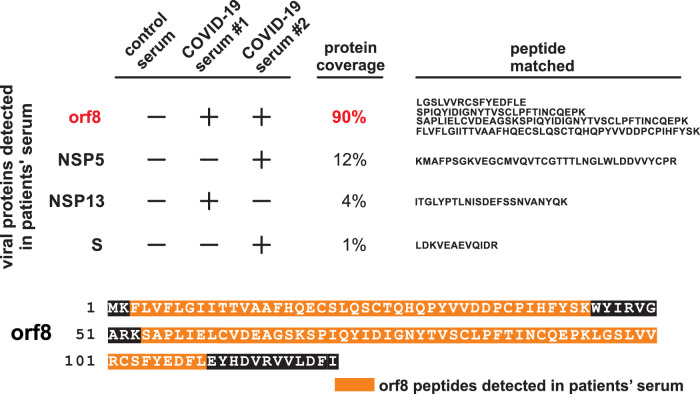
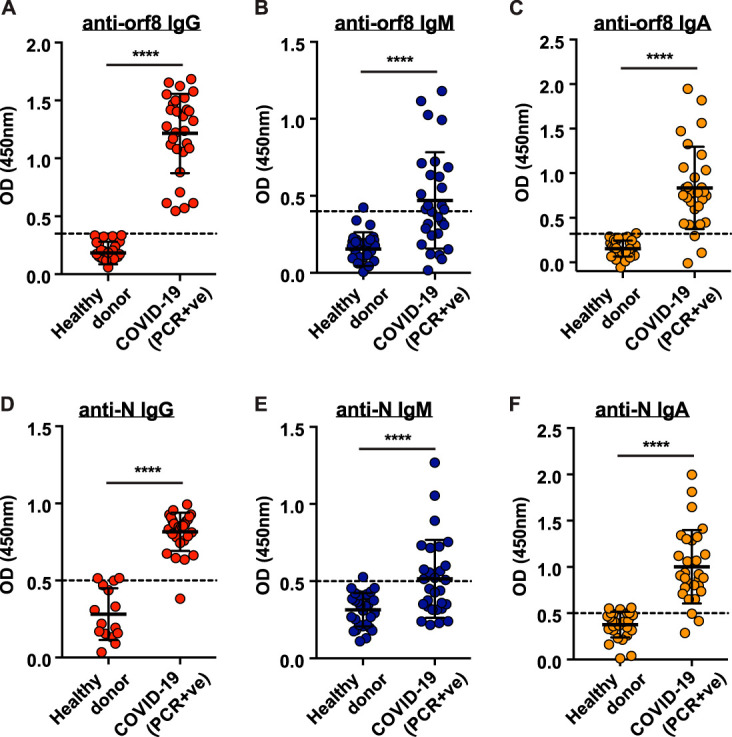
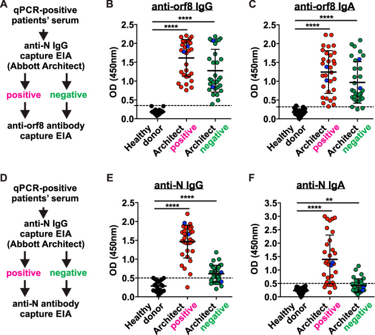
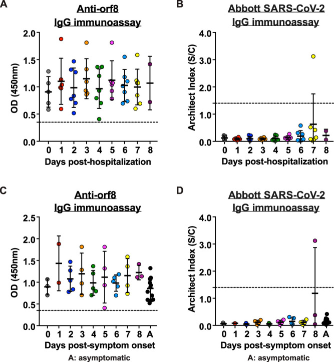
An accurate diagnostic test for early severe acute respiratory syndrome coronavirus 2 (SARS-CoV-2) infection is the key weapon to control the coronavirus disease 2019 (COVID-19) pandemic. We previously reported that the SARS-CoV-2 genome contains a unique orf8 accessory gene absent from other human-pathogenic coronaviruses. Here, we characterized the SARS-CoV-2 orf8 as a novel immunogenic secreted protein and utilized it for the accurate diagnosis of COVID-19. Extracellular orf8 protein was detected in cell culture supernatant and in sera of COVID-19 patients. In addition, orf8 was found highly immunogenic in COVID-19 patients, who showed early seropositivity for anti-orf8 IgM, IgG, and IgA. We hypothesize that orf8 secretion during SARS-CoV-2 infection facilitates early mounting of B cell response. The serological test detecting anti-orf8 IgG antibody can be used for the early and accurate diagnosis of COVID-19.IMPORTANCE Current commercially available serological tests for COVID-19 patients are detecting antibodies against SARS-CoV-2 nucleoprotein and spike glycoprotein. The antinucleoprotein and antispike antibodies can be accurately detected in patients during the mid or late stage of infection, and therefore, these assays have not been widely used for early diagnosis of COVID-19. In this study, we characterized the secretory property of a SARS-CoV-2 orf8 protein and proposed that orf8 secretion during infection facilitates early mounting of the B cell response. We demonstrated the presence of anti-orf8 antibodies in both symptomatic and asymptomatic patients during the early stage of infection, while the anti-N antibody is not detected. Our serological test detecting anti-orf8 antibodies may facilitate the development of early and accurate diagnosis for COVID-19.

Keywords: COVID-19; SARS-CoV-2; diagnosis; orf8.


Figures

FIG 1
FIG 1
Characterization of SARS-CoV-2 orf8 protein. SARS-CoV-2 orf8 protein is unique in human-pathogenic coronaviruses. (A) Phylogenetic analysis of orf8 amino acid sequences. There are three clades of orf8 sequences originating from bat SARS-like coronaviruses (clade 1 [pink]), human SARS-CoV-2 and its closely related bat SARS-like coronaviruses (clade 2 [orange]), and human SARS-CoV and its closely related paguma and bat SARS-like coronaviruses (clade 3 [green]). (B) Multiple alignment of orf8 amino acid sequences. The signal peptide (black bar) is predicted in all clade 2 orf8 sequences. Four cysteine (C25, C37, C83, and C90) sites are predicted to form two disulfide bonds. The leucine (L)-to-serine (S) amino acid substitution at site 84 of SARS-CoV-2 orf8 is highlighted in red. Predicted strands (a to g) are highlighted (green bars). (C) In silico structural prediction of mature orf8 protein. The mature orf8 starts from the amino acid position 16 (N terminus) and end at the amino acid position 121 (C terminus). The predicted structure contains seven strands (wide green arrows) and loops (orange or blue lines) which are linked by two disulfide bonds. The unknown external domain is shown in blue.
FIG 2
FIG 2
SARS-CoV-2 orf8 is actively secreted into culture supernatant. (A) Schematic diagram illustrating the method of orf8 detection in cell and culture supernatant. Plasmid DNA encoding the SARS-CoV-2 orf8 or orf9b with Flag tag was transfected into A549 and 293FT cells. At 48 h posttransfection, cell lysate (CL) and culture supernatant (SN) were harvested and orf8/orf9b proteins were immunoprecipitated (IP) and detected by Western blotting. (B and C) Orf8 is actively secreted into culture supernatant of A549 (B) and 293FT (C) cells. Secreted orf8 protein detected in culture supernatant is indicated by a red asterisk. α-Flag, anti-Flag antibody. (D) Diagram illustrating the N-terminal signal peptide mutants of orf8 used for the study of protein secretion. Three mutants include the following: (i) a signal peptide deletion mutant (ΔSP), (ii) mutation of internal hydrophobic leucine into hydrophilic arginine (mut 1), and (iii) mutation of three small uncharged amino acids near the proposed cleave sites into arginine (mut 2). (E) Plasmid DNA expressing Flag-tagged orf8 wild-type and signal peptide mutants were transfected into 293FT cells for 48 h. Intracellular orf8 (expressed in CL) and extracellular orf8 (secreted in SN) were detected as described above for panels B and C. Cellular expression of orf8 transcripts (mRNA) were detected by RT-PCR. (F) Intracellular and extracellular expression of two genotypes of orf8 were detected as described above for panels B and C. (G and H) Subcellular localization of orf8 protein. A549 cells were transfected with Flag-tagged orf8 and stained with anti-Flag (green in panels G and H), anti-α-tubulin (red in panel G), or anti-calnexin antibodies (red in panel H). The nucleus was stained and shown in blue color. (I) Z-stack visualization of A549 cells transfected with Flag-tagged orf8. Green signal represents Flag-tagged orf8, red signal represents GM130, and blue signal represents 4′,6′-diamidino-2-phenylindole (DAPI).
FIG 3
FIG 3
SARS-CoV-2 orf8 peptides found in the sera of COVID-19 patients. The orf8 peptide was abundantly detected in SARS-CoV-2-infected patient serum. Serum samples were subjected to liquid chromatography-mass spectrometry analysis. Sequence coverage of SARS-CoV-2 orf8 in the serum samples of two COVID-19 patients are shown. The peptides identified in both samples are highlighted in orange.
FIG 4
FIG 4
Abundant anti-orf8 antibodies detected in the sera of COVID-19 patients ≥28 days post-symptom onset. (A to C) Titers of antibodies against SARS-CoV2 orf8 in 29 hospitalized patients’ sera collected ≥28 days post-symptom onset and healthy control donors (n = 30) were examined by ELISA. OD, optical density; PCR+ve, PCR positive. (D to F) Serum titers of antibodies against SARS-CoV-2 nucleoprotein (N) in the patients and healthy blood donors (A to C) were examined by ELISA. Serum samples were tested at a dilution of 1:100. P values were calculated by Mann-Whitney U test. The dashed lines represent cutoff values (the mean absorbance at 450 nm of serum samples obtained from healthy control donors plus two times the standard deviation). Data were presented as means ± standard deviations (SD) (error bars). ****, P < 0.0001.
FIG 5
FIG 5
Detection of anti-orf8 IgG in anti-N IgG-negative serum samples from COVID-19 patients. Anti-orf8 antibodies are detectable in RT-qPCR-confirmed but Abbot Architect seronegative COVID-19 cases. (A to C) orf8-specific IgG and IgA in 64 serum samples from PCR-confirmed COVID-19 cases and healthy control donors (n = 20) were quantified by ELISA. The patient sera were divided into two groups: seropositive (n = 32) and seronegative (n = 32) based on the results of Abbott Architect SARS-CoV-2 N IgG assay. EIA, enzyme immunoassay. (D to F) SARS-CoV-2 N-specific IgG and IgA in serum samples in panels A and B were examined using in-house N ELISA. The dashed lines represent cutoff values. Data were presented as means ± SD. **, P < 0.01; ****, P < 0.0001.
FIG 6
FIG 6
Detection of serum anti-orf8 IgG during early hospitalization of COVID-19 patients. Detection of anti-orf8 antibodies in RT-qPCR confirmed symptomatic and asymptomatic COVID-19 patients. (A) Anti-orf8 IgG was quantitated in serial serum samples obtained from 14 RT-qPCR-confirmed COVID-19 patients. The dashed lines represent cutoff values. (B) Anti-N IgG was quantitated by Abbott Architect SARS-Cov-2 IgG assay in the same set of serum samples tested in panel A. (C and D) Data generated from panels A and B were represented as days post-symptom onset for 10 symptomatic and 4 asymptomatic COVID-19 patients. Data were statistically analyzed and presented as means ± SD.

Similar articles

Cited by

References

    1. World Health Organization. 2020. WHO coronavirus disease (COVID-19) dashboard. World Health Organization, Geneva, Switzerland: https://covid19.who.int/.
    1. Amanat F, Stadlbauer D, Strohmeier S, Nguyen THO, Chromikova V, McMahon M, Jiang K, Arunkumar GA, Jurczyszak D, Polanco J, Bermudez-Gonzalez M, Kleiner G, Aydillo T, Miorin L, Fierer DS, Lugo LA, Kojic EM, Stoever J, Liu STH, Cunningham-Rundles C, Felgner PL, Moran T, Garcia-Sastre A, Caplivski D, Cheng AC, Kedzierska K, Vapalahti O, Hepojoki JM, Simon V, Krammer F. 2020. A serological assay to detect SARS-CoV-2 seroconversion in humans. Nat Med 26:1033–1036. doi:10.1038/s41591-020-0913-5. - DOI - PMC - PubMed
    1. Zhao J, Yuan Q, Wang H, Liu W, Liao X, Su Y, Wang X, Yuan J, Li T, Li J, Qian S, Hong C, Wang F, Liu Y, Wang Z, He Q, Li Z, He B, Zhang T, Fu Y, Ge S, Liu L, Zhang J, Xia N, Zhang Z. 2020. Antibody responses to SARS-CoV-2 in patients of novel coronavirus disease 2019. Clin Infect Dis 28:ciaa344. doi:10.1093/cid/ciaa344. - DOI - PMC - PubMed
    1. To KK, Tsang OT, Leung WS, Tam AR, Wu TC, Lung DC, Yip CC, Cai JP, Chan JM, Chik TS, Lau DP, Choi CY, Chen LL, Chan WM, Chan KH, Ip JD, Ng AC, Poon RW, Luo CT, Cheng VC, Chan JF, Hung IF, Chen Z, Chen H, Yuen KY. 2020. Temporal profiles of viral load in posterior oropharyngeal saliva samples and serum antibody responses during infection by SARS-CoV-2: an observational cohort study. Lancet Infect Dis 20:565–574. doi:10.1016/S1473-3099(20)30196-1. - DOI - PMC - PubMed
    1. Reference deleted.

Publication types

MeSH terms