. 2020 Nov 27;370(6520):1089-1094.
doi: 10.1126/science.abe1502. Epub 2020 Oct 20.

Structural analysis of full-length SARS-CoV-2 spike protein from an advanced vaccine candidate

Affiliations

Structural analysis of full-length SARS-CoV-2 spike protein from an advanced vaccine candidate

Sandhya Bangaru et al. Science. .

Abstract

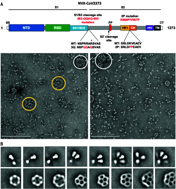
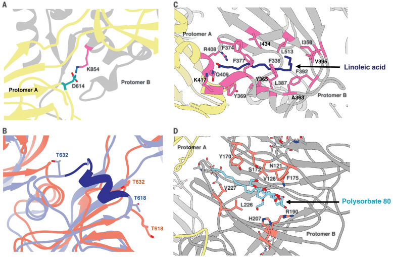
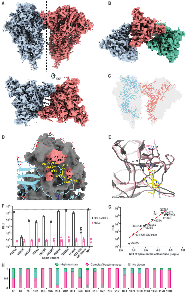
Vaccine efforts to combat the severe acute respiratory syndrome coronavirus 2 (SARS-CoV-2), which is responsible for the current coronavirus disease 2019 (COVID-19) pandemic, are focused on SARS-CoV-2 spike glycoprotein, the primary target for neutralizing antibodies. We performed cryo-election microscopy and site-specific glycan analysis of one of the leading subunit vaccine candidates from Novavax, which is based on a full-length spike protein formulated in polysorbate 80 detergent. Our studies reveal a stable prefusion conformation of the spike immunogen with slight differences in the S1 subunit compared with published spike ectodomain structures. We also observed interactions between the spike trimers, allowing formation of higher-order spike complexes. This study confirms the structural integrity of the full-length spike protein immunogen and provides a basis for interpreting immune responses to this multivalent nanoparticle immunogen.


Figures

Fig. 1
Fig. 1. Evaluation of SARS-CoV-2 3Q-2P-FL spike glycoprotein.
(A) Linear diagram of the sequence and structure elements of the FL SARS-CoV-2 spike protein showing the S1 and S2 ectodomain. Structural elements include a cleavable signal sequence (SS, white), NTD (blue), RBD (green), SD1 and SD2 (light blue), protease cleavage site 2′ (S2′, arrow), fusion peptide (FP, red), heptad repeat 1 (HR1, yellow), central helix (CH, brown), heptad repeat 2 (HR2, purple), TM domain (black), and CT (white). The native furin cleavage site was mutated (RRAR→QQAQ) to be protease resistant and stabilized by introducing two proline (2P) substitutions at positions K986P and V987P to produce SARS-CoV-2 3Q-2P-FL spike. A, Ala; D, Asp; E, Glu; K, Lys; L, Leu; N, Asn; P, Pro; Q, Gln; R, Arg; S, Ser; V, Val. (B) Representative negative-stain EM images and 2D classes of SARS-CoV-2 3Q-2P-FL, formulated in PS 80 detergent in the presence of Matrix-M adjuvant. In the raw micrograph, spike rosettes are circled in yellow and Matrix-M adjuvant cages are circled in white. 2D classes showing individual spikes, higher-order spike nanoparticles, and Matrix-M cages of different sizes. Matrix-M does not appear to interact with the spike nanoparticles.
Fig. 2
Fig. 2. Cryo-EM analysis of SARS-CoV-2 3Q-2P-FL spikes.
(A) Representative electron micrograph and 2D class averages of 3Q-2P-FL spikes showing free trimers and complexes of trimers. (B) Side and top views of the B factor–sharpened cryo-EM map of 3Q-2P-FL free trimers showing the spike in prefusion state, with the RBDs in closed conformation. The protomers are colored in blue, green, and coral for clarity. (C) Side and top view of the atomic model of free trimer represented as a ribbon diagram fit into the map density. The protomers are colored in blue, green, and coral, and the map is shown as a transparent gray density. (D) Comparison of 3Q-2P-FL spike with published structures (PDB IDs 6VXX and 6VSB) on a subunit level. PDB 6VXX is shown in cyan, PDB 6VSB in blue, and 3Q-2P-FL spike in coral.
Fig. 3
Fig. 3. Structural features of the SARS-CoV-2 3Q-2P-FL spike trimer.
(A) Interprotomeric salt-bridge interaction between D614 and K854 in 3Q-2P-FL spike trimer. (B) Comparison of the 615 to 635 loop between 3Q-2P-FL spike shown in coral and PDB 6X6P shown in blue. The residues that were built in 6X6P model but not in our model are shown in dark blue. Threonines at positions 618 and 632 flanking the gap in the 3Q-2P-FL trimer model are shown on both models to highlight their relative positions. T, Thr. (C) Linoleic acid (dark blue) binding within a hydrophobic pocket of one RBD where the fatty acid head group reaches out to interact with the closed RBD of the adjacent protomer. The interacting residues are shown in pink. F, Phe; I, Ile; Y, Tyr. (D) PS 80 detergent (blue) binding within the NTD with potential hydrogen bonding with R190 and H207. The interacting residues are shown in orange. Adjacent protomers are shown in yellow and gray in (A), (C), and (D). H, His.
Fig. 4
Fig. 4. Trimer-trimer interactions and glycan analysis.
(A) Side and top views of the sharpened cryo-EM map of 3Q-2P-FL dimers of spike trimers. Individual spike trimers are shown in blue and coral along a twofold axis of symmetry (dotted line). (B) Top view of the B factor–sharpened cryo-EM map of trimer-of-trimers complex with individual trimers colored in blue, coral, and green. (C) Ribbon representation of a protomer from one trimer (blue) interacting with the protomer from the adjacent trimer (coral) docked into the dimers-of-trimers density. (D) A close-up view of the interaction between the protomers of adjacent trimers. One protomer is shown as a ribbon diagram in blue, and its binding partner is shown as surface in gray. Residues 621-PVAIHADQ-628 in the loop with potential interactions to the neighboring NTD are colored yellow, and the residues in the NTD binding pocket are highlighted in coral. Residue D614 at the start of the loop is highlighted in dark blue. Glycosylation at residue 616 is not shown for clarity. G, Gly. (E) Changes occurring in the binding pocket in the bound state (gray) versus the free trimer (pink). Y145 and H146 switch positions to accommodate the loop better, also resulting in salt-bridge formation between H146 and D627. It also results in stacking between W152 and H146. W, Trp. (F) Pseudoviruses expressing SARS-CoV-2 WT or mutant spikes were used to infect HeLa or HeLa-ACE2 cells for 42 to 48 hours. Infection was measured by luciferase intensity RLU (relative light unit) in the lysed cells after infection. (G) Correlation between pseudovirus infection (RLU) and surface expression of SARS-CoV-2 spike variants in 293T cells measured by MFI (mean fluorescence intensity). (H) Site-specific glycan analysis of 3Q-2P-FL spike protein expressed in Sf9 insect cell line. Proportions shown for no occupancy, oligomannose, and complex or paucimannose potential N-linked glycosylation sites (PNGS) are the average and SEM of 3 to 32 distinctive peptides for each glycosite except for sites 17, 709, and 717, where only a single peptide was observed.

Update of

Similar articles

Cited by

References

    1. Cherry J. D., Krogstad P., SARS: The first pandemic of the 21st century. Pediatr. Res. 56, 1–5 (2004). 10.1203/01.PDR.0000129184.87042.FC - DOI - PMC - PubMed
    1. Zhu N., Zhang D., Wang W., Li X., Yang B., Song J., Zhao X., Huang B., Shi W., Lu R., Niu P., Zhan F., Ma X., Wang D., Xu W., Wu G., Gao G. F., Tan W., China Novel Coronavirus Investigating and Research Team , A novel coronavirus from patients with pneumonia in China, 2019. N. Engl. J. Med. 382, 727–733 (2020). 10.1056/NEJMoa2001017 - DOI - PMC - PubMed
    1. Amanat F., Krammer F., SARS-CoV-2 vaccines: Status report. Immunity 52, 583–589 (2020). 10.1016/j.immuni.2020.03.007 - DOI - PMC - PubMed
    1. Lurie N., Saville M., Hatchett R., Halton J., Developing Covid-19 vaccines at pandemic speed. N. Engl. J. Med. 382, 1969–1973 (2020). 10.1056/NEJMp2005630 - DOI - PubMed
    1. Salvatori G., Luberto L., Maffei M., Aurisicchio L., Roscilli G., Palombo F., Marra E., SARS-CoV-2 spike protein: An optimal immunological target for vaccines. J. Transl. Med. 18, 222 (2020). 10.1186/s12967-020-02392-y - DOI - PMC - PubMed

Publication types

Substances